Ultrasonographic Imaging of Ovarian Masses Surgery or Surveillance


")


 MI Total 1 2, 349 1 0. 04")


: U. S.")


- Slides: 36
Ultrasonographic Imaging of Ovarian Masses Surgery or Surveillance? Frederick R. Ueland, MD Professor and Director Division of Gynecologic Oncology University of Kentucky
Disclosure I have no financial disclosures
Wind River Range, WY July, 2017
Ovarian Tumor Overview Past § 1980’s “palpable ovary syndrome” § 2000’s observation of unilocular cysts § 2010’s observation of septate cysts Present § 10% of women undergo surgery for adnexal mass in their lifetime 1 § 13%-21% of these masses are malignant 2 1) Moore, Mc. Meekin, Brown et al. Gynecol Oncol, 2009 2) Jordan. Current Biomarker Findings, 2013
Ovarian Tumor Overview Premenopausal Postmenopausa l n Many tumors, few cancers n Few tumors, many cancers n 15% are malignant n 50% are malignant - n Benign tumors - n 70% functional cysts 20% neoplastic 10% endometriomas Other - - Germ cell tumors LMP tumors Epithelial cancers Inflammatory - n Epithelial ovarian cancer Metastatic cancer Granulosa cell tumors Benign tumors - Cystadenoma Fibroma Thecoma
Ovarian Tumor Overview N=39, 337 v 21% Low risk v 9% High risk 6% solid 24% cystic+solid 35% septate Pavlik E, Ueland F, Miller R, et al. Obstet Gynecol, 2013 35% unilocular
Ultrasound Lessons Learned Reducing Subjectivity IOTA: Simple Rules, ADNEX Model Kentucky Morphology Index Comparison
Lessons Learned Tumor morphology helps stratify cancer risk Screening trial Surgeries per cancer - UKCTOCS 35. 2 - PLCO 19. 5 - Kentucky n first decade (1990’S) 12. 5 n second decade (2000’S) 5. 2 n third decade (2010’S) 4. 0 8 18 -Sep 21
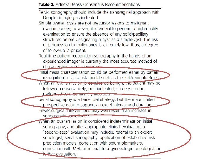
Reducing Subjectivity ■ First International Consensus Report 1 ■ International Ovarian Tumor Analysis (IOTA) o Simple Rules 2 o ADNEX 3 ■ Kentucky Morphology index 4 ■ Serial ultrasonography 5 1) J Ultrasound Med 2017; 2) Ultrasound Obstet Gynecol 2008; 3) BMJ 2014; 4) Gynecol Oncol 2003; 5) Gynecol Oncol 2014
IOTA Simple Rules ■ ■ ■ ■ ■ M 1 Irregular solid M 2 Presence of ascites M 3 At least 4 papillary projections M 4 Irregular multilocular solid, largest diameter ≥ 10 cm M 5 Very strong blood flow B 1 Unilocular B 2 Solid component < 7 mm B 3 Presence of acoustic shadows B 4 Smooth multilocular tumor, largest diameter < 10 cm B 5 No blood flow Timmerman et al. Simple ultrasound-based rules for the diagnosis of ovarian cancer. Ultrasound Obstet Gynecol 31; 681 -690, 2008
Simple Rules Malignant If one or more M-rules apply in the absence of a B-rule, the mass is classified as malignant Benign If one or more B-rules apply in the absence of an M-rule, the mass is classified as benign. Indeterminate If both M-rules and B-rules apply, the mass cannot be classified. If no rule applies, the mass cannot be classified Timmerman et al. Simple ultrasound-based rules for the diagnosis of ovarian cancer. Ultrasound Obstet Gynecol 31; 681 -690, 2008
ADNEX Risk Model Belgium, Italy, Czech Republic, Poland, UK, Sweden Van Calster et al. BMJ, 2014 _________ _ __________________ _____ _____ 13
Kentucky Morphology Index Ueland F, De. Priest P, Pavlik E, et al. Gynecol Oncol, 2003
Kentucky MI Malignan t ROM (%) MI Total 1 2, 349 1 0. 04 2 2, 365 0 0. 00 3 2, 635 3 0. 11 4 1, 579 7 0. 44 5 1, 061 29 2. 73 6 241 9 3. 73 7 87 11 8 30 8 26. 67 9 18 5 27. 78 10 3 1 33. 33 85% Total 10, 368 Ueland F, De. Priest P, Pavlik E, et al. Gynecol 74 Oncol, 2003 12. 64 0. 71 Sensitivity: 86% Specificity: 98%
Comparing Models ADNEX Model Kentucky MI n ROM n 52% n 15% n Clustering n MI n Misses n Identifies has non-linear correlation with CA risk of cancers in lowest ROM groups limits utility as clinical aid stage 1 cancers has linear correlation with CA risk of cancers in lowest ROM groups useful for decisions of surgery vs. surveillance Lefringhouse J, Ueland F, Ore R, et al. 16 SGO, 2016 stage 1 cancers 18 -Sep 21
Biomarkers Diagnostic Triage Comparison
‘Diagnostic’ Biomarkers • CEA • CA 19 -9 • LDH • β-h. CG • AFP • HE-4 • CA 125 *Multivariate + MIA 2 G Triage Biomarkers • OVA 1* • ROMA • Overa+ Index Assay 18
CA 125 Performance Myers et al. Management of adnexal mass. Rockville (MD): U. S. Department of Health and Human Services, 19 2006
Triage Biomarker Tests OVA 1 • FDA-cleared September, 2009 • Multivariate Index Assay Range 0 -10 Premenopausa l Post Low Risk < 5. 0 < 4. 4 High Risk ≥ 5. 0 ≥ 4. 4 ROMA • FDA-cleared September, 2011 • Dual marker test HE 4 Range 0 -10 Premenopausa Post l Low Risk < 1. 31 < 2. 77 High Risk ≥ 1. 31 ≥ 2. 77 20 CA 125 +
Triage Biomarker Tests Overa • FDA-cleared September, 2016 • Multivariate Index Assay-2 G • CA 125, HE-4, FSH, Apolipoprotein A 1, Transferrin Range 0 -10 Result Low Risk < 5. 0 High Risk ≥ 5. 0 21
Comparing Biomarkers Sensitivity Overa 1 OVA 12, 3 ROMA 4 CA 125 -II 2, 5 All malignancies 91% 93% 89% 69% Epithelial ovarian cancers 95% 99% 94% 82% Early stage EOC 89% 98% 75% 66% Premenopausal women 90% 94% 76% 36% Postmenopausal women 92% 100% 92% 80% 69% 54% 75% 87% Specificity All malignancies OVA 1 detected 76% of malignancies missed by CA 1251 1. 2. 3. 4. 5. Coleman R, Herzog T, Chan D, et al. Am J Obstet Gynecol, 2016 Ueland F, De. Simone C, Seamon L, et al. Obstet Gynecol, 2011 Bristow R, Smith A, Zhang Z, et al. Gynecol Oncol, 2013 Moore R, Mc. Meekin S, Brown A, et al. Gynecol Oncol, 2009 Myers et al. Management of adnexal mass. Rockville (MD): U. S. Department of HHS, 2006
Recommended Evaluation Determine Malignant Risk with Ultrasound 1. Low risk- surveillance 2. Indeterminate- secondary testing 3. High risk- refer to Gyn Oncologist for surgery
Recommended Evaluation Malignant Risk Low Indeterminate High Distribution 65% 25% 10% US morphology Unilocular or septate Partly solid, small wall abnormalities Mostly solid, papillary projections Secondary testing No YES No Surgery No Maybe YES 24
Low Risk • Smooth-walled • Unilocular or septate Septate cyst 3 Unilocular cyst 1, 2 1 Modesitt et al. Gynecol Oncol, 2003; 2 Bailey et al. Gynecol Oncol, 1998; 3 Saunders B. et al. Gynecol Oncol, 2010
Malignant Potential for Low Risk Summary of Valentin et al, 2013 33% unilocular 1% malignant • 0. 54% Premenopausal • 2. 76% Postmenopausal 7/11 had solid or papillary component on visual surgical inspection Valentin, Ameye, Franchi et al. Ultrasound Obstet Gynecol, 2013 26 18 -Sep 21
Resolution for Low Risk Resolution Time Type of Abnormality Cyst & Septae Cyst & Solid Scans 6, 239 1790 581 154 Abnormalities 1, 288 366 122 24 Average Scans 4. 8 4. 9 4. 8 6. 4 Mean (mo) 31. 0 26. 5 23 26. 4 17 14. 1 8. 3 12. 7 38. 4 36. 0 33. 8 38. 7 70. 9 64. 5 64. 3 Median (mo) 75 th percentile (mo) 90 th percentile (mo) 93. 8 Ore R, Ueland F, Lefringhouse J, et al. SGO Annual Meeting abstract, 201627
Septate Ovarian Tumors N= 1114 spontaneously resolving septate cysts Saunders B, Podzielinski I, Ware R, et al. Gynecologic Oncology 118; 278– 282, 2010
Recommended Evaluation Malignant Risk Low Indeterminate High Distribution 65% 25% 10% US morphology Unilocular or septate Partly solid, small wall abnormalities Mostly solid, papillary projections Secondary testing No YES No Surgery No Maybe YES 29
Indeterminate Risk • Small, irregular wall abnormalities • Partly solid • Atypical, non-papillary projections 30 18 -Sep 21
Serial Ultrasound Tumor type • Malignant • Non-malignant • Resolving MI score Increase Stable or gradual rise Decrease Elder J, Pavlik E, Long A et al. Gynecol Oncol 135; 8 -12, 31
N ∆MI P-value ∆MI per month P-value Surgery for epithelial ovarian malignancy 50* 1. 9 P<0. 001 0. 9 P<0. 001 Surgery for nonmalignancy 272 0. 7 P<0. 001 0. 2 P<0. 001 Resolved ovarian cysts 5811 -2. 7 P<0. 001 -1. 1 P<0. 001 *24 subjects had 1 scan only
Recommended Evaluation Malignant Risk Low Indeterminate High Distribution 65% 25% 10% US morphology Unilocular or septate Partly solid, small wall abnormalities Mostly solid, papillary projections Secondary testing No YES No Surgery No Maybe YES 33
High Risk • Irregular, solid • Papillations • Ascites • ROM >25% • Refer to Gyn Oncologist 34 18 -Sep 21
Summary 1. Ultrasound is the preferred test to evaluate an ovarian tumor 2. Risk of malignancy § Low: monitor without surgery o 6 months, then annually for 5 years § Indeterminate: secondary testing o Serial ultrasound o Biomarker testing (OVA 1, ROMA, Overa) § High: surgery o Refer to a Gynecologic Oncologist 35
Questions?