ULCER Definition A break in the epithelial continuity

ULCER Definition : • A break in the epithelial continuity • Discontinuity of the skin or mucous membrane which occurs due to the microscopic death of the tissues • Usually chronic
 • Arterial Disease ; Large vessel (Atherosclerosis)")
Etiology : • Venous Disease (Varicose Veins) • Arterial Disease ; Large vessel (Atherosclerosis) or Small vessel (Diabetes) • Arteritis : Autoimmune (Rheumatoid Arthritis, Lupus) • Trauma • Chronic Infection : TB/Syphilis • Neoplastic : Squamous or BCC, Sarcoma

Wagner’s Grading of ulcers • Grade 0 - Preulcerative lesion/healed ulcer • Grade 1 - Superficial ulcer • Grade 2 - Ulcer deeper to Subcutaneous tissue exposing soft tissue or bone • Grade 3 - Abscess formation or osteomyelitis • Grade 4 - Gangrene of part of tissues/limb/foot • Grade 5 - Gangrene of entire one area/foot

classification A. Clinical B. Pathological
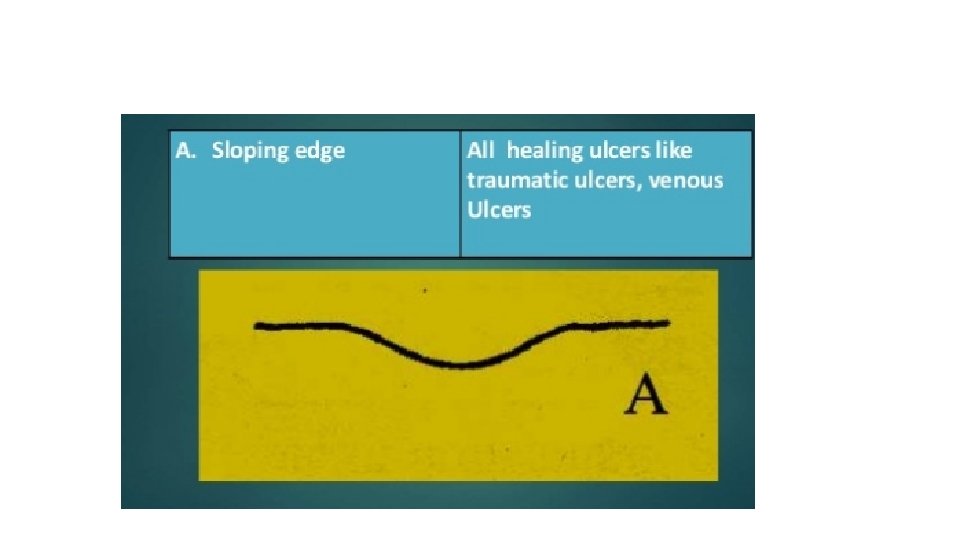
 • Healing : (Edge is")
Clinical • Spreading : (Edge - Inflamed & Edematous) • Healing : (Edge is sloping with healthy red granulation tissue & serous discharge) • Callous : (Floor contains pale unhealthy granulation tissue with indurated edge)

Pathological • 1. Nonspecific • 2. Specific • 3. Malignant

Non specific • Traumatic Ulcer • Arterial Ulcer • Venous Ulcer • Trophic ulcer pressure ulcer • Neuropathic Ulcer • Infective Ulcer • Diabetic Ulcer
 2. Physical-")
Traumatic ulcer 1. Mechanical- Dental ulcer on tongue ( jagged tooth ) 2. Physical- Electrical burn 3. Chemical- Application of caustics Acute, Superficial, Painful, Tender

Arterial Ulcer • Caused due to peripheral vascular disease • LL : Atherosclerosis • UL : Cervical Rib, Raynaud's • Chief complaint : Severe Pain • Toes, Feet, Legs & UL Digits

Venous ulcers • Medial aspect of lower 3 rd of lower limb • Ankle ( Gaiters Zone ) : Chronic Venous HTN • Ulcers are Painless • Varicose Veins or Post thrombotic(Phlebitic) limb ( PTS )

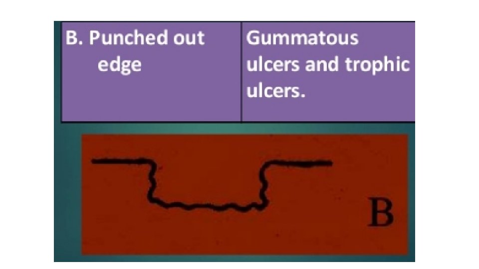
Trophic Ulcer • Pressure Sore or Decubitus Ulcer • Punched out edge with slough on the floor • Ex: Bed Sores & Perforating ulcers • Develop as a result of Prolonged Pressure • Sites : Ischial Tuberosity > Greater Trochanter > Sacrum > Heel > Malleolus > Occiput

Tropical ulcer • Tropical regions : Africa, India, S. America • Trauma or Insect Bite • Fusobacterium fusiformis & Borrelia vincentii • Abrasions, Redness, Papules & Pustules • Severe Pain

Diabetic Ulcer • It may be caused due to Diabetic Neuropathy Diabetic Microangiopathy • Increased Glucose : Increased Infection • Foot ( Plantar ), Leg, Back, Scrotum, Perineum • Ischemia, Septicemia, Osteomyelitis

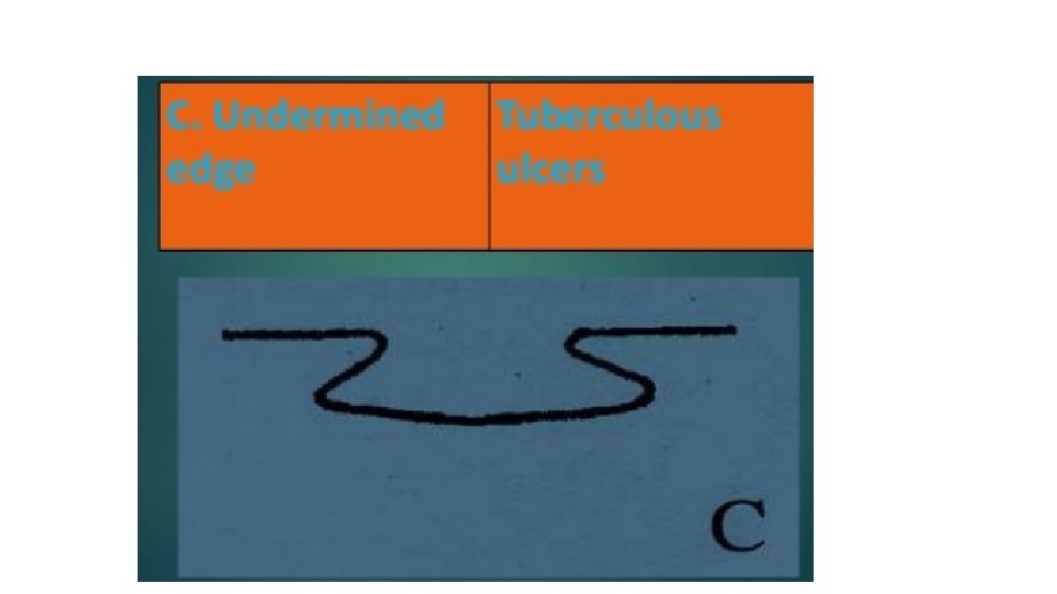
Specific • Tuberculosis • Syphilis • Actinomycosis

Malignant • Squamous cell ca • Basal cell ca • Malignant melanoma

Examination Inspection Palpation Examination of lymph nodes Vascular insufficiency Nerve lesions

• INSPECTION Location, size, shape, floor, edge, discharge, surrounding area. • PALPATION Tenderness, local rise of temperature, bleeding on touch, consistency of the ulcer, edge, surrounding area - edema, mobility. • REGIONAL LYMPH NODES • SENSATIONS • PULSATIONS • FUNCTION OF THE JOINT • SYSTEMIC EXAMINATION

INSPECTION LOCATION OF THE ULCER FLOOR OF THE ULCER DISCHARGE FROM THE ULCER EDGE SURROUNDING AREA

Location of the ulcer

FLOOR OF THE ULCER • Definition : This is the part of the ulcer which is exposed or seen.

Floor of the ulcer

Discharge from the ulcer

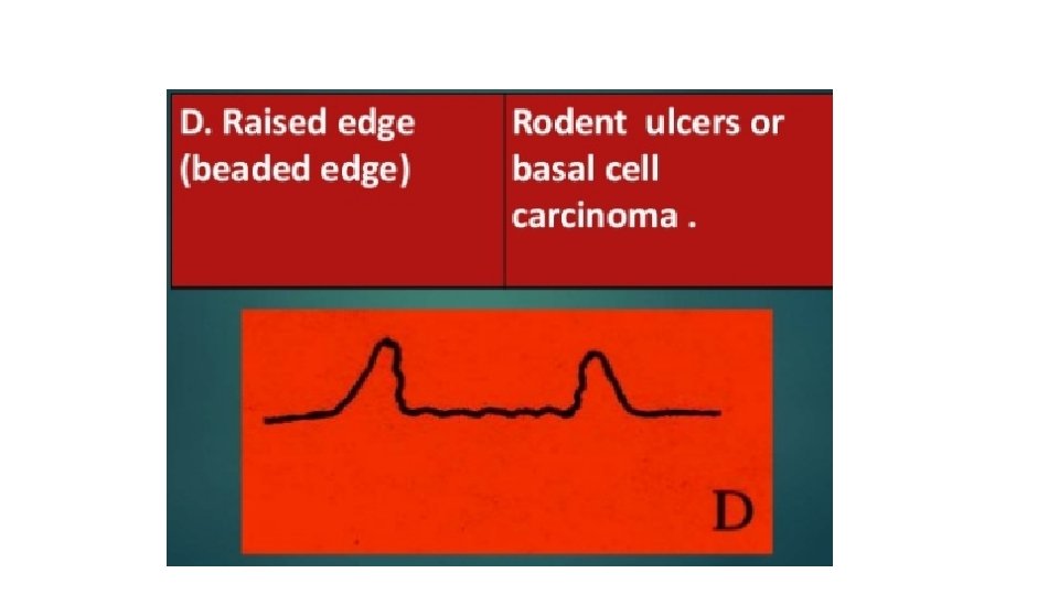
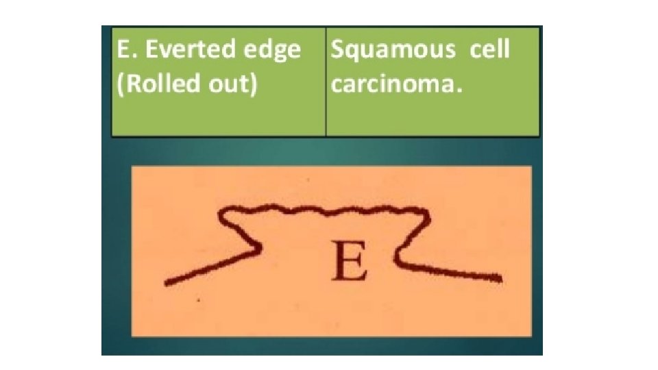
EDGE • This is between the floor of the ulcer and the margin. • The margin : is the junction between the normal epithelium and the ulcer. • These two parts represent areas of maximum activity.

PALPATION • EDGE • BASE • MOBILITY • BLEEDING • SURROUNDING AREA
 of the edge is very characteristic of squamous cell carcinoma.")
EDGE • Induration (hardness) of the edge is very characteristic of squamous cell carcinoma. • It is said to be a host defense mechanism. • Tenderness of the edge is characteristic of infected ulcers and arterial ulcers

BASE • It is the area on which ulcer rests. • Marked induration at the base is diagnostic of squamous cell carcinoma.

INDURATION.

MOBILITY • Gentle attempt is made to move the ulcer to know its fixity to the underlying tissues. • Malignant ulcers are usually fixed, benign ulcers are not.

BLEEDING • Malignant ulcer is friable like a cauliflower. • On gentle palpation, it bleeds • Granulation tissue as in a healing ulcer also causes bleeding.

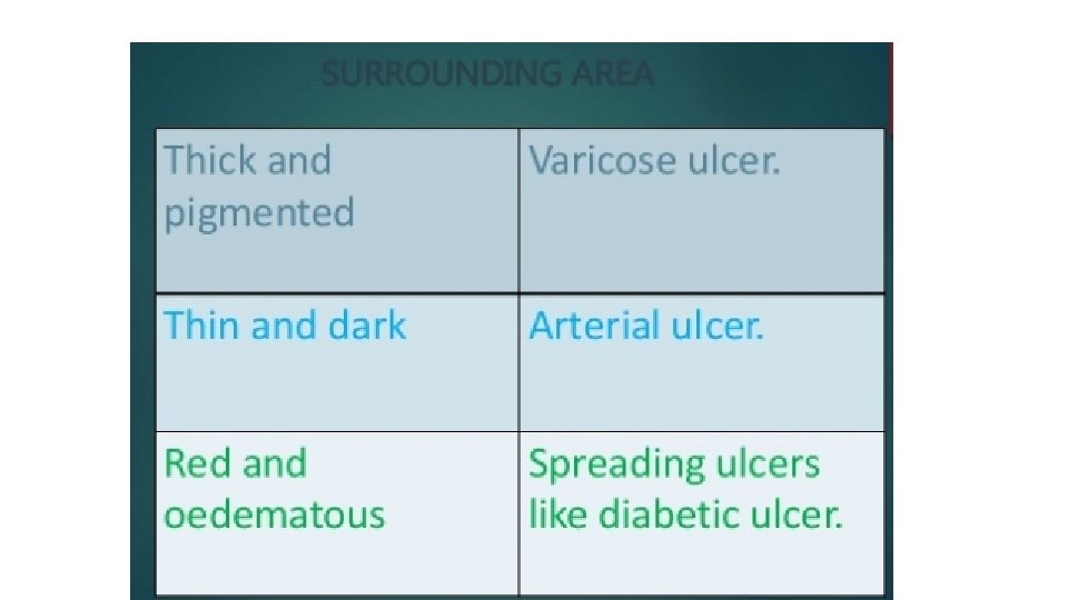
SURROUNDING AREA • Thickening and induration is found in squamous cell carcinoma. • Tenderness and pitting on pressure indicates spreading inflammation surrounding the ulcer.

RELEVANT CLINICAL EXAMINATION • REGIONAL LYMPH Painful , enlarge >> infection

MANAGEMENT Venous ulcer Squamous cell carcinoma BCC PUNCHED OUT BCC

GRANULATION TISSUE

GRANULATION TISSUE
 CBC, ESR 2) Urine and blood examination to rule out diabetes 3)")
Investigations 1) CBC, ESR 2) Urine and blood examination to rule out diabetes 3) Chest X-ray - PA. view to rule out TB 4) Pus for culture/sensitivity 5) Lower limb angiography in cases of arterial diseases 6) X-ray of the part to see for Osteomyelitis 7) Biopsy: Non-healing/malignant ulcers

Treatment • Address cause • Correct deficiencies • Control pain, infection • Debridement, dressing • Closure of defect

TREATMENT OF THE ULCERS • Treatment of Spreading Ulcers • Treatment of Healing Ulcers • Treatment of Chronic Ulcers • Treatment of The Underlying Disease

TREATMENT OF SPREADING ULCERS • Pus Culture/Sensitivity report • Appropriate Antibiotics • Solutions to treat the Slough : H₂O₂ & EUSOL - Edinburgh University Solution (Hypochlorite solution) • Excessive Granulation Tissue : Excision or Application of Copper Sulphate or Silver Nitrate • Repeated Dressings

TREATMENT OF HEALING ULCER • Regular dressings are done for a few days • Antiseptic creams like , Zinc Oxide or Silver Sulphadiazine. • Culture swab is taken to rule out Streptococcus Haemolyticus ( contraindication for skin grafting ) • Ulcer is small - Heals by itself ( Epithelialization ) • Large - Free Split Skin Graft applied • Closure : primary , secondary , delayed primary , graft , flap

TREATMENT OF CHRONIC ULCERS • These do not respond to conventional methods of treatment. • The following are tried: • Infrared radiation, short-wave therapy, ultraviolet rays decrease the size of the ulcer. • Amnion helps in epithelialization. • Chorion helps in granulation tissue. • These ulcers ultimately may require skin grafting.
")
Vaccum therapy ( -ve pressure )

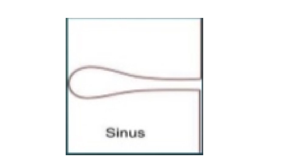
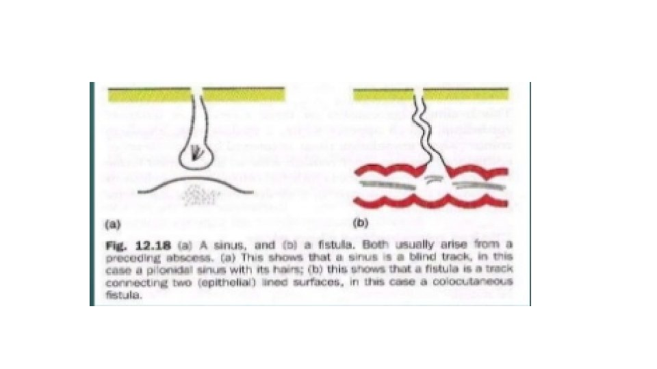
SINUS • tract lined with granulation tissue “GT” connecting an abnormal cavity “an abscess” to an epithelial surface. • Blind track lined by granulation tissue leading from epithelial surface down into the tissues. • Latin: Hollow (or) a bay • Symptoms: recurrent infection and discharge

Factors that can lead to sinus formation • Inadequate drainage of an abscess • Chronic inflammation, TB, Syphilis, fungal and Crohn’s disease. • Foreign body stimulates prolonged and recurrent infection e. g. suture material • Stitches sinus : common with non absorbable suture
")
CAUSES • CONGENITAL : Preauricular sinus, branchial cyst sinus ( anterior to sternocleidomastoid ) • ACQUIRED : Pilonidal sinus Malignant(Paget’s disease of the nipple and sister Joseph’s nodule from stomach ca) Actinomycosis TB sinus

FISTULA • A fistula is an abnormal tract between two epithelial surfaces. • ABNORMAL communication between lumen of one viscus and lumen of another (INTERNAL FISTULA) (or) • between lumen of one hollow viscus to the exterior (EXTERNAL FISTULA) (or) • between any two vessels • Most commonly, It occurs when an abscess breaks into two adjacent epithelial surfaces
 a tube.")
• Latin : pipe (or) a tube.

• Etiological factors as in sinuses Inadequate drainage of an abscess • Chronic inflammation. • Foreign body. • Congenital. • Malignant disease.

Types of fistulae • CONGENITAL Branchial fistula Tracheo-esophageal Umbilical Congenital AV fistula Thyroglossal fistula • ACQUIRED Traumatic Inflammatory Malignancy Iatrogenic
 following surgery : eg. , intestinal fistulas (faecal, biliary, pancreatic)")
ACQUIRED • TRAUMATIC: (A) following surgery : eg. , intestinal fistulas (faecal, biliary, pancreatic) (B) following instrumental delivery (or) difficult labour e. g. , vesicovaginal, rectovaginal, ureterovaginal fistula

• INFLAMMATORY: Intestinal actinomycosis, TB with crohns disease • MALIGNANCY: when growth of one organ penetrates into the nearby organ. e. g. , Rectovesical fistula in carcinoma rectum • IATROGENIC: AVF for hemodialysis • Non healed fistula indicate malignancy
 fistula • Presence of a foreign body. e.")
Causes for persistence of sinus (or) fistula • Presence of a foreign body. e. g. , suture material • Presence of necrotic tissue underneath. e. g. , sequestrum • Insufficient (or) non-dependent drainage. e. g. , TB sinus • Distal obstruction. e. g. , faecal (or) biliary fistula • Persistent drainage like urine/faeces/CSF
 endothelisation of the track. e. g. , AVF • Malignancy.")
• Epithelialisation (or) endothelisation of the track. e. g. , AVF • Malignancy. • Dense fibrosis • Irradiation poor vascularity , slow healing • Malnutrition • Specific causes. e. g. , TB, actinomycosis • Ischemia , Drugs. e. g. , steroids • interference by the patient

CLINICAL FEATURES • Usually asymptomatic but when infected manifest as Recurrent/ persistent discharge. • Pain. • Constitutional symptoms if any deep seated origin.

CLINICAL EXAMINATION • INSPECTION: • 1. Location: usually gives diagnosis in most of the cases. SINUS: pre-auricular, TB- neck. • FISTULA: branchial- sternomastoid ant border. parotid- parotid region thyroglossal- midline of neck below hyoid.
 actinomycosis.")
2. Number • usually single but multiple seen in HIV patients (or) actinomycosis.

3. Discharge • White thin caseous, cheesy like- TB sinus • Faecal- faecal fistula • Yellow sulphur granules- actinomycosis • Bony granules- osteomyelitis • Yellow purulent- staph. infections • Thin mucous like- brachial fistula • Saliva- parotid fistula
 Temperature and tenderness b) Discharge: after application of pressure over the surrounding")
Palpation a) Temperature and tenderness b) Discharge: after application of pressure over the surrounding area. c) Induration: present in chronic fistulae/sinus as in actinomycosis, OM d) Fixity: e) Palpation at deeper plane: lymph nodes- TB Thickening of bone underneath- OM

INVESTIGATIONS • CBP- Hb, TLC, DLC, ESR. • Discharge for C/S , AFB, cytology, Gram staining. • X-RAY of the part to rule out OM, foreign body. • X-RAY KUB and USG abdomen in cases of lumbar fistula to rule out staghorn calculi. • MRI • BIOPSY from edge of sinus • CT Sinusogram
fistula. • Water soluble")
FISTULOGRAPHY/ SINUSOGRAPHY • For knowing the exact extent/origin of sinus (or)fistula. • Water soluble or ultrafluid lipoidal iodine dye is used. • Lipoidal iodine is poppy seed oil containing 40% iodine.

TREATMENT BASIC PRINCIPLES • Antibiotics • Adequate excision • Adequate drainage.

• After excision specimen SHOULD be sent for HPE. • Treating the cause. e. g. , ATT for TB sinus. • removal of any foreign body. • sequestrectomy for OM.

PNS

Thank you
- Slides: 72