
Ukrainian Medical Stomatological Academy Department of obstetric and

Ukrainian Medical Stomatological Academy Department of obstetric and gynecology № 2 Lections for the students of the ІV course of stomatologycal faculty Basically questions of perinatology. Risk factors of perinatologycal period
Lecture plan Perinatal period Perinatal mortality The critical periods of fetal development Adverse perinatal factors The stages of pregnancy planning Modern methods of diagnostics of the fetal condition
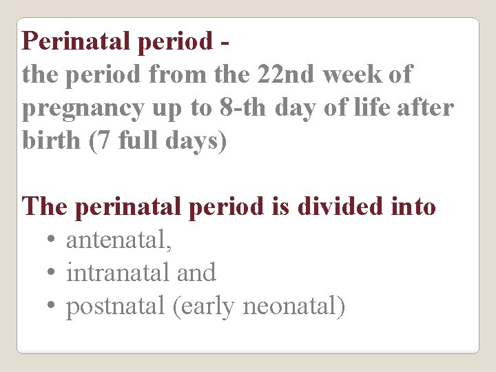
Perinatal period the period from the 22 nd week of pregnancy up to 8 -th day of life after birth (7 full days) The perinatal period is divided into • antenatal, • intranatal and • postnatal (early neonatal)
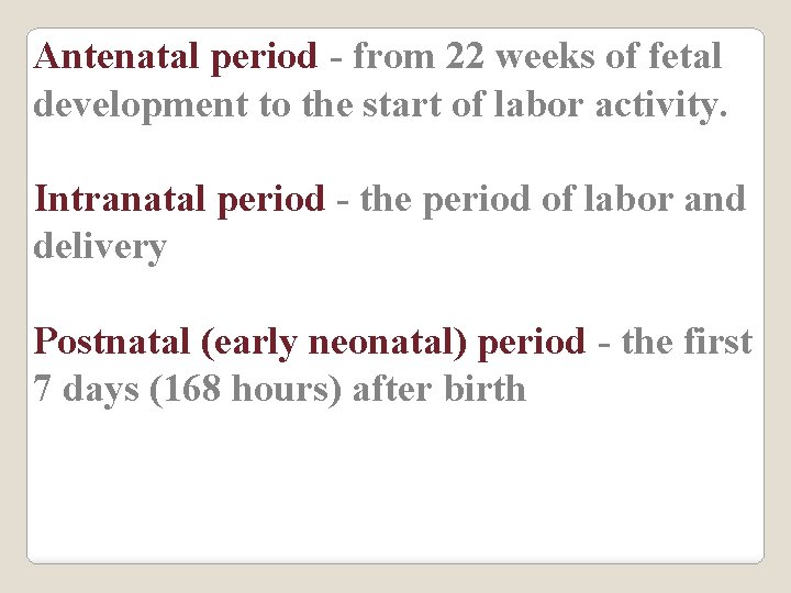
Antenatal period - from 22 weeks of fetal development to the start of labor activity. Intranatal period - the period of labor and delivery Postnatal (early neonatal) period - the first 7 days (168 hours) after birth
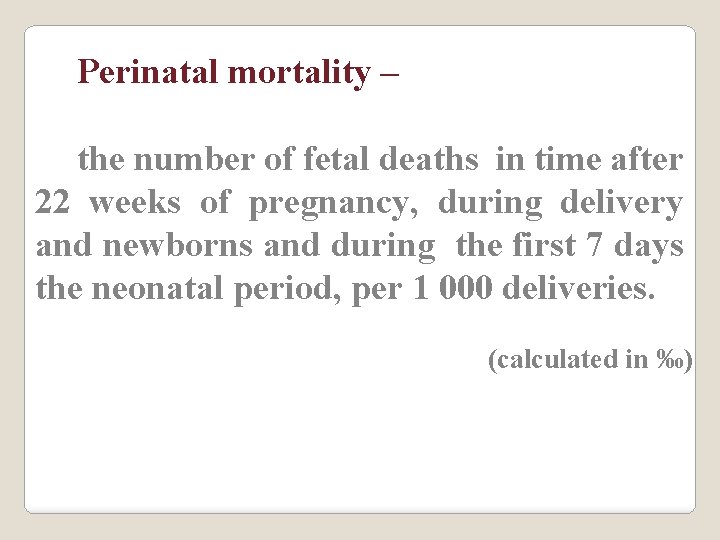
Perinatal mortality – the number of fetal deaths in time after 22 weeks of pregnancy, during delivery and newborns and during the first 7 days the neonatal period, per 1 000 deliveries. (calculated in ‰)
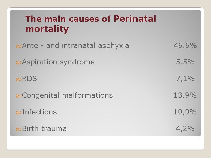
The main causes of Perinatal mortality Ante - and intranatal asphyxia Aspiration syndrome RDS 5. 5% 7, 1% Congenital malformations Infections Birth 46. 6% trauma 13. 9% 10, 9% 4, 2%
Perinatology - the science about the development and protection of the fetus and the newborn child in the perinatal period
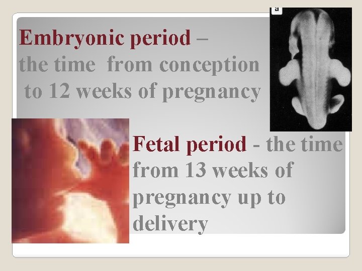
Embryonic period – the time from conception to 12 weeks of pregnancy Fetal period - the time from 13 weeks of pregnancy up to delivery
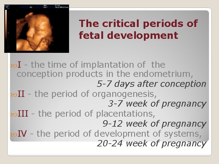
The critical periods of fetal development I - the time of implantation of the conception products in the endometrium, 5 -7 days after conception II - the period of organogenesis, 3 -7 week of pregnancy III - the period of placentations, 9 -12 week of pregnancy IV - the period of development of systems, 20 -24 week of pregnancy
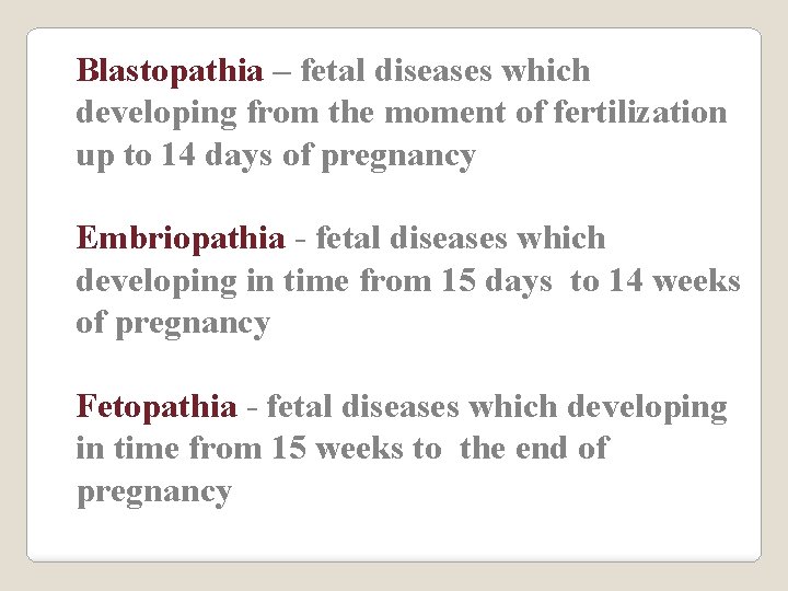
Blastopathia – fetal diseases which developing from the moment of fertilization up to 14 days of pregnancy Embriopathia - fetal diseases which developing in time from 15 days to 14 weeks of pregnancy Fetopathia - fetal diseases which developing in time from 15 weeks to the end of pregnancy
Adverse perinatal factors Exogenous Genetic Parent Fetal which are in the process of labor which are in the early neonatal period

Factors of perinatal risk Which have a place before start of pregnancy the birth of the dead children in anamnesis; the birth of children with congenital diseases; the death of the preceding children in the neonatal age; abortions and pre-time labor; the birth of children with chromosomal pathology in anamnesis; genetically diseases in the family; mother's age more than 35 years.
Factors perinatal risk (continued) Which have a place during pregnancy extra-genital mother’s pathology; dysfunction of the placenta; threat of abortions and pre-time labor; acute and chronically urogenital infections TORCH-infections in the mother gestosis; bleeding during pregnancy; Rh- incapability, ABO conflict; poly – and oligohydroamnion; delay the development of the fetus; disorders of fetal position; pre-time rupture of amniotic sac; detachment of the placenta; fetal distress pathological delivery.
Physiological development of the fetus and newborn provide: planning of pregnancy, the conscious attitude to the birth of the child; monitoring of the fetal development; definition of perinatal risk of pregnancy with intensive perinatal observation in case of need; medico-genetic counseling; the timely examination of the leading agents of TORCH-infections, which increase the risk of perinatal pathology psychological preparation of mother to delivery; physiological childbirth; contact «skin-to-skin» not less than 30 minutes, early attachment of the child to the mother's breast in the first 30 minutes after birth; stay of mother and newborn in the hospital together; breastfeeding infants; effective primary reanimation of newborns.
The stages of pregnancy planning Improvement of adolescent girls and women of reproductive age Diagnosis / treatment of STDs Screening for infection TORСH-group In the absence of antibodies to rubella vaccination Readjustment of the foci of chronic infection and compensation of chronic diseases Folic acid at a dose of 0. 8 - 1. 0 mg/day and multivitamins for 2 -3 months before the planned pregnancy
Modern methods of diagnostics of the fetal condition USG Biophysical profile of the fetus Cardiography Amnioscopia Amniocentesis Cordocentesis The chorion and placental biopsy
Diagnostic capabilities of ultrasound Diagnosis of malformations Measurement of the size of of the fetus (fetometry) Assessment of the status of the placenta Assessment of the quantity and quality of amniotic fluid Assessment of the status of the umbilical cord Evaluation of blood circulation in the arteries of the umbilical cord and of the fetus (Doppler effect)
The timing of routine ultrasound examination from 11+1 day to 13 weeks +6 days (definition of the term of pregnancy, measuring neck zone and detect ultrasonic markers of congenital and chromosomal pathology) 2. from 18 weeks to 20 weeks and 6 days (determination of structural abnormalities of the fetus). 3. 32 -34 weeks - definition of conformity of parameters of development of the fetus and placenta pregnancy, measurement of the fetal size (fetometry), 1.
The first trimester of pregnancy Restricting the reception of medicines with proven / possible teratogenic effect Intake of folic acid at a dose of 0. 8 - 1. 0 mg/day and multivitamins The conduct of the prenatal diagnosis of groups at risk of congenital malformations double biochemical test (free-h. CG and Papp (A) First screening ultrasound investigation
Indications for referral of pregnant at the medicogenetic counseling The age of the pregnant 35 years and more. Age of men of 40 years and more. The presence of one of the spouses hereditary pathology, chromosomal adjustment or congenital(s) malformations. The presence of a pregnant woman phenylketonuria, cystic fibrosis and other hereditary diseases The presence in the family of the living or the dead children of: - hereditary or chromosomal pathology; - congenital malformations - isolated or multiple; - mental retardation; the death of children. The presence of the above pathology among relatives. Marriage between the relative. 2 and more miscarriage of unknown genesis in the first trimester of pregnancy. Adverse effects in early pregnancy (diseases, diagnostic and therapeutic procedures, medications). Complicated course of pregnancy (threat of interruption of the early period, which is not amenable to therapy, poly – and oligohydroamnion). Pregnant after ECF. Pathology of the fetus, detected during the prenatal screening (USG, biochemical markers, high individual genetic risk of chromosomal and some congenital pathology). The presence of the spouses of the harmful factors, connected with the profession. Medico-genetic counseling should be conducted outside of pregnancy, or in her early with a detailed statement of the previous pregnancies, births, the status of the newborn, the results of the survey, data pathological-anatomical studies.
The main tasks of USG in the first trimester: Detection of pregnancy, which is developed or not developed; Assessment of the conformity of the embryo sizes and term of the pregnancy; Place of implantation and localization of the chorion; Anembrionia; Stage of spontaneous abortion; Multiple pregnancy; Ectopic pregnancy; Abnormalities of the uterus and appendages.
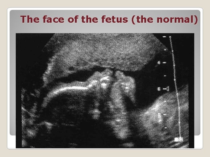
The face of the fetus (the normal)
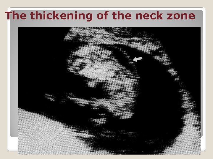
The thickening of the neck zone
The second trimester of pregnancy II-nd screening ultrasound investigation for detect congenital malformations and fetometria triple biochemical test in the 16 -17 weeks: h. CG and estriol, β-fetoprotein
The main tasks of USG in the second trimester: Accurate measurement of the size of the fetus - fetometria Define: Biparietal the size of the The average diameter of abdomen; The length of the femur. head; the chest and
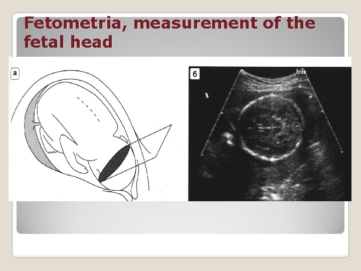
Fetometria, measurement of the fetal head
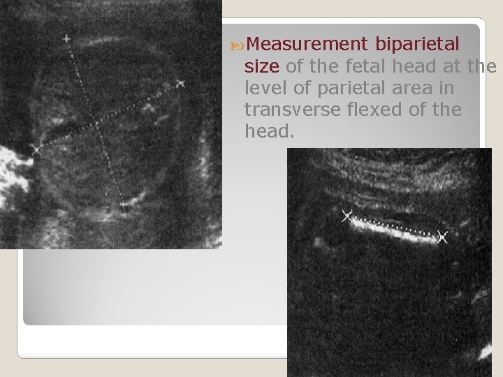
Measurement biparietal size of the fetal head at the level of parietal area in transverse flexed of the head.
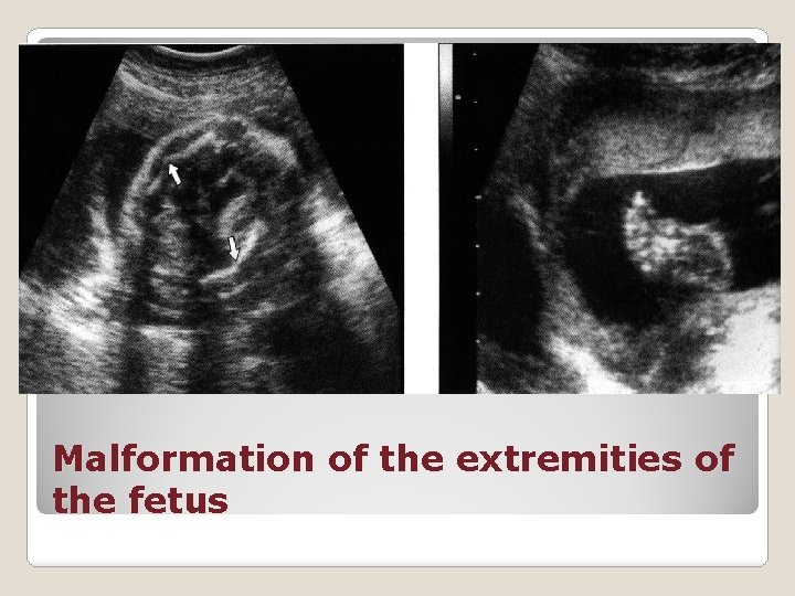
Malformation of the extremities of the fetus
The third trimester of pregnancy Ultrasound pregnancy investigation in 32 -34 weeks of ◦ for determine the status of the placenta, ◦ for determine the status of fetus ◦ Biophysical type of fetus (BTF) (from the 30 weeks of pregnancy) ◦ for the diagnostic of intrauterine fetal growth restriction and distress of fetus By indications- special methods of diagnostics of the condition of the fetus
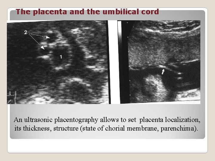
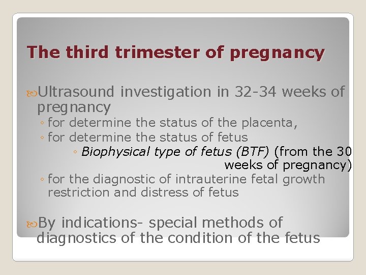
The placenta and the umbilical cord An ultrasonic placentography allows to set placenta localization, its thickness, structure (state of chorial membrane, parenchima).
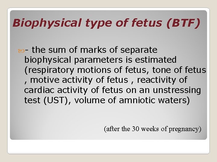
Biophysical type of fetus (BTF) - the sum of marks of separate biophysical parameters is estimated (respiratory motions of fetus, tone of fetus , motive activity of fetus , reactivity of cardiac activity of fetus on an unstressing test (UST), volume of amniotic waters) (after the 30 weeks of pregnancy)
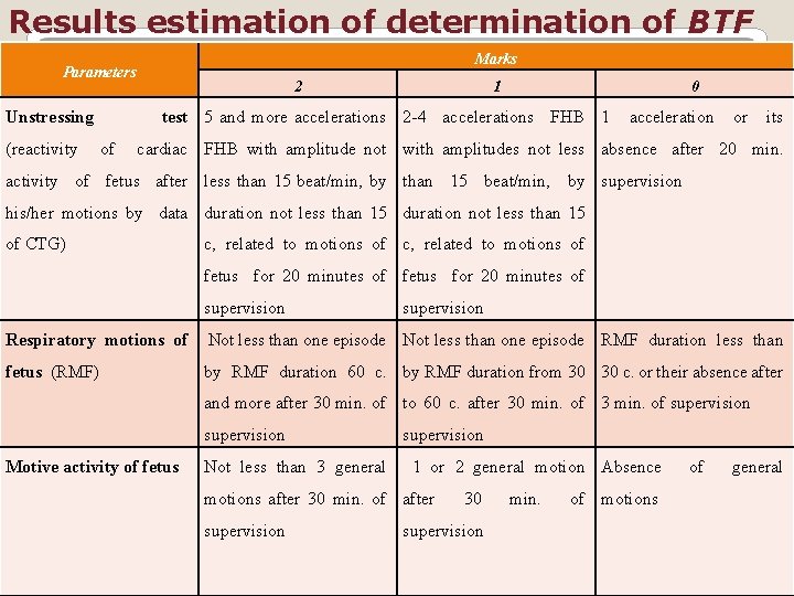
Results estimation of determination of BTF Marks Parameters Unstressing (reactivity 2 1 0 test 5 and more accelerations 2 -4 accelerations FHB 1 of acceleration or its cardiac FHB with amplitude not with amplitudes not less absence after 20 min. activity of fetus after less than 15 beat/min, by supervision his/her motions by data duration not less than 15 of CTG) с, related to motions of fetus for 20 minutes of supervision Respiratory motions of Not less than one episode RMF duration less than fetus (RMF) by RMF duration 60 с. by RMF duration from 30 30 с. or their absence after and more after 30 min. of to 60 с. after 30 min. of 3 min. of supervision Motive activity of fetus Not less than 3 general supervision 1 or 2 general motion Absence motions after 30 min. of after supervision 30 supervision min. of motions of general
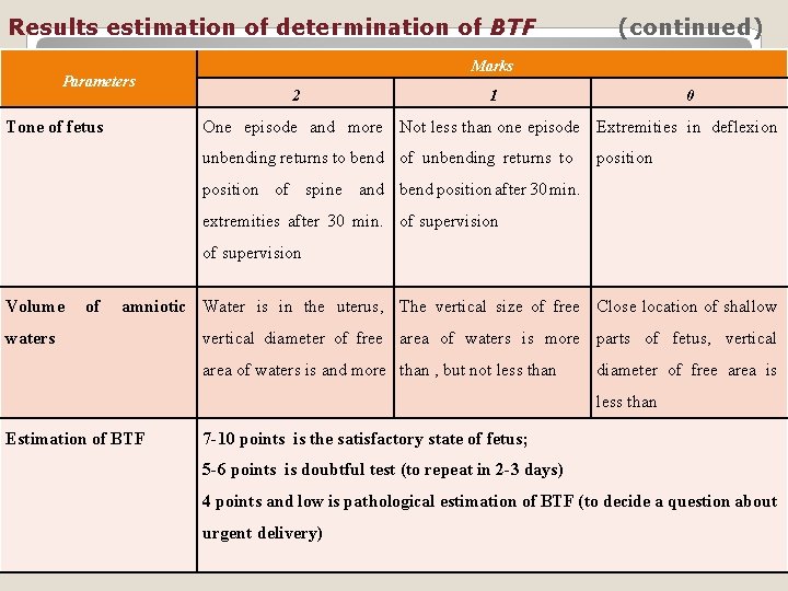
Results estimation of determination of BTF Parameters Tone of fetus (continued) Marks 2 1 0 One episode and more Not less than one episode Extremities in deflexion unbending returns to bend of unbending returns to position of spine and bend position after 30 min. extremities after 30 min. of supervision Volume of amniotic Water is in the uterus, The vertical size of free Close location of shallow waters vertical diameter of free area of waters is more parts of fetus, vertical area of waters is and more than , but not less than diameter of free area is less than Estimation of BTF 7 -10 points is the satisfactory state of fetus; 5 -6 points is doubtful test (to repeat in 2 -3 days) 4 points and low is pathological estimation of BTF (to decide a question about urgent delivery)
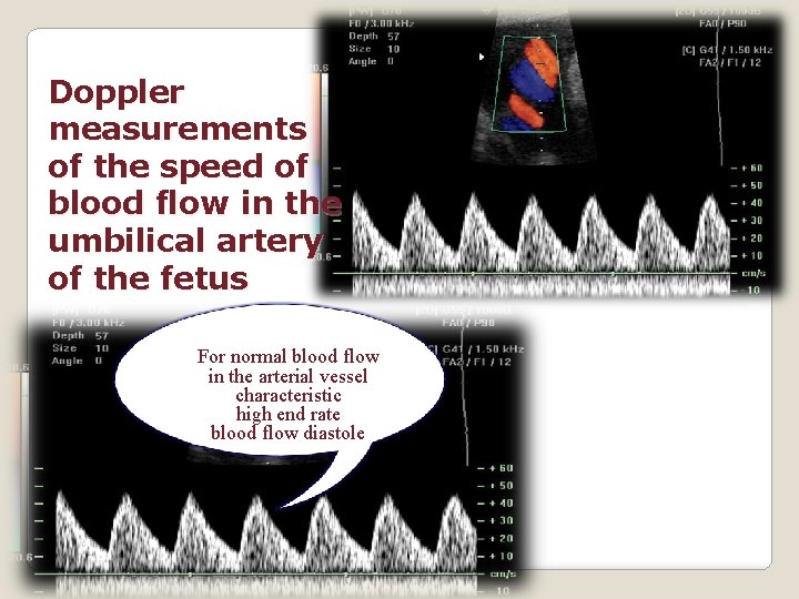
Doppler measurements of the speed of blood flow in the umbilical artery of the fetus For normal blood flow in the arterial vessel characteristic high end rate blood flow diastole
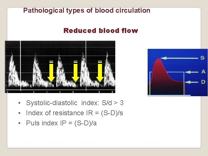
Pathological types of blood circulation Reduced blood flow • Systolic-diastolic index: S/d > 3 • Index of resistance IR = (S-D)/s • Puls index IP = (S-D)/a Повторне дослідження в динаміці - лікування не проводиться
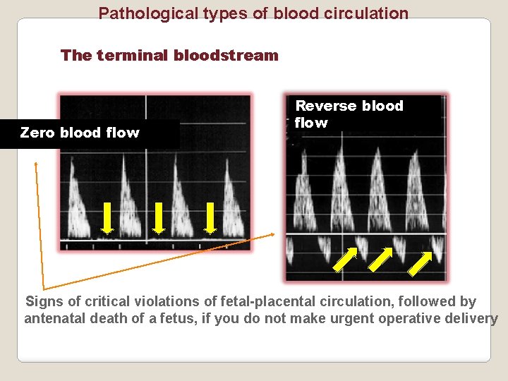
Pathological types of blood circulation The terminal bloodstream Zero blood flow Reverse blood flow Signs of critical violations of fetal-placental circulation, followed by antenatal death of a fetus, if you do not make urgent operative delivery
Cardiotocography (CTG) is synchronous record of cardiac rhythm of fetus and uterine contractions in prolong 10 -15 minutes by a vehicle cardiotocographe.
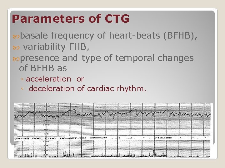
Parameters of CTG basale frequency of heart-beats (BFHB), variability FHB, presence and type of temporal changes of BFHB as ◦ acceleration or ◦ deceleration of cardiac rhythm.
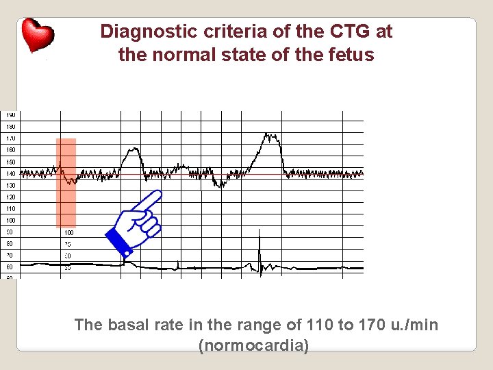
Diagnostic criteria of the CTG at the normal state of the fetus The basal rate in the range of 110 to 170 u. /min (normocardia)
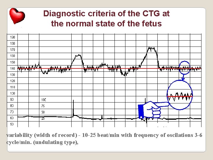
Diagnostic criteria of the CTG at the normal state of the fetus variability (width of record) - 10 -25 beat/min with frequency of oscilations 3 -6 cycle/min. (undulating type),
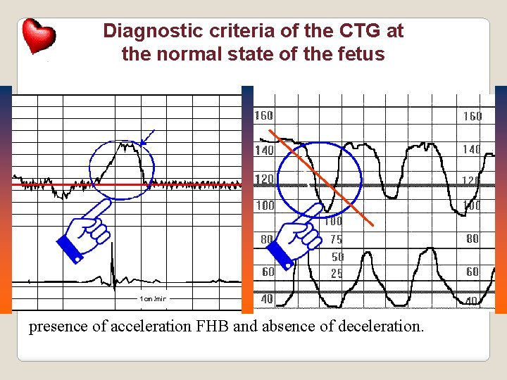
Diagnostic criteria of the CTG at the normal state of the fetus presence of acceleration FHB and absence of deceleration.
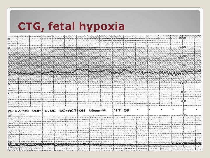
CTG, fetal hypoxia
Amnioscopia method of visualization of amniotic fluid The ability to determine The presence or absence of membranes The vascular membranes The color of amniotic fluid The presence of impurities in the amniotic waters

Amniocentesis is receipt of amniotic waters by the method of punction of amniotic cavity through an abdominal wall (trancabdominal access), or through vagina (transvaginalis). Get amniotic water for biochemical, hormonal, immunological, citological, genetic research, depending on the purpose of amniocentesis.
Test by Salling - method for determination of ph of the blood with the presenting part of the fetus during birth, after the rupture of the membranes
Chorion and placenta biopsy taking chorionic (or placental) tissue through the cervical canal or through the front abdominal wall, allows you to diagnose genetic and chromosomal diseases of the fetus
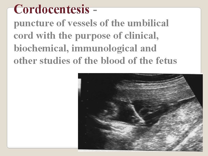
Cordocentesis puncture of vessels of the umbilical cord with the purpose of clinical, biochemical, immunological and other studies of the blood of the fetus
Fetoscopythe method of direct imaging of the fetus and intrauterinal environment with the help of a special flexible endoscope
Studies of hormonal function of the placenta Human chorionic gonadotropin (h. CG); Placental lactogen (PL); Estriol; Cortisol; α-fetoprotein (αFP); Placental α-1 microglobulin (PAMG).
Questions What is the perinatal mortality? How long is the early neonatal period? What parameters does cardiotocography evaluate? What is the test by Selling?

Thank you for your attention
- Slides: 51