typically referred to as contractions In concentric contraction


 is brief, involuntary twitching of a muscle or a group of")


, a transcriptional coactivator of nuclear receptors important to")
- Slides: 51
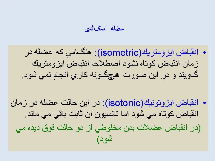
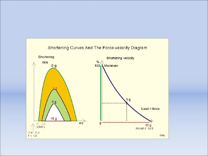
typically referred to as "contractions". In concentric contraction, the force generated is sufficient to overcome the resistance, and the muscle shortens as it contracts. This is what most people think of as a muscle contraction. In eccentric contraction, the force generated is insufficient to overcome the external load on the muscle and the muscle fibers lengthen as they contract. An eccentric contraction is used as a means of decelerating a body part or object, or lowering a load gently rather than letting it drop. In isometric contraction, the muscle remains the same length. An example would be holding an object up without moving it; the muscular force precisely matches the load, and no movement results. In isotonic contraction, the tension in the muscle remains constant despite a change in muscle length. This can occur only when a muscle's maximal force of contraction exceeds the total load on the muscle. In isovelocity contraction (sometimes called "isokinetic"), the muscle contraction velocity remains constant, while force is allowed to vary. True isovelocity contractions are rare in the body, and are primarily an analysis method used in experiments on isolated muscles that have been dissected out of the organism
Various Properties of Different Fiber Types Properties Type I fibers Type IIA fibers Type IIX fibers Motor Unit Type Slow Oxidative (SO) Fast Oxidative/Glycolytic (FOG) Fast Glycolytic (FG) Twitch Speed Slow Fast Twitch Force Small Medium Large Resistance to fatigue High Low Glycogen Content Low High Capillary Supply Rich Poor Myoglobin High high Low Red Color Dark Pale Mitochondrial density High Low Capillary density High Intermediate Low Oxidative Enzyme Capacity High Intermediate-high Low Z-Line Width Intermediate Wide Narrow Alkaline ATPase Activity Low High Acidic ATPase Activity High Medium-high Low
Prisoner of war exhibiting muscle loss as a result of malnutrition
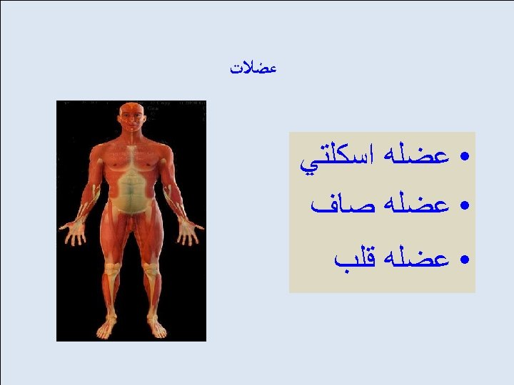
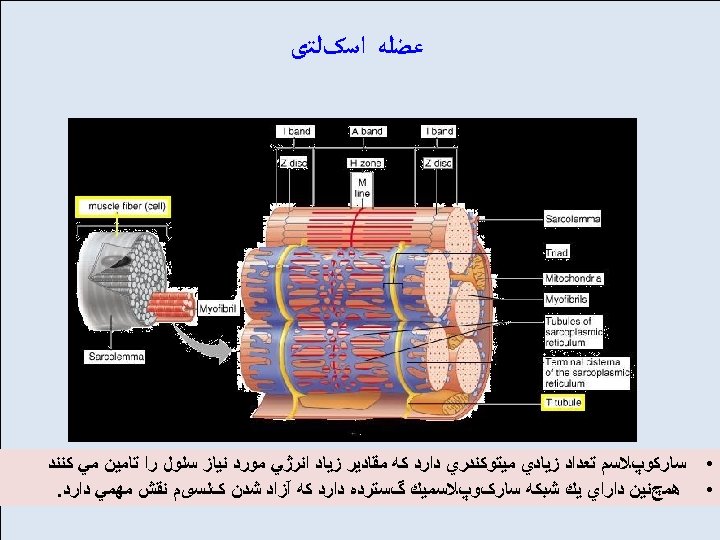
Cramps ﻋﻀﻠﻪ ﺍﺳکﻠﺘی are unpleasant, often painful • sensations caused by muscle contraction or overshortening. The common causes of skeletal muscle cramps are muscle fatigue and a sodium imbalance. Smooth muscle cramps may be due to menstruation or Gastroenteritis.
Causes of cramping include hyperflexion, hypoxia, exposure to large changes in temperature, dehydration, or low blood salt. Muscle cramps may also be a symptom or complication of pregnancy, kidney disease, thyroid disease, hypokalemia, hypomagnesemia or hypocalcemia (as conditions), restless-leg syndrome, varicose veins and multiple sclerosis ]
Dystonia is a neurological movement disorder, in which sustained muscle contractions cause twisting and repetitive movements or abnormal postures. [1] The disorder may be hereditary or caused by other factors such as birthrelated or other physical trauma, infection, poisoning (e. g. , lead poisoning) or reaction to pharmaceutical drugs, particularly neuroleptics. Treatment is difficult, since there is no cure available.
Fasciculation or "muscle twitch", is a small, local, involuntary muscle contraction and relaxation visible under the skin arising from the spontaneous discharge of a bundle of skeletal muscle fibers (muscle fascicle). Fasciculations have a variety of causes, the majority of which are benign, but can also be due to disease of the motor neurons
Myoclonus (pronounced /maɪˈɒklənəs/) is brief, involuntary twitching of a muscle or a group of muscles. It describes a medical sign and, generally, is not a diagnosis of a disease. The myoclonic twitches are usually caused by sudden muscle contractions; they also can result from brief lapses of contraction. Contractions are called positive myoclonus; relaxations are called negative myoclonus. The most common time for people to encounter them is while falling asleep (hypnic jerk), but myoclonic jerks are also a sign of a number of neurological disorders. Hiccups are also a kind of myoclonic jerk specifically affecting the diaphragm. Also when a spasm is caused by another person it is known as a "provoked spasm". Shuddering attacks with babies also fall in this category
spasm is a sudden, involuntary contraction of a muscle, a group of muscles or a hollow organ, or a similarly sudden contraction of an orifice. It is sometimes accompanied by a sudden burst of pain, but is usually harmless and ceases after a few minutes. Spasmodic muscle contraction may also be due to a large number of medical conditions, including the dystonias spasm is a temporary burst of energy, activity, emotion, stress, or anxiety
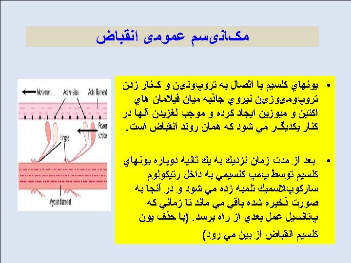
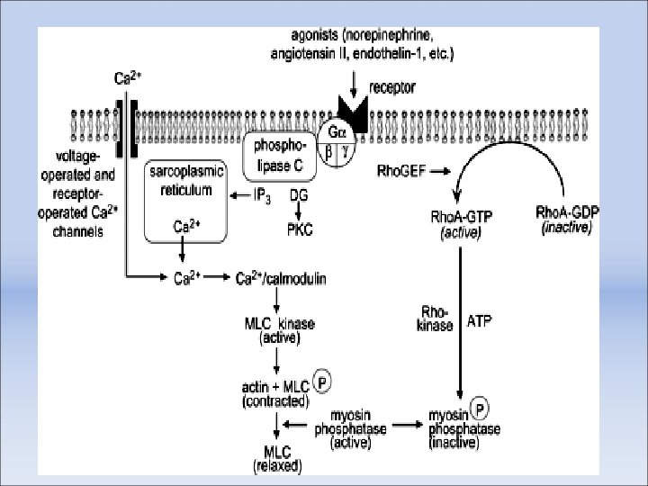
latch-bridges" During contraction of muscle, rapidly cycling crossbridges form between activated actin and phosphorylated myosin, generating force. It is hypothesized that the maintenance of force results from dephosphorylated "latch-bridges" that slowly cycle and maintain force. A number of kinases such as Rho kinase, Zip kinase, and Protein Kinase C are believed to participate in the sustained phase of contraction, and calcium flux may be significant.
Signal transduction pathways Skeletal muscle fiber-type phenotype in adult animals is regulated by several independent signaling pathways. These include pathways involved with the Ras/mitogen-activated protein kinase (MAPK) pathway, calcineurin, calcium/calmodulin-dependent protein kinase IV, and the peroxisome proliferator γ coactivator 1 (PGC-1). The Ras/MAPK signaling pathway links the motor neurons and signaling systems, coupling excitation and transcription regulation to promote the nerve-dependent induction of the slow program in regenerating muscle. Calcineurin, a Ca 2+/calmodulin-activated phosphatase implicated in nerve activity-dependent fiber-type specification in skeletal muscle, directly controls the phosphorylation state of the transcription factor NFAT, allowing for its translocation to the nucleus and leading to the activation of slow-type muscle proteins in cooperation with myocyte enhancer factor 2 (MEF 2) proteins and other regulatory proteins. Ca 2+/calmodulin-dependent protein kinase activity is also upregulated by slow motor neuron activity, possibly because it amplifies the slow-type calcineurin-generated responses by promoting MEF 2 transactivator functions and enhancing oxidative capacity through stimulation of mitochondrial biogenesis. Contraction-induced changes in intracellular calcium or reactive oxygen species provide signals to diverse pathways that include the MAPKs, calcineurin and calcium/calmodulin-dependent protein kinase IV to activate transcription factors
PGC 1 -α (PPARGC 1 A), a transcriptional coactivator of nuclear receptors important to the regulation of a number of mitochondrial genes involved in oxidative metabolism, directly interacts with MEF 2 to synergistically activate selective ST muscle genes and also serves as a target for calcineurin signaling. A peroxisome proliferatoractivated receptor δ (PPARδ)-mediated transcriptional pathway is involved in the regulation of the skeletal muscle fiber phenotype. Mice that harbor an activated form of PPARd display an “endurance” phenotype, with a coordinated increase in oxidative enzymes and mitochondrial biogenesis and an increased proportion of ST fibers. Thus—through functional genomics—calcineurin, calmodulin-dependent kinase, PGC-1α, and activated PPARδ form the basis of a signaling network that controls skeletal muscle fiber-type transformation and metabolic profiles that protect against insulin resistance and obesity. The transition from aerobic to anaerobic metabolism during intense work requires that several systems are rapidly activated to ensure a constant supply of ATP for the working muscles. These include a switch from fat-based to carbohydrate-based fuels, a redistribution of blood flow from nonworking to exercising muscles, and the removal of several of the by-products of anaerobic metabolism, such as carbon dioxide and lactic acid. Some of these responses are governed by transcriptional control of the FT glycolytic phenotype. For example, skeletal muscle reprogramming from an ST glycolytic phenotype to an FT glycolytic phenotype involves the Six 1/Eya 1 complex, composed of members of the Six protein family. Moreover, the hypoxia-inducible factor 1 -α (HIF 1 A) has been identified as a master regulator for the expression of genes involved in essential hypoxic responses that maintain ATP levels in cells. Ablation of HIF-1α in skeletal muscle was associated with an increase in the activity of ratelimiting enzymes of the mitochondria, indicating that the citric acid cycle and increased fatty acid oxidation may be compensating for decreased flow through the glycolytic pathway in these animals. However, hypoxia-mediated HIF-1α responses are also linked to the regulation of mitochondrial dysfunction through the formation of excessive reactive oxygen species in mitochondria. Other pathways also influence adult muscle character. For example, physical force inside a muscle fiber may release the transcription factor serum response factor (SRF) from the structural protein titin, leading to altered