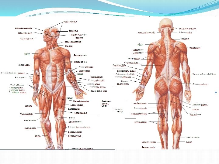
Types of Muscle Skeletal Striated Voluntary Multinucleated Smooth


 and thin")


- Slides: 21
Types of Muscle � Skeletal � Striated � Voluntary � Multinucleated � Smooth � Non-striated � Involuntary � Cardiac � Striated � Involuntary � Intercalated disks ~ 50% of body weight Work in groups to perform a function
General Functions of Muscular System �Movement �Heat Production �Posture �Continued partial contraction of muscle in order to perform many functions
Characteristics of all Muscles �Excitability �Respond to stimuli �Contractility �Actively shorten to exert a pull �Tension that can be harnessed �Extensibility �Continue to contract over range of resting lengths � Ex: smooth muscle can be stretched to several times its original length and still contract on stimulation �Elasticity �Return to original length after contraction
Muscle Microanatomy
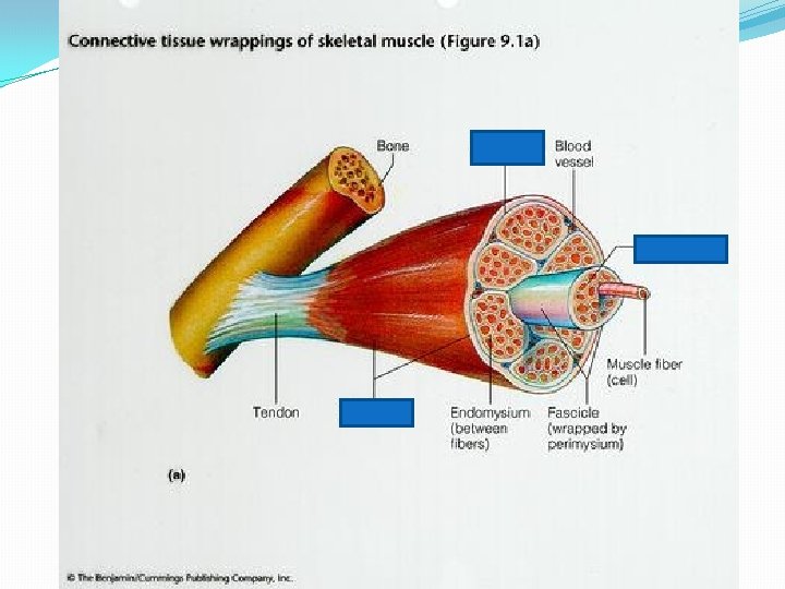
The Coverings �Epimysium is CT that covers the entire muscle
Coverings continued �Perimysium covers a fascicle �A fascicle is a bundle of muscle cells aka muscle fibers
Coverings continued �Endomysium- covers a muscle cell aka muscle fiber
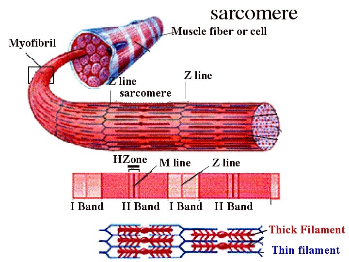
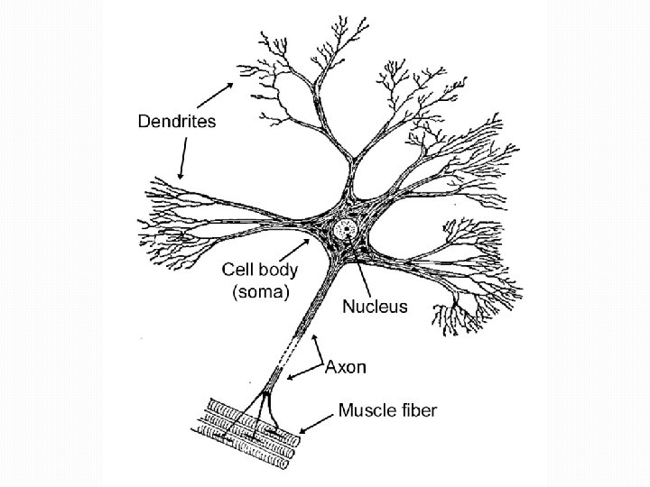
The parts of a muscle cell �Sarcolemma- plasma membrane �Sarcoplasmic reticulum- endoplasmic reticulum �Sarcoplasm- cytoplasm �Myofibrils= rod-like unit. Have sarcomeres
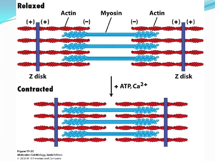
Going further…. the Sarcomere �Dark bands are “A bands” �Thick filaments (myosin) and thin filaments (actin) �Light bands are “I bands” �Thin filaments (actin)
Still further with the Sarcomere �H Zone �Found within the Dark band, made up of only thick filament (central region of the Dark) �Z lines- create the boundary of each sarcomere. Formed by connection of actin filaments to each other
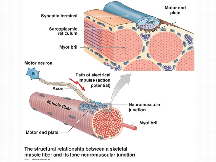
Muscle Contraction �Requires: �sarcoplasmic reticulum- produces calcium ions �transverse tubules- tubes that connect SR deeper into the cell and aid in conducting the signal �Happens because a motor neuron releases the neurotransmitter, acetylcholine (Ach), into the synaptic cleft
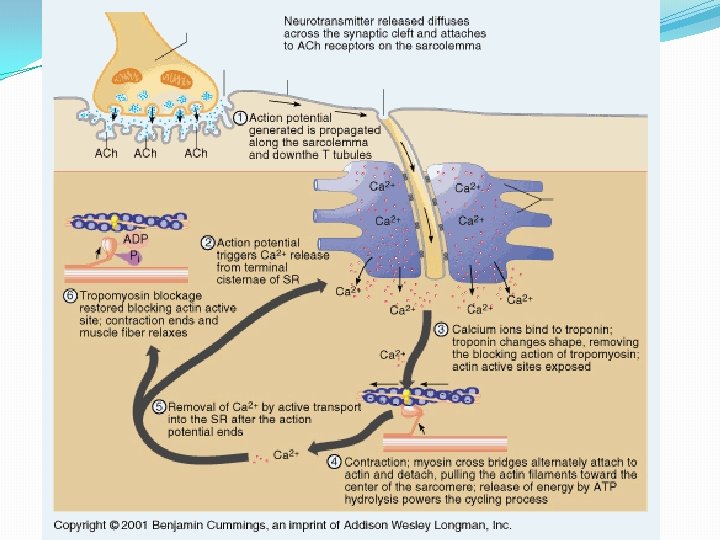
Action Potential! �Absolutely necessary for a muscle to contract Acetylcholine is released by a motor neuron Acetylcholine bonds to receptors in the sarcolemma The receptors open, allowing Na+ to flow into the area, temporarily causing it to become positively charged 4. A wave reaction opens as the local positive charge allows the next gate to open. 5. The action potential continues down the T-tubules to the sarcoplasmic reticulum 6. The positive charge allows calcium to be released from the SR 7. Calcium binds to troponin (one blocker against myosin) 8. Troponin removes tropomyosin (the other blocker) 9. Myosin is able to bond with actin in a stroke-like motion with the aid of ATP 10. Another ATP molecule is used to put myosin back in relaxed state 1. 2. 3.
Rigor Mortis �No more ATP to release myosin �After 16 -24 hours, rigor mortis disappears due to the muscle proteins dissolving