Types Of Bone Fracture Amal Jameel Hamzah Alajarma

Types Of Bone Fracture Amal Jameel Hamzah Alajarma Omar Mobideen

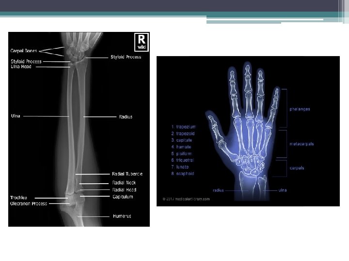
Normal X ray of Upper limb

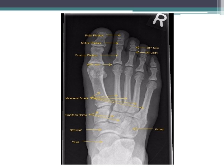
Normal X ray of the lower limb

Fractures • Fracture: is a break in the continuity of bone or cartilage. Closed fracture: Fracture with intact skin. Open fracture: Fracture with skin and soft tissue wound connecting the fracture to the external environment.

Fibula Tibia Broken skin Closed fracture Open fracture

Causes and Symptoms • Causes: • 1 - Most fractures are caused by a bad fall or automobile accident. • 2 - People with underlying illnesses and conditions that may weaken their bones. • 3 -Repeated stresses and strains, commonly found among professional sports people.

• Symptoms: • • • 1 -pain 2 -swelling 3 -bruising 4 -discolored skin around the affected area 5 -angulation – the affected area may be bent at an unusual angle 6 -the patient is unable to put weight on the injured area 7 -the patient cannot move the affected area 8 -the affected bone or joint may have a grating sensation 9 -if it is an open fracture, there may be bleeding

Types of Fractures • • • Linear Fracture. Comminuted Fracture. Avulsion Fracture. Pathological Fracture. Greenstick Fracture. Compression Fracture. Depressed Fracture. Epiphyseal Plate Fracture. Stress Fracture. Spiral Fracture. Oblique Displaced/Non Displaced Fracture.
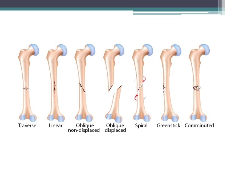

• 1 -Linear fracture: A fracture that extends over part or the entire length of a bone. Linear fracture in the 3 rd metacarpal bone

• Linear skull fractures: are breaks in the bone that transverse the full thickness of the skull from the outer to inner table.

Linear fracture lateral view of skull for child along parital bone

• 2 - Comminuted Fracture: : a fracture with multiple fragments (the bone is shattered into many pieces). Comminuted olecranon fracture Lateral view Frontal view
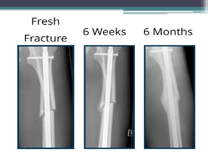
 A 28 -year-old male presented femur shaft comminuted fracture and tibia shaft fracture.")
A) A 28 -year-old male presented femur shaft comminuted fracture and tibia shaft fracture. (B) He undertook closed reduction and internal fixation with intramedullary nail

• 3 -Avulsion Fracture: a muscle or ligament pulls on the bone, fracturing it (a fragment of bone is detached from the site of a ligament or tendon insertion).

Avulsion fracture of the quadriceps tendon with retraction of the muscle

• 4 -Pathological Fracture: Bone fracture caused by an underlying disease/condition that weakened the bone). Pathological Fracture in the neck of the humeurs Pthological Fracture in the femur

Pathological Fracture in the Humerus before and after fixation

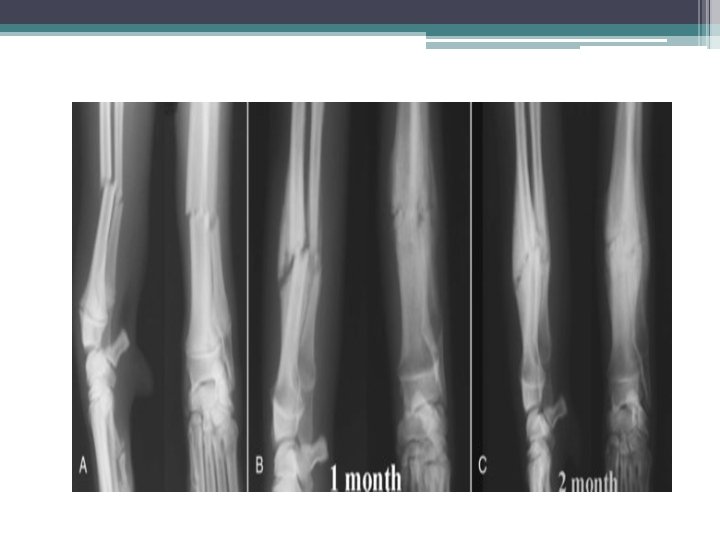
• 5 -Greenstick Fracture: : Incomplete fracture that usually occurs in children (the bone partly fractures on one side, but does not break completely because the rest of the bone can bend).

Greenstick fracture in the radius Greenstick fracture in the ulna

• 6 -Compression Fracture: : force is applied in the longitudinal axis of bone, generally occurs in the spongy bone in the spine.

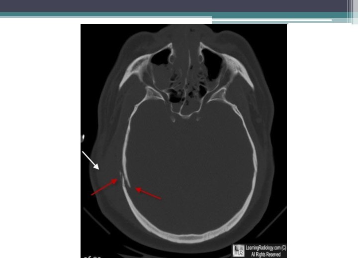
• 7 -Depressed Fracture: occurs in the skull, usually resulting from blunt force trauma.

• 8 -Epiphyseal Plate Fracture: It is a form of child bone fracture that involves the epiphyseal plate or growth plate.
 and the yellow arrow to a fracture of")
Fracture of the epiphysis (white arrow) and the yellow arrow to a fracture of the distal tibial metaphysis in this Salter-Harris IV fracture of the ankle. Epiphyseal Plate in the neck of the Radius

• 9 - Stress Fracture: A bone breaks because of repeated stresses and strains, more common among athletes.

Stress Fracture in the 5 th metatarsal

Ø March Fracture: is a type of stress fracture, also known as fatigue fracture of second and third metatarsal bones caused by recurrent overstress, is more common in soldiers. March Fracture in the 3 rd metatarsal

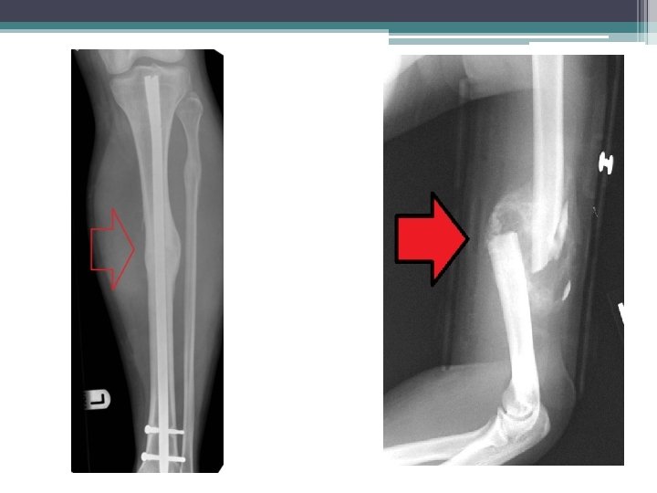
• 10 -Spiral Fracture: a fracture where at least one part of the bone has been twisted. Spiral Fracture in the femur Spiral Fracture in the humerus

Spiral fracture in fibula

• 11 -Oblique Fractures: a fracture that is diagonal to a bone’s long axis (the break is on an angle). Oblique Displaced Non-displaced

Oblique Fracture in the 2 nd proximal phalanx Oblique Fracture in the 5 th metatarsal

Other Fractures: • -Burst Fracture:

• Fracture Dilocation:

• Sternal Fracture:

**Clinical Case: v. Presentation: v 21 years old Male was presented with motor vehicle accident as he crashed into a tree. v. On examination findings were: v open wound, neuro-vascularly intact. X ray was done for him (frontally)

v Case Discussion: 1. Fracture of the mid shaft of the humerus with absent distal humeral shaft. 2. Fracture dislocation of the elbow joint with displaced humeral fragments. 3. No evidence of fracture within the proximal radius or ulna. 4. The ambulance service found a 12 cm fragment of humerus in a tree. 5. Despite the proximity of the radial nerve to the humeral shaft, he still had all sensation and motor function in his hand. v Management: v His hand is planned for reconstruction
- Slides: 43