Types of Blood Vessels Layers of blood vessels

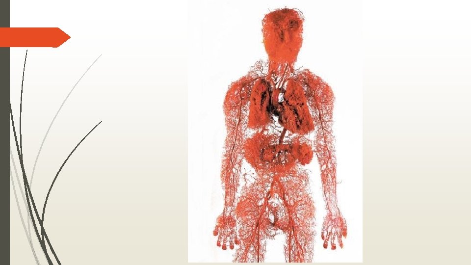
Types of Blood Vessels

Layers of blood vessels Tunica intima- inner Contains endothelium- simple squamous lining Tunica media- smooth muscle and elastin Regulated by sympathetic nervous system Often thickest layer Tunica Externa- collagen layer with nerves and lymph vessels Vasa vasorum – tiny blood vessels for large blood vessels
 AKA Conducting arteries")
Arteries Elastic- Thick walled near the heart (aorta and major branches) AKA Conducting arteries Allow blood flow here to be continuous Muscular- Deliver blood to organs AKA Distributing arteries Most of the named arteries Thickest tunica media Do vasoconstriction Arterioles- Determine blood flow to capillaries.

Blood vessel types

Capillaries Microscopic, consist of just a thin layer of endothelium. Form capillary beds- interwoven networks of capillaries
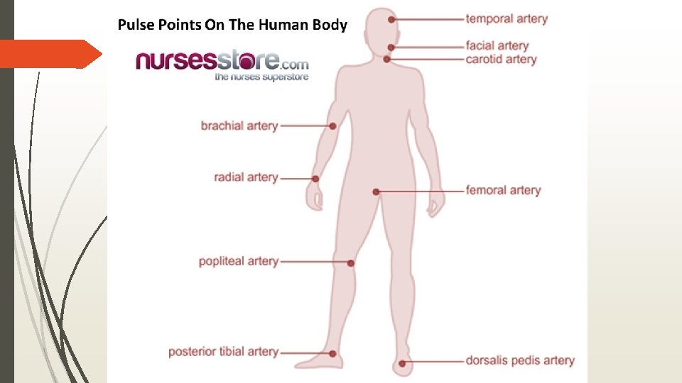

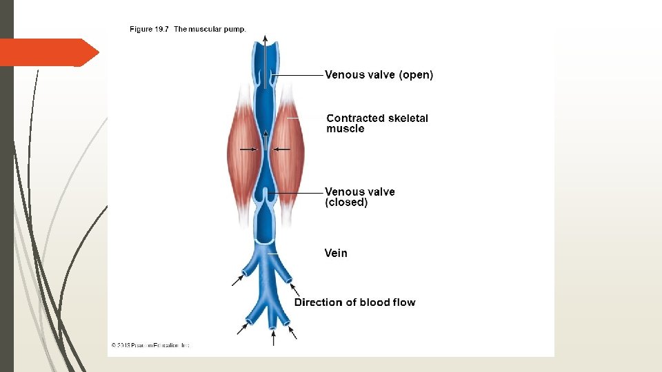
Venous System Carry blood from capillaries to the heart Get progressively larger Venules Veins Have all 3 layers, but thinner than arteries Very little smooth muscle Mostly tunica externa Blood reservoirs- can hold up to 65% of bodies blood Venous system much more interconnected than arteries Veins are more superficial than arteries Valves- Mostly in limbs Demonstrate

Heart Sounds

Heart Problems Arrhythmias- uncoordinated atrium and ventricles Defibrillation – electrically shocking heart to depolarize whole heart and hope SA node takes back over. Heart Murmur- Often due to acquired valve defects. Valve Stenosis- narrow valves, hard to get blood through. Valve regurgitation- hard to completely close. Endocarditis, valve calcification, and rheumatic fever Valve prolapse- valve is weak and is pushed in the atrium during contraction

Heart Sounds https: //www. youtube. com/watch? v=42 Iah. K-zxj 0&list=PLsvmwq 3 f 7 zb. VGJCD 7 Uix. V 6 t 4 ie. EOMt. Br

Coronary Arteries
 Part of the heart does not receive enough blood flow. 1")
Heart Attack (MI) Part of the heart does not receive enough blood flow. 1 of 5 is “silent” Most common cause is coronary artery plaque

11/10 What are the 3 layers of an artery? Which is thickest in muscular artery? Which is the only one in a capillary? Which do veins have? What is different between veins and arteries What is Mitral Valve Regurgitation? Objective:

Myocardial infarction

Heart Problems Angina Pectoralis
 Tachycardia-resting")
Cardiac Cycle Systole-period of contraction of the ventricles Diastole- period of relaxation (filling) Tachycardia-resting heart rate over 100 Stress, elevated body temperature, drugs, and disease can cause it Promotes fibrillation Bradycardia- under 60

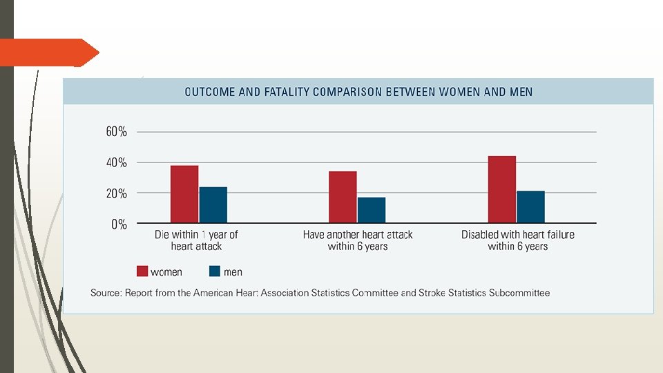
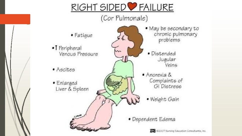
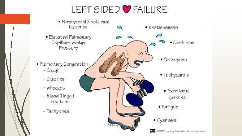
Congestive Heart Failure Progressive inability of heart to pump blood efficiently Persistent High Blood pressure is the main cause Heart must exert more force to open aortic valve MI can cause Atherosclerosis of coronary arteries Left sided most common

Stroke Blood supply to portion of brain impacted Brain cells begin dying within minutes 85% due to thrombus or embolus 15% Hemorrhagic - blood vessel burst Transient Ischemic attack- (TIA) temporary decrease of blood flow to area of brain Still should seek care , high risk of future stroke Risks: BP, obesity, Older men, Family history, heart disease, diabetes, cigarettes.

Stroke

WBC diseases Leukemia- Group of cancers of WBCs Usually affect only one cell type Causes a flood of immature WBCs Nonfunctional Sx: fever(night sweats), weight loss, bone pain, prolonged fatigue Die from internal hemorrhage and infection

Lymphatic System

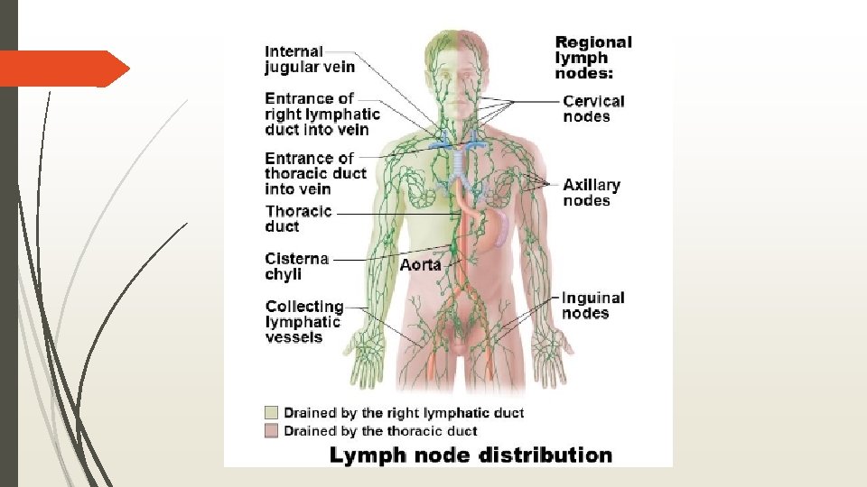
Lymphatic System 2 functionally different but structurally overlapping systems Lymphatic system- network of vessels, lymph, and nodes. Lymphoid organs and tissue- structural basis of the immune system Spleen, thymus, tonsils, nodes, other tissues.

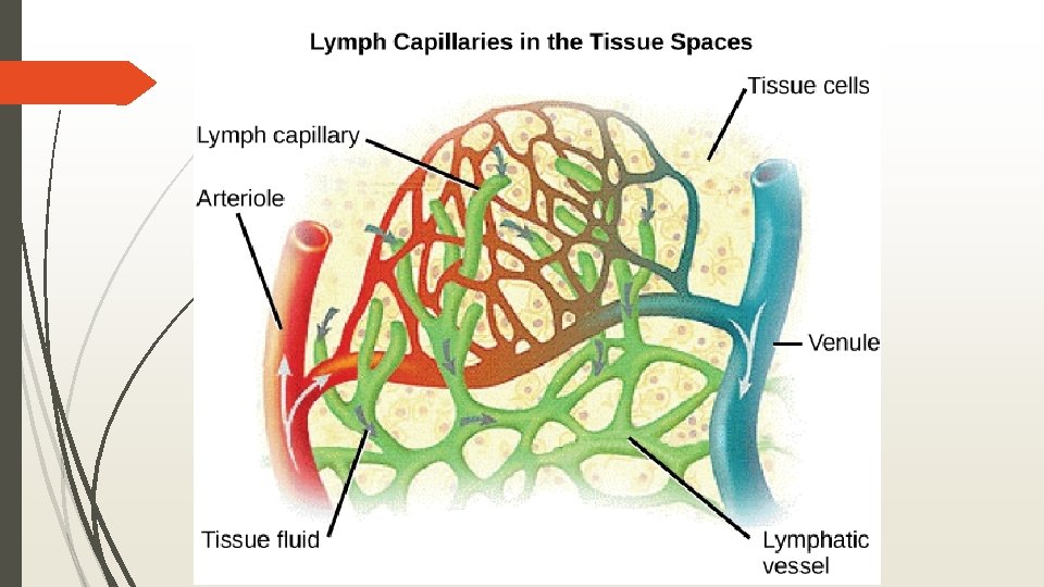
Lymphatic System Collect leaked fluid from capillaries Return it to blood to ensure volume Also return proteins from blood “Interstitial fluid” Have valves like veins No pumping system- rely on nearby muscles to milk it back to heart or nearby arteries. Lymphedema- anything that blocks the normal flow causes buildup of fluid. Can eventually regrow vessels around obstruction

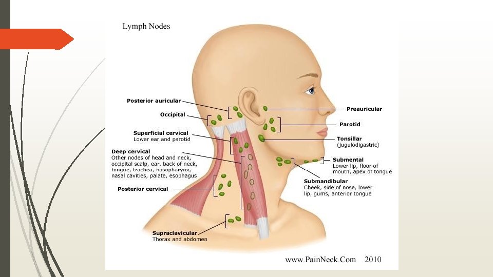
Lymph Organs- nodes Cluster in inguinal, axillary, cervical regions Places where vessels converge to form ducts Functions: Filtration- macrophages remove debris and microorganisms. Immune System activation Cancerous- swollen and non-tender, stuck to tissue Infection- swollen and tender, moveable
 Functions Blood cleaning- moves defective")
Spleen Size of fist, located just beneath diaphragm (left) Functions Blood cleaning- moves defective and old cells. Stores material from RBC breakdown- iron Stores platelets and monocytes for release when needed Can rupture Used to be removed, but now they try to let it repair itself. Liver and bone marrow will take over most of its function if removed.

4/27 Describe the steps of decreasing blood pressure in response to elevated blood pressure.

Thymus Anterior to the neck Produces T lymphocytes Most important in younger. Completely atrophies in older age.
 Help protect against never ending pathogens that try to enter")
Mucosa-associated Lymphoid Tissue (MALT) Help protect against never ending pathogens that try to enter body. Tonsils- Entrance to throat Remove particles in air and food Trap bacteria and allow local infection so that they can develop a ‘memory’ for later years Why at risk for infection early on Peyer’s Patches- like tonsils but on distal small intestine. Appendix- Off of Large Intestine
- Slides: 37