Tumors Tumors Adenoma Benign Carcinomas malignant Others q

Tumors
 � Carcinomas (malignant) � Others")
ØTumors � Adenoma (Benign) � Carcinomas (malignant) � Others

q Pelomorphic adenoma q Warthin tumor q Basal cell adenoma q Oncocytoma q Ductal papilomas q Canalicular adenoma q Sabaceous adenoma q Cyst adenoma

� Mucoepidermoid � Acinic ca cell ca � Adenoid cystic ca � Ca arising in Pelomorphic adenoma � Polymorphous low grade adenoca � Basal cell carcinoma � Sebaceous carcinoma � Cystadenocarcinoma

� Soft tissue tumors Haemangioma � Lymphoma Hodgkin lymphoma Diffuse large B-cell lymphoma Extranodal marginal B –cell lymphoma

Ø Diverse Ø histopathology 1% of all the tumors of human body Ø 2% to 5% of head and neck neoplasms
 75% benign Pleomorphic adenoma 10% other benign 15% Malignant")
�Parotid: 78% overall; (85%) 75% benign Pleomorphic adenoma 10% other benign 15% Malignant

�Submandibular: 12% overall; 30% Malignant

�Sublingual: 0. 3% overall; 14% benign 86% Malignant

� Minor S Gland: 10% Overall; 55% Benign 45% Malignant

�Most common of all salivary gland neoplasms 70 -80% of parotid tumors 50% of Submandibular tumors 45% of minor salivary gland tumors 6% of sublingual tumors �Age above 50 years (5 th, 6 th Decade) �Female more common
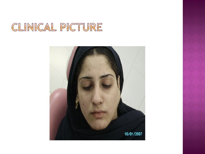
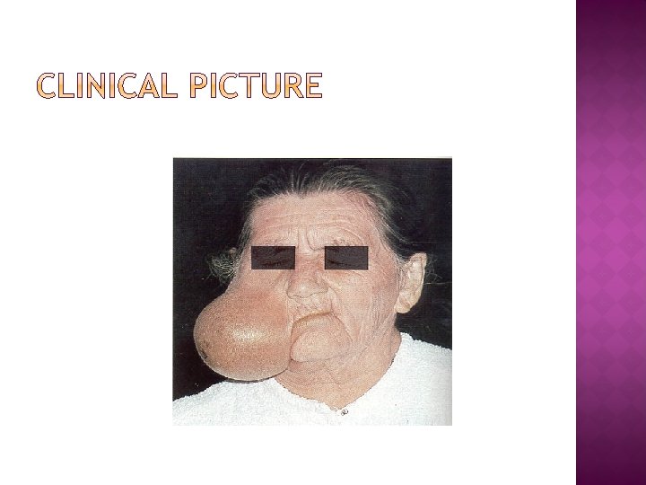
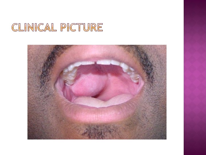
 � Painless mass � Mostly")
� Slow growing (may be present since many yrs) � Painless mass � Mostly unilateral � On palpation tumor is smooth, round and mobile. � The growth is rubbery in consistency with overlying skin or mucosa intact

� 90% in superficial lobe, most in tail of gland. �Facial nerve is never paralyzed �“Dumb bell tumor” when arise from deep lobe.

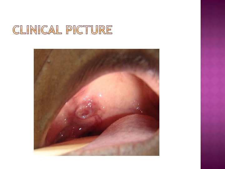
MINOR SALIVARY GLAND: � lateral palate � submucosal mass


HISTOLOGY � Tumor is typically well circumscribed � Some times may be uncapsulated or infltarated with tumor cells � Mixture of epithelial, myopeithelial and stromal components �Epithelial cells: nests, sheets, ducts, trabeculae �Stroma: myxoid, chrondroid, fibroid, osteoid �Tumor pseudopods

HISTOLOGY

� History � Clinical examination � Investigation Biopsy CT scan or MRI

�The tumor is highly implantable, �Recurrence is about 5%. �If rate after primary surgery simple enucleation is performed , the recurrence rate is between 20 -30%.

� 6 -8% of salivary gland tumors �Arise from striated duct cells. � 6 -10% of parotid neoplasms � 3% with associated neoplasms

CLINICAL PRESENTATION �Slow-growing, painless mass �Usually in the tail of parotid gland �Fluctuant , smooth soft and compressible �Cystic once contain mucoid fluid �Or some time solid in nature �Older Males � 10% bilateral

�The tumor, at the right of the image, is welldemarcated from the adjacent parotid tissue and tends to shell out from it.

HISTOLOGY � Composed of ductal epithelium & lymphoid stroma � Inner luminal layer is consist of tall columnar cells with centrally placed hyperchromatic nuclei � second layer is cuboidal cells �Stroma: � mature lymphoid follicles with germinal centers

HISTOLOGY
 Grading (Prognosis)")
q � Staging (Treatment plan) Grading (Prognosis)

STAGING �TNM STAGING OF Salivary gland tumors

TNM STAGING �T = Tumor size �N = Lymph node involvement �M = metastasis

� T 0 - No primary tumor � Tis - Carcinoma in situ � T 1 - Tumor 2 cm or smaller � T 2 - Tumor 4 cm or smaller � T 3 - Tumor larger than 4 cm without the involvement of Nerve � T 4 - Tumor larger than 4 cm and deep invasion and nerve involvement

N - REPRESENTS LYMPHATIC NODE INVOLVEMENT �N 0 - No nodes �N 1 - Single homolateral node smaller than 3 cm �N 2 - Nodes(s) homolateral smaller than 6 cm �N 3 - Nodes(s) larger than 6 cm and/or bilateral

M- REPRESENTS TUMOR METASTASIS �M 0 - No metastasis �M 1 - Metastasis noted
")
� High Grade � Low Grade � High/low grade � (Well, Moderate, Poor Differentiated)

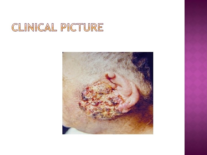
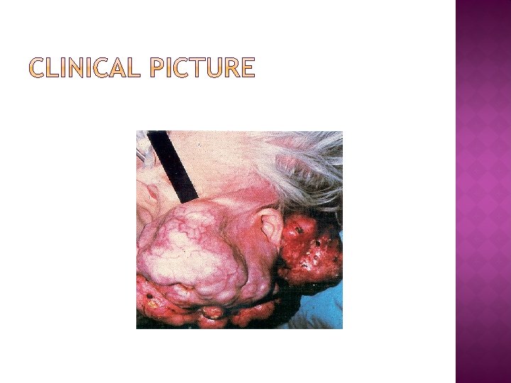
�Usually tumor grows rapidly �Pain, Ulcerated, �Involvement of facial nerve �Involvement of skin �Involvement of L node �Metastasis: L, L, L �(Lung, Liver, Long Bone)

MUCOEPIDERMOID CA

Major gland: � 90% arise in parotid, � 8% in the Submandibular, � 1% in the sublingual. Minor gland: more common: � 41% palate, � 14% buccal, � 9% tongue, � 5% lip.

� 4 -9% of salivary gland tumors � 3 rd-8 th decades, � peak in 5 th decade � Females more common � Most common salivary gland in children

PRESENTATION �Low-grade: slow growing, painless mass �High-grade: rapidly enlarging, +/- pain, Ulceration

GROSS PATHOLOGY Well-circumscribed to partially encapsulated to unencapsulated Solid, cystic, mixed

� Mixture of mucous secrecting cells which are Cubodail or columnar in nature & squamous cells

� Overall 2 nd most common malignancy � Most common in minor salivary gland, � Parotid gland, � Submandibular gland, � sublingual �M = F � 5 th decade

PRESENTATION �Asymptomatic enlarging mass �Ulcerated 50 % to 60 % �Pain, paresthesias, �Facial weakness/paralysis �Perinural invasion

GROSS PATHOLOGY Well-circumscribed Solid, rarely with cystic spaces Infiltrative

HISTOLOGY � Tubular Pattern � Solid Pattern � Cribriform Pattern

� Basloid epithelial cells contain multiple cylindrical, cyst like spaces resemblimg swiss cheese appearance � The tumor cells are small and cubiodal

TUBULAR PATTERN Layered cells forming multiple duct- or tubules like within hyalinized stroma

� Solid nests of cells without cystic or tubular spaces � Cellular phelomorphism and mitotic activity � Focal necrosis in the centre of tumor island may be observed
- Slides: 56