
Tumor Progression Md Abdur Rakib Ph D Associate
Tumor Progression Md. Abdur Rakib, Ph. D Associate Professor Department of Biochemistry and Molecular Biology University of Rajhahi, Bangladesh.
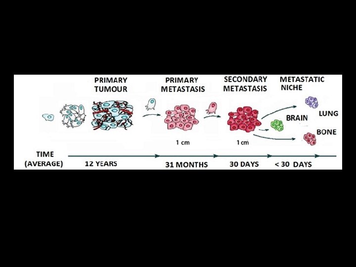
Tumor Progression Tumor formation is a complex process that usually proceeds over a period of decades. Normal cells evolve into cells with increasingly neoplastic phenotypes through a process termed tumor progression. This process takes place at myriad sites throughout the normal human body, advancing further and further as we get older.
Tumor Progression Cancers Usually Originate in Proliferating Cells In order for most oncogenic mutations to induce cancer, they must occur in dividing cells so that the mutation is passed on to many progeny cells. When such mutations occur in non-dividing cells (e. g. , neurons and muscle cells), they generally do not induce cancer, which is why tumors of muscle and nerve cells are rare in adults.
Tumor Progression Cancer is not a static disease In many tumors (e. g, colon, breast, cervical, pancreatic, melanoma), there appears to be an orderly progression from benign tissue to premalignant lesion to malignancy. In other tumors, premalignant lesions may not have been identified, but it is likely the tumor has passed through less-malignant stages before detection. The pathological and clinical criteria for tumor progression are often specific to a given type of tumor, but include local spread along tissue planes and into various tissue spaces and cavities. Tumors also have the capacity to invade and spread from their origins to other organs in the body; This process is referred to as metastasis. Increasing numbers and types of genetic abnormalities accompany tumor progression and metastasis.
Tumor Progression What Drives Tumorogenesis Tumor progression is driven by a sequence of randomly occurring mutations and epigenetic alterations of DNA that affect………. -the genes controlling cell proliferation, survival, and -other traits associated with the malignant cell phenotype
Most human cancers develop over many decades of time (Cancer formation is a long process) Epidemiologic studies have shown that age is a surprisingly large factor in the incidence of cancer. In the United States, the risk of dying from colon cancer is as much as 1000 times greater in a 70 -year-old man than in a 10 -year-old boy. This fact, on its own, suggests that this type of cancer and, by extension, many other cancers common in adults require years if not decades to develop.
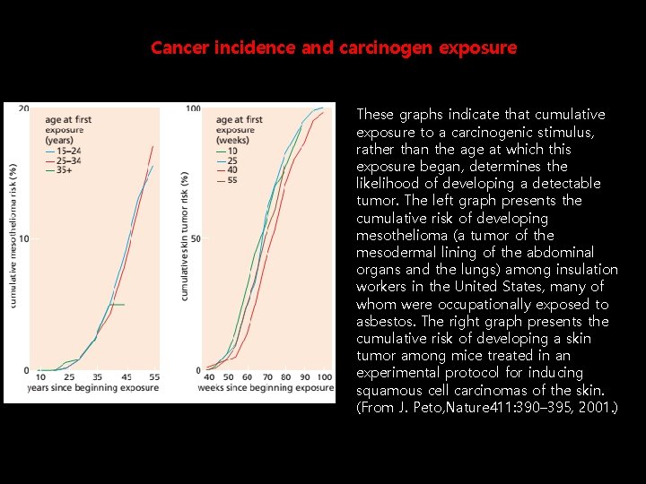
Cancer incidence and carcinogen exposure These graphs indicate that cumulative exposure to a carcinogenic stimulus, rather than the age at which this exposure began, determines the likelihood of developing a detectable tumor. The left graph presents the cumulative risk of developing mesothelioma (a tumor of the mesodermal lining of the abdominal organs and the lungs) among insulation workers in the United States, many of whom were occupationally exposed to asbestos. The right graph presents the cumulative risk of developing a skin tumor among mice treated in an experimental protocol for inducing squamous cell carcinomas of the skin. (From J. Peto, Nature 411: 390– 395, 2001. )
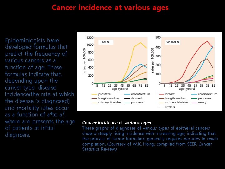
Cancer incidence at various ages Epidemiologists have developed formulas that predict the frequency of various cancers as a function of age. These formulas indicate that, depending upon the cancer type, disease incidence(the rate at which the disease is diagnosed) and mortality rates occur as a function of a 4 to a 7, where are presents the age of patients at initial diagnosis. Cancer incidence at various ages These graphs of diagnoses of various types of epithelial cancers show a steeply rising incidence with increasing age, indicating that the process of tumor formation generally requires decades to reach completion. (Courtesy of W. K. Hong, compiled from SEER Cancer Statistics Review. )
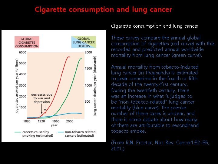
Cigarette consumption and lung cancer These curves compare the annual global consumption of cigarettes (red curve) with the recorded and predicted annual worldwide mortality from lung cancer (green curve). Annual mortality from tobacco-induced lung cancer (in thousands) is estimated to peak sometime in the fourth or fifth decade of the twenty-first century. During the twentieth century, there was an increase in what is judged to be “non-tobacco-related” lung cancer mortality (blue curve). The precise number of these cases is unclear, and there is some debate about how many of them are attributable to secondhand tobacco smoke. (From R. N. Proctor, Nat. Rev. Cancer 1: 82– 86, 2001. )
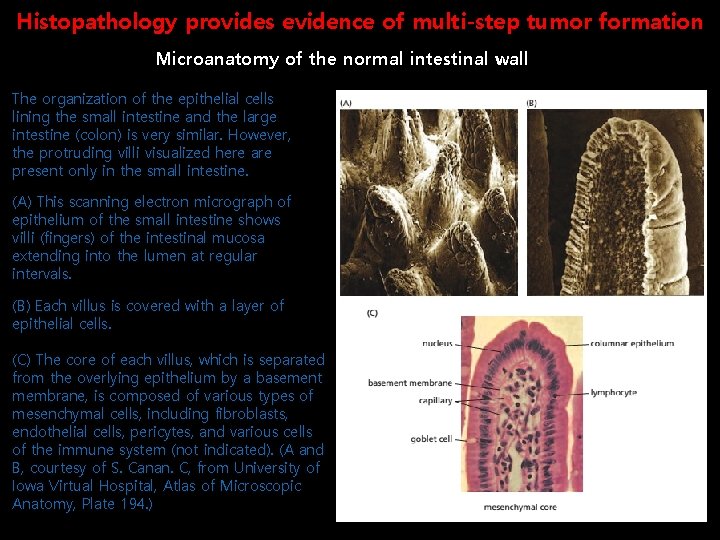
Histopathology provides evidence of multi-step tumor formation Microanatomy of the normal intestinal wall The organization of the epithelial cells lining the small intestine and the large intestine (colon) is very similar. However, the protruding villi visualized here are present only in the small intestine. (A) This scanning electron micrograph of epithelium of the small intestine shows villi (fingers) of the intestinal mucosa extending into the lumen at regular intervals. (B) Each villus is covered with a layer of epithelial cells. (C) The core of each villus, which is separated from the overlying epithelium by a basement membrane, is composed of various types of mesenchymal cells, including fibroblasts, endothelial cells, pericytes, and various cells of the immune system (not indicated). (A and B, courtesy of S. Canan. C, from University of Iowa Virtual Hospital, Atlas of Microscopic Anatomy, Plate 194. )
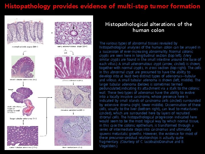
Histopathology provides evidence of multi-step tumor formation Histopathological alterations of the human colon The various types of abnormal tissues revealed by histopathological analyses of the human colon can be arrayed in a succession of ever-increasing abnormality. Normal colonic crypts are seen here in longitudinal section (top left). (Very similar crypts are found in the small intestine around the base of each villus. ) A small adenomatous crypt (arrow, circled) is shown, together with normal crypts, in cross section (top right). The cells in this abnormal crypt are presumed to have the ability to develop into at least two distinct types of adenomas—tubular and villous. A small tubular adenoma is shown (left, middle). The larger tubular adenoma (below) is sometimes termed pedunculated, indicating its attachment via a stalk to the colonic wall. These two types of adenomas have the ability to evolve into a locally invasive carcinoma, whose presence here is indicated by small islands of carcinoma cells (circled) surrounded by extensive stroma (right, lower middle). Dissemination of these cells, usually to the liver (bottom right), can lead to metastases (circled), which are surrounded here by layers of recruited stromal cells. The histopathological progression indicated here would seem to be the most logical way by which normal tissue, in this case the colonic epithelium, is transformed through a series of intermediate steps into carcinomas and ultimately spawns metastatic growths. However, the evidence for most of these precursor–product relationships is actually quite fragmentary. (Courtesy of C. Iacobuzio. Donahue and B. Vogelstein. )
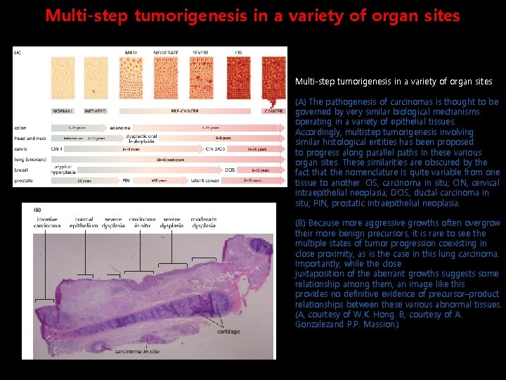
Multi-step tumorigenesis in a variety of organ sites (A) The pathogenesis of carcinomas is thought to be governed by very similar biological mechanisms operating in a variety of epithelial tissues. Accordingly, multistep tumorigenesis involving similar histological entities has been proposed to progress along parallel paths in these various organ sites. These similarities are obscured by the fact that the nomenclature is quite variable from one tissue to another. CIS, carcinoma in situ; CIN, cervical intraepithelial neoplasia; DCIS, ductal carcinoma in situ; PIN, prostatic intraepithelial neoplasia. (B) Because more aggressive growths often overgrow their more benign precursors, it is rare to see the multiple states of tumor progression coexisting in close proximity, as is the case in this lung carcinoma. Importantly, while the close juxtaposition of the aberrant growths suggests some relationship among them, an image like this provides no definitive evidence of precursor–product relationships between these various abnormal tissues. (A, courtesy of W. K. Hong. B, courtesy of A. Gonzalezand P. P. Massion. )
Molecular Genetics of Tumor Progression Tumor progression is driven by a sequence of randomly occurring mutations and epigenetic alterations of DNA that affect the genes controlling cell proliferation, survival, and other traits associated with the malignant cell phenotype. Epigenetic changes such as methylation of cytosine bases in DNA or modifications to chromatin structure (eg, by methylation, acetylation, sumoylation, or phosphorylation) can modify the expression of genes and are an important mechanism for “silencing” genes during normal differentiation. Genetic changes may occur by point mutation, deletion, gene amplification, chromosomal translocation or other mechanisms.
A cell is continually exposed to both external and internal stresses, such as reactive oxygen species, which may cause DNA damage. Moreover, there are inherent errors made by DNA polymerases whenever DNA is being replicated. Normally such damage is either repaired by the various DNA repair mechanisms in the cell or damaged cells undergo apoptosis. However, these mechanisms are not perfect, leading to a natural frequency of mutation in cells.
Loss of heterozygosity (LOH)
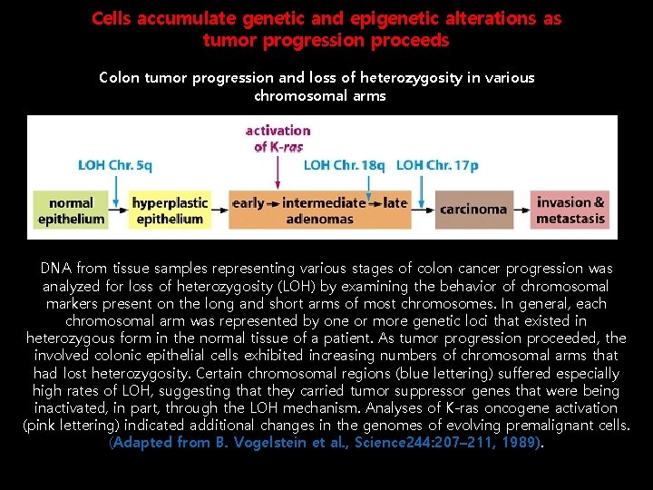
Cells accumulate genetic and epigenetic alterations as tumor progression proceeds Colon tumor progression and loss of heterozygosity in various chromosomal arms DNA from tissue samples representing various stages of colon cancer progression was analyzed for loss of heterozygosity (LOH) by examining the behavior of chromosomal markers present on the long and short arms of most chromosomes. In general, each chromosomal arm was represented by one or more genetic loci that existed in heterozygous form in the normal tissue of a patient. As tumor progression proceeded, the involved colonic epithelial cells exhibited increasing numbers of chromosomal arms that had lost heterozygosity. Certain chromosomal regions (blue lettering) suffered especially high rates of LOH, suggesting that they carried tumor suppressor genes that were being inactivated, in part, through the LOH mechanism. Analyses of K-ras oncogene activation (pink lettering) indicated additional changes in the genomes of evolving premalignant cells. (Adapted from B. Vogelstein et al. , Science 244: 207– 211, 1989).
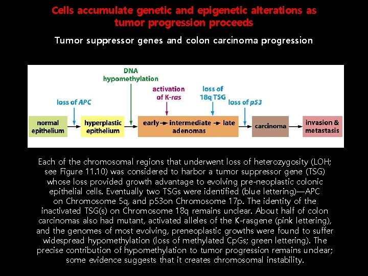
Cells accumulate genetic and epigenetic alterations as tumor progression proceeds Tumor suppressor genes and colon carcinoma progression Each of the chromosomal regions that underwent loss of heterozygosity (LOH; see Figure 11. 10) was considered to harbor a tumor suppressor gene (TSG) whose loss provided growth advantage to evolving pre-neoplastic colonic epithelial cells. Eventually two TSGs were identified (blue lettering)—APC on Chromosome 5 q, and p 53 on Chromosome 17 p. The identity of the inactivated TSG(s) on Chromosome 18 q remains unclear. About half of colon carcinomas also had mutant, activated alleles of the K-rasgene (pink lettering), and the genomes of most evolving, preneoplastic growths were found to suffer widespread hypomethylation (loss of methylated Cp. Gs; green lettering). The precise contribution of hypomethylation to tumor progression remains unclear; some evidence suggests that it creates chromosomal instability.
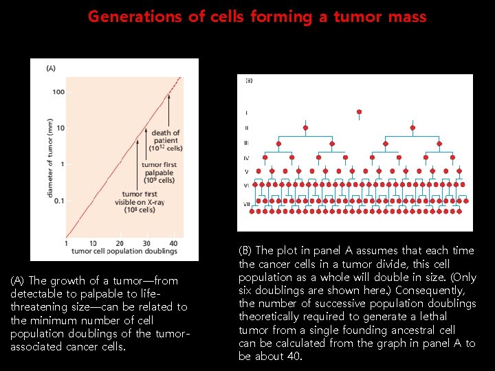
Generations of cells forming a tumor mass (A) The growth of a tumor—from detectable to palpable to lifethreatening size—can be related to the minimum number of cell population doublings of the tumorassociated cancer cells. (B) The plot in panel A assumes that each time the cancer cells in a tumor divide, this cell population as a whole will double in size. (Only six doublings are shown here. ) Consequently, the number of successive population doublings theoretically required to generate a lethal tumor from a single founding ancestral cell can be calculated from the graph in panel A to be about 40.
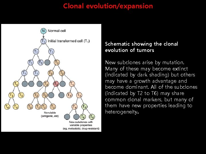
Clonal evolution/expansion Schematic showing the clonal evolution of tumors New subclones arise by mutation. Many of these may become extinct (indicated by dark shading) but others may have a growth advantage and become dominant. All of the subclones (indicated by T 2 to T 6) may share common clonal markers, but many of them have new properties leading to heterogeneity.
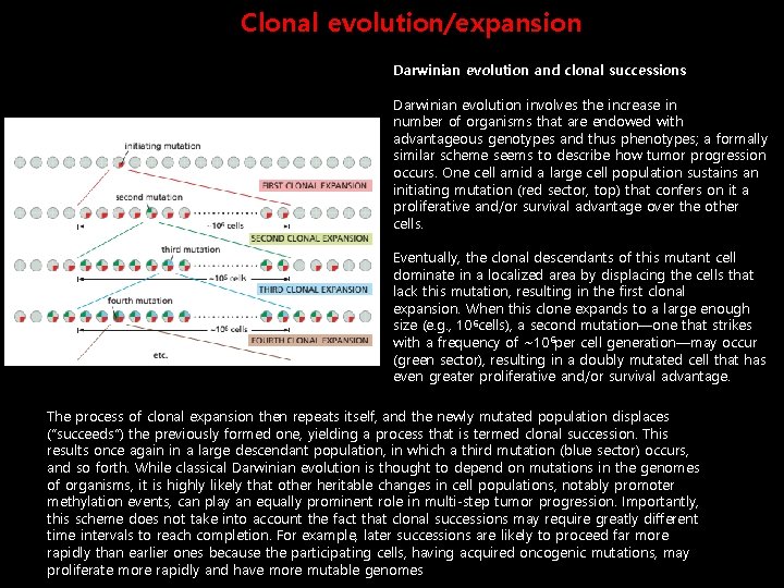
Clonal evolution/expansion Darwinian evolution and clonal successions Darwinian evolution involves the increase in number of organisms that are endowed with advantageous genotypes and thus phenotypes; a formally similar scheme seems to describe how tumor progression occurs. One cell amid a large cell population sustains an initiating mutation (red sector, top) that confers on it a proliferative and/or survival advantage over the other cells. Eventually, the clonal descendants of this mutant cell dominate in a localized area by displacing the cells that lack this mutation, resulting in the first clonal expansion. When this clone expands to a large enough size (e. g. , 10 6 cells), a second mutation—one that strikes with a frequency of ~10 6 per cell generation—may occur (green sector), resulting in a doubly mutated cell that has even greater proliferative and/or survival advantage. The process of clonal expansion then repeats itself, and the newly mutated population displaces (“succeeds”) the previously formed one, yielding a process that is termed clonal succession. This results once again in a large descendant population, in which a third mutation (blue sector) occurs, and so forth. While classical Darwinian evolution is thought to depend on mutations in the genomes of organisms, it is highly likely that other heritable changes in cell populations, notably promoter methylation events, can play an equally prominent role in multi-step tumor progression. Importantly, this scheme does not take into account the fact that clonal successions may require greatly different time intervals to reach completion. For example, later successions are likely to proceed far more rapidly than earlier ones because the participating cells, having acquired oncogenic mutations, may proliferate more rapidly and have more mutable genomes
Clonal evolution: the changing nature of myeloma https: //youtu. be/Ve. Uid-tz. Yuo
- Slides: 22