Trypanosomiasis Sleeping sickness African trypanosomiasis Sleeping sickness protozoan

 • – protozoan disease transmitted to human beings by the")
, Kingdom Protista, Phylum Protozoa. • East African")

 is located on the posterior end of")
 is more centrally located, usually just anterior")
 is towards the anterior end a free")

 a slowly developing")


 cause CNS demyelination. Immunosuppression by the parasite facilitates")


, Kingdom Protista, Phylum Protozoa. • Trypanosoma cruzi.")

")


- Slides: 50
Trypanosomiasis. Sleeping sickness.
African trypanosomiasis (Sleeping sickness) • – protozoan disease transmitted to human beings by the bite of infected tsetse flies.
Classification: • Eukaryota (organisms with nucleated cells), Kingdom Protista, Phylum Protozoa. • East African trypanosomiasis is caused by the parasite Trypanosoma brucei rhodesiense. • West African trypanosomiasis is caused by Trypanosoma brucei gambiense. The parasites are spread by tsetse flies, found only in Africa.
Morphology Trypanosomes have a single central nucleus and a single flagellum originating at the kinetoplast and joined to the body by an undulating membrane. The outer surface of the organism is densely coated with a layer of glycoprotein, the variable surface glycoprotein (VSG). From the point of view of functional and physiologic complexity, a protozoan is more like an animal than like a single cell.
Morphological Forms TRYPOMASTIGOTE • The kinetoplast (kt) is located on the posterior end of the parasite. The flagellum emerges from the posterior end and folds back along the parasite's body. This attachment of the flagellum to the body forms an undulating membrane (um) that spans the entire length of the parasite and the free flagellum emerges from the anterior end. This is considered the anterior end since the flagellum pulls the organism and the end with the free flagellum is the front in reference to the direction of movement. The undulating membrane functions like a fin and increases the motility of the organism.
Morphological Forms EPIMASTIGOTE • The kinetoplast (kt) is more centrally located, usually just anterior to nucleus (Nu). The flagellum (fg) emerges from the middle of the parasite and forms a shorter undulating membrane (um) than observed in trypomastigotes. Epimastigotes are noticeably less motile than trypomastigotes.
Morphological Forms PROMASTIGOTE • The kinetoplast (kt) is towards the anterior end a free flagellum (fg) with no undulating membrane emerges. The end that the free flagellum emerges from in all three motile forms is designated as the anterior end because they swim in that direction. In other words, the flagellum pulls the organism.
Morphological Forms AMASTIGOTE • The parasite is more spherical in shape and has no free flagellum. A basal body (bb) and the base of the flagellum is still present. The kinetoplast (kt) is usually detectable as a darkly staining body near the nucleus (Nu). This form is a non-motile intracellular stage.
Etiology. There are two clinical forms of African trypanosomiasis: • 1) a slowly developing disease caused by Trypanosome brucei gambiense and • 2) a rapidly progressing disease caused by T. brucei rhodesiense.
• The Rhodesian type of sleeping sickness evolves more acutely to death and its neurologic effects are less characteristic. • The Gambian form tends to be more chronic and sometimes takes several years to develop central nervous system (CNS) involvement.
Major Differences Between African Trypanosome Species Attribute T. rhodesiense T. gambiense tsetse vector G. morsitans group G. palpalis group ecology dry bush, woodland rainforest, riverine, lakes transmission cycle ungulate-fly-human-fly-human non-human reservoir wild animals domestic animals epidemiology sporadic, safaris endemic, some epidemics disease progression rapid, often fatal slow (~1 yr) acute ⇒ chronic parasitemia high low asymptomatic carriers rare common
Life cycle of Trypanosoma brucei parasites
Epidemiology • Human trypanosomiasis has caused massive epidemics in the past. At the turn of the century, in Zaire and around Lake Victoria, large epidemics caused at least threequarters of a million deaths. Although only approximately 20000 cases are reported each year to the World Health Organization, gross under-reporting, reduced surveillance, and recent epidemics in Zaire, Uganda, Tanzania, Mozambique, and Sudan underline the continuing importance of human trypanosomiasis in public health. Trypanosomiasis of domestic animals and man remains an important deterrent to development in endemic areas and shows increased prevalence associated with war, civil disturbance, and refugee groups as well as deteriorating health services and diseasespecific control programmes.
Pathogenesis • Inflammatory changes (possibly autoimmune) cause CNS demyelination. Immunosuppression by the parasite facilitates secondary infections.
Clinical Symptoms • A tsetse fly bite is often painful and can develop into a red sore, called a chancre.
Clinical Symptoms • • • fever, severe headache, irritability, extreme tiredness, swollen lymph glands, aching muscles and joints.
Clinical Symptoms • Weight loss and a body rash are also common. • Infection of the central nervous system causes confusion, personality changes, slurred speech, seizures, and difficulty in walking and talking. If left untreated, the illness becomes worse, and death occurs within several weeks to months
Laboratory Diagnostics: • In the early stages of the disease, the parasites can be demonstrated in lymph nodes and blood; later, they appear in the cerebrospinal fluid. In the Rhodesian type, lumbar puncture is indicated because of early CNS invasion. Culture or laboratory animal inoculations can be useful. Serologic tests, such as indirect immunofluorescence, direct card agglutination, and indirect hemagglutination, are used successfully for diagnosis.
Treatment • Medicine for the treatment of African trypanosomiasis is available. Treatment should be started as soon as possible and is based on the infected person's symptoms and laboratory tests results. • Patients need to be hospitalized for treatment and require periodic follow-up exams for 2 years. • The current standard treatment for first stage disease is: Intravenous pentamidine (for T. b. gambiense); or suramin (for T. b. rhodesiense). • The current standard treatment for second stage disease is: Intravenous melarsoprol. • In areas with melarsoprol resistance or in patients who have relapsed after melarsoprol monotherapy, the treatment should be: melarsopsol and nifurtimox, or eflornithine.
Prevention • There is no vaccine or drug to prevent African trypanosomiasis. • When traveling in areas where the disease occurs, take these precautions against bites from tsetse flies and other insects.
• Trypanosoma cruzi belongs to the subkingdom Protozoa. They are flagellar organisms that have one nucleus and an organelle, the kinetoplast, that gives rise to one mitochondrion and mitochondrial DNA.
Classification • Eukaryota (organisms with nucleated cells), Kingdom Protista, Phylum Protozoa. • Trypanosoma cruzi. • T. cruzi reproduce asexually by binary fission. • Like all other trypanosomes, T. cruzi live one stage of their lives in the blood and/or tissues of vertebrate hosts and during other stages they live in the digestive tracts of invertebrate vectors (temporary hosts).
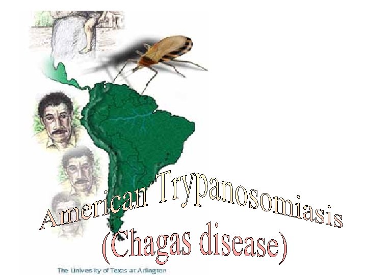
Chagas disease, named for Carlos Chagas, the Brazilian doctor who first described the disease in 1909, is caused by Trypanosoma cruzi, a flagellate protozoan parasite. Case of human trypanosomiasis have been reported in almost all countries of the Americas, including the southern United States, but the main foci are in poor rural areas of Latin America.
Life cycle of Trypanosoma cruzi. • An infected triatomine insect vector (or “kissing” bug) takes a blood meal and releases trypomastigotes in its feces near the site of the bite wound. • Inside the host, the trypomastigotes invade cells, where they differentiate into intracellular amastigotes. • The amastigotes multiply by binary fission and differentiate into trypomastigotes, and then are released into the circulation as bloodstream trypomastigotes
• Trypomastigotes infect cells from a variety of tissues and transform into intracellular amastigotes in new infection sites. • The bloodstream trypomastigotes do not replicate. • Replication resumes only when the parasites enter another cell or are ingested by another vector. • The “kissing” bug becomes infected by feeding on human or animal blood that contains circulating parasites. • The ingested trypomastigotes transform into epimastigotes in the vector’s midgut. • The parasites multiply and differentiate in the midgut.
Epidemiology • Chagas disease is transmitted by cone-nosed triatomine bugs of several genera (Triatoma, Rhodnius, Panstrongylus). • Trypanosoma cruzi can also be transmitted through blood transfusions, organ transplantation, transplacentally, breast milk and in laboratory accidents.
Transmission Methods of T. cruzi • Contamination through the insect's feces is the primary mechanism by which vinchucas pass T. cruzi to humans.
Transmission Methods of T. cruzi • Blood Transfusions and Organ Transplants Blood transfusions are the second most common mechanism of transmission of Chagas' disease to people in Latin America, Europe, and the United States.
Transmission Methods of T. cruzi • Transmission Through Birth Mothers pass T. cruzi on to their children as T. cruzi travels through the placenta, birth canal, and possibly maternal milk. This type of transmission occurs less frequently than other methods. Possibilites include diffusion of the parasite across the extra-embryonic membranes, or through the maternal blood supply.
Geographic • Chagas disease is found only in Latin America
• Natural foci of Chagas disease exist among wild mammals and their associated triatomines. • Humans and domestic animals became involved in the epidemiologic chain several centuries ago, when insects living under wild conditions began adapting to households. • Opossums, armadillos, and wild rodents are reservoirs of the parasite, linking the wild and domestic cycles
Pathogenesis Inflammatory reactions at the sites of rupturing pseudocysts can lead to pathologic manifestations, such as acute myocarditis and destruction of parasympathetic ganglia of the heart and myenteric plexus. An autoimmune reaction may develop.
Clinical Symptoms. The incubation period is 7 -14 days. The human disease occurs in 3 stages: • the acute stage shortly after the infection; • the indeterminate stage; • the chronic stage that may develop over 10 years.
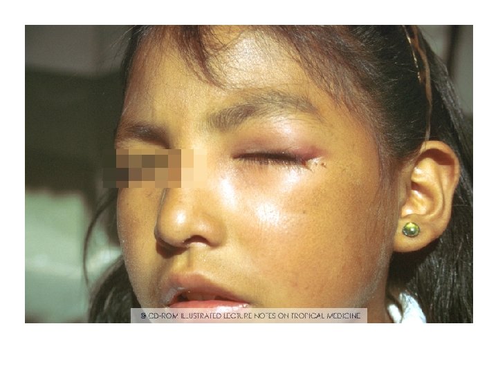
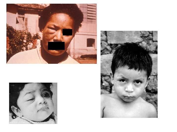
Acute phase of Chagas disease • A local skin nodule called a chagoma can appear at the site of inoculation. • When the inoculation site is the conjunctival mucous membranes, the patient may develop unilateral periorbital edema, conjunctivitis, and preauricular lymphadenitis. (Romaña's sign). • The acute phase is usually asymptomatic, but may present symptoms of fever, anorexia, lymphadenopathy, mild hepatosplenomegaly, and myocarditis.
• • Other symptoms are: tiredness, sometimes a rash, loss of appetite, diarrhea, and vomiting. Infants and very young children can get an often-fatal swelling of the brain.
Indeterminate stage • During the indeterminate stage, about 8 to 10 weeks after infection, infected persons have no symptoms.
Chronic stage of Chagas disease • • • The disease affects the nervous system, digestive system and heart: dementia, damage to the heart muscle (cardiomyopathy), altered heart rate or rhythm, sometimes dilation of the digestive tract (megacolon and megaesophagus), Weight loss. Swallowing difficulties may be the first symptom of digestive disturbances and may lead to malnutrition. Left untreated, Chagas disease can be fatal, in most cases due to the cardiomyopathy component.
Gross anatomy of a heart
The risk factors for getting Chagas disease • • • International travel Undeveloped countries Poorly constructed houses Rural areas Mud houses Adobe houses Thatch houses Assassin bugs Persons with weakened immune systems are at risk of severe infections and complications.
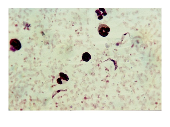
Laboratory Diagnostics • microscopic blood examination, • Xenodiagnosis; • by culturing the blood. • serologic tests : Ш indirect hemagglutination, Ш indirect immunofluorescence, Ш enzyme-linked immunosorbent assay (ELISA)]
Xenodiagnosis • In this test, uninfected vinchucas are placed in a jar and tucked under the armpit of a patient suspected of being infected. • The vinchucas are allowed to consume blood for thirty minutes, and their feces are examined for T. cruzi thirty and sixty days afterward. • This technique is rarely used on children, and many adults have are hesitant in being willfully bitten by vinchucas.
ELISA testing • People without noticeable signs of chagas who live in chagasic areas should be encouraged to have an ELISA test.
Culturing the blood. Amastigotes infecting cells of muscle tissue Trypanosoma cruzi in cultured He. La cells (Giemsa)
Treatment • No effective treatment. • Available drugs only kill extracellular parasites. • Benznidazole and Nifurtinox: current drugs of choice. Required daily for up to 2 months or more. • Hospitalization may be needed because of adverse effects
Preventation • There is no vaccine or drug to prevent Chagas disease. When traveling to areas where Chagas disease occurs, follow these precautions: • Avoid sleeping in poorly constructed thatch, mud, or adobe houses. If that is not possible, use a bednet. • Use insecticides to kill insects and reduce the risk of transmission. • Be aware of the risk of contracting Chagas disease through blood transfusions. In many countries, the blood supply is not well screened.