Trypanosoma spp Trypanosoma pass their life cycle in


 It is found in Eastern and Central Africa.")
 Edema, myocarditis,")


, which are excreted")


 is the simplest")
 test and ELISA. Histopathology: Biopsy examination of lymph")
- Slides: 34
Trypanosoma spp. - Trypanosoma pass their life cycle in 2 hosts vertebrate hosts (definitive hosts) and insect vectors (intermediate hosts). The vector becomes infective to the vertebrate host only after an extrinsic incubation period, during which the parasite undergoes development and multiplication. - In the vector, the trypanosomes follow one or two modes of development and are accordingly classified into 2 groups: Salivaria and Stercoraria. 1. Salivaria (anterior station): In salivaria, the trypanosomes migrate to mouth parts of the vectors, so that infection is transmitted by their bite (inoculative transmission).
Examples are T. gambiense and T. rhodesiense causing African trypanosomiasis, which are transmitted by the bite of tsetse flies. 2. Stercoraria (posterior station): In stercoraria, the trypanosomes migrate to the hindgut and are passed in feces (stercorian transmission), e. g. T. cruzi causing Chagas’ disease, which is acquired by rubbing the feces of the vector bug into the wound caused by its bite.
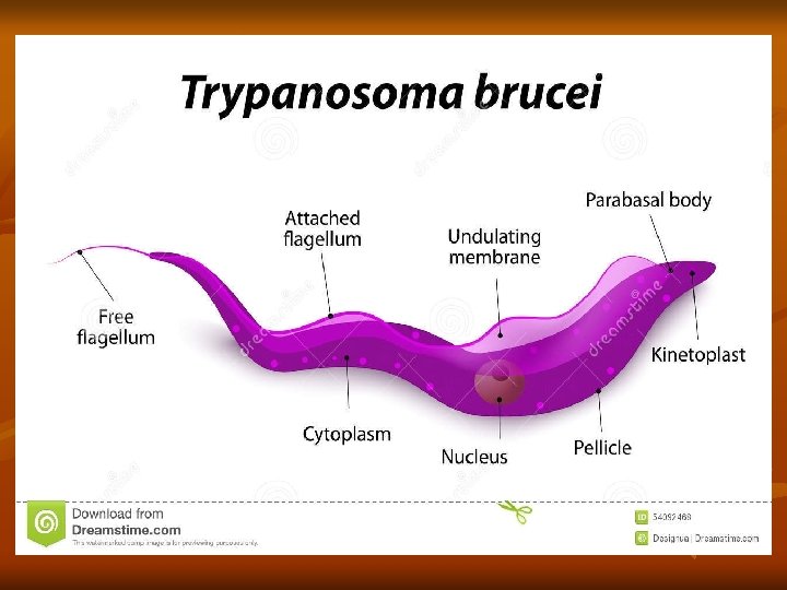
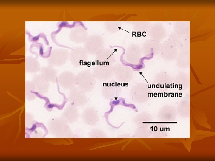
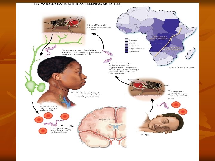
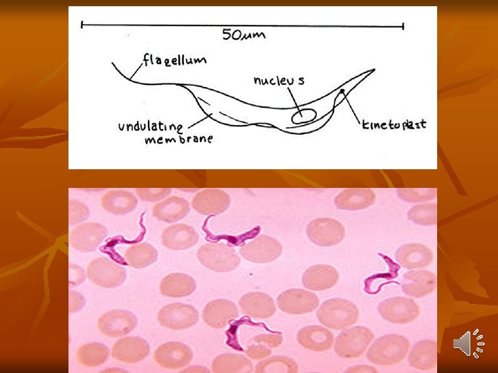
Trypanosoma Brucei Gambiense West African Trypanosomiasis Distribution: - It is endemic in scattered foci in West and Central Africa between 15°N and 18°S latitudes. Habitat: They are essentially a parasite of connective tissue, where they multiply rapidly and then invade regional lymph nodes, blood, and finally may involve central nervous system. Morphology: In the blood of vertebrate host, T. brucei gambiense exists as trypomastigote form, which is highly pleomorphic.
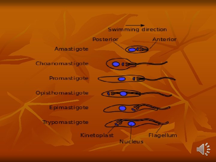
1 -long slender form: 30μ, active motile with free flagellum. 2 -Short stumpy short broad form: 15μ sluggish with attenuated or absent flagellum. 3 -Intermediate form: 20μ with a short free flagellum. In insects, it occurs in 2 forms: - Epimastigotes - Metacyclic trypomastigote forms. Life Cycle: Vertebrate host: Man, game animals, and other domestic animals. Invertebrate host: Tsetse fly. Both male and female tsetse fly of Glosina species (G. palpalis) are capable of transmitting the disease to humans.
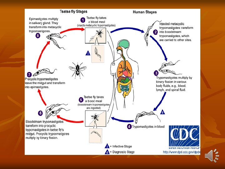
Definitive host: Man Reservoirs: Pigs and others domestic animals. Infective form: Metacyclic trypomastigote forms are infective to humans. Mode of transmission: - By bite of tsetse fly - Congenital transmission. Development in Man : Metacyclic stage (infective stage) of trypomastigotes are inoculated into a man through skin when an infected tsetse fly takes a blood meal. Transforms into short slender form that multiply asexually for 1– 2 days before entering the peripheral blood and lymphatic circulation.
These become ‘stumpy’ via intermediate forms and enter the blood stream. In chronic infection, the parasite invades the central nervous system. Trypomastigotes (short stumpy form) are ingested by tsetse fly (male or female) during blood meal. Development in Tsetse Fly: In the midgut of the fly, short stumpy trypomastigotes develop into long, slender forms and multiply. After 2– 3 weeks, they migrate to the salivary glands, where they develop into epimastigotes, which multiply and fill the cavity of the gland eventually transform into the infective metacyclic trypomastigotes.
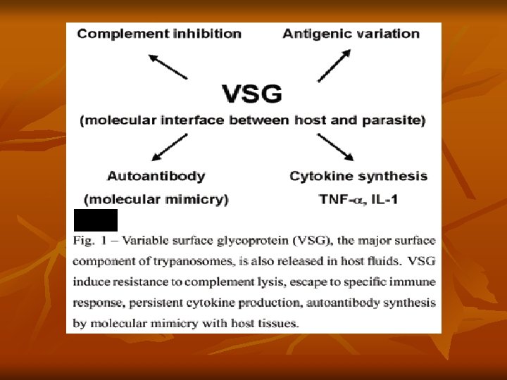
Development of the infective stage within the tsetse fly requires 25– 50 days (extrinsic incubation period). Thereafter, the fly remains infective throughout its life of about 6 months. Antigenic Variation: Trypanosomes exhibit unique antigenic variation of their glycoproteins. - There is a cyclical fluctuation in the trypanosomes in the blood of infected persons after every 7– 10 days. - Each successive wave represents a variant antigenic type (VAT) of trypomastigote posssesing variant surface specific antigens (VSSA) or variant surface glycoprotein (VSG) coat antigen. - It is estimated that a single trypanosome may have as many as 1, 000 or more VSG genes, that help to evade immune response.
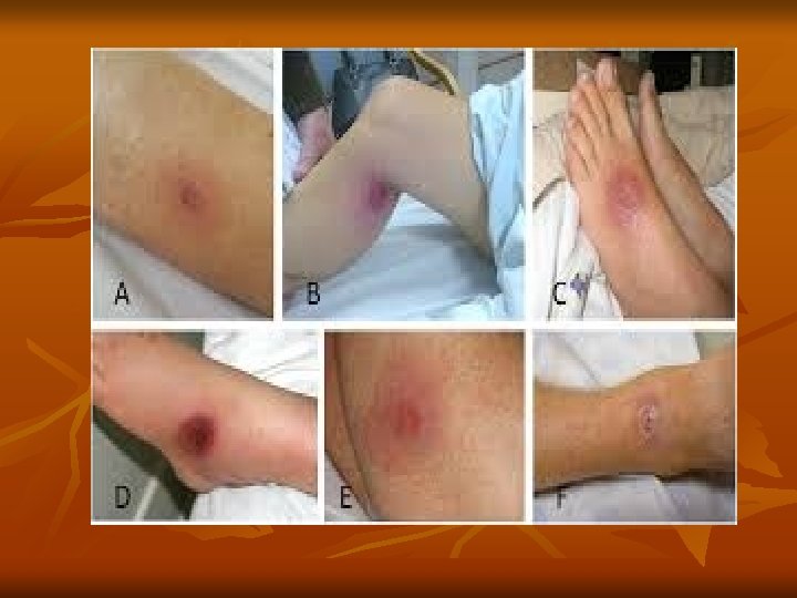
Besides this, trypanosomes have other mechanisms also that help them to evade host immune responses. Pathogenesis and Clinical Features: T. brucei gambiense causes African trypanosomiasis (West African sleeping sickness). The illness is chronic and can persist for many years. There is an initial period of parasitemia, following which parasite is localized predominately in the lymph nodes. A painless chancre (trypanosomal chancre) appears on skin at the site of bite by tsetse fly (it is a small subcutaneous inflammatory nodule, elevated, tender and indurated), followed by intermittent fever, chills, rash, anemia, weight loss, and headache.
Systemic trypanosomiasis without central nervous system involvement is referred to as stage I disease. In this stage, there is hepatosplenomegaly and lymphadenopathy, particularly in the posterior cervical region (Winterbottom’s sign). Myocarditis develops frequently in patients with stage I disease and is especially common in T. brucei rhodesiense infections. Hematological manifestations seen in stage I include anemia, moderate leucocytosis, and thrombocytopenia. High levels of immunoglobulins mainly immunoglobulin (Ig)M are a constant feature. Stage II disease involves invasion of central nervous system. This occurs after several months, the ‘sleeping sickness’ starts.
There is severe headache, mental dullness, apathy, and day time sleeping. Patients falls into profound coma followed by death from asthenia Histopathology shows chronic meningoencephalitis. The meninges are heavily infiltrated with lymphocytes, plasma cells, and morula cells, which are atypical plasma cells containing mulberry-shaped masses of Ig. A. Brain vessels show perivascular cuffing. This is followed by infiltration of the brain and spinal cord, neuronal degeneration, and microglial proliferation. Abnormalities in cerebrospinal fluid include raised pressure, pleocytosis, and raised total protein concentrations.
Trypanosoma Brucei Rhodesiense (East African Trypanosomiasis) It is found in Eastern and Central Africa. The principal vector is G. morisitans which live in the open savannah countries. Although the disease is usually transmitted by the vector from man to man, the disease is actually a zoonosis, with the reservoir being wild game animals like bush buck, antelope and domestic animals like cattle. Morphology and life cycle is similar to T. brucei gambiense. Pathogenesis and Clinical Feature: T. brucei rhodesiense causes East African sleeping sickness. East African trypanosomiasis is more acute than the Gambian form and appears after an incubation period of 4 weeks.
Pathological features are similar in both diseases with some variations. a) Edema, myocarditis, and weakness are more prominent in East African sickness. b) Lymphadenitis is less prominent. c) Febrile paroxysms are more frequent and severe. d) There is a larger quantity of parasite in the peripheral blood. e) Central nervous system involvement occurs early. f) Mania and delusions may occur but the marked somnolence, which occurs in T. brucei gambiense infection is lacking
Diagnosis : 1 - Clincal picture: Chancre, Winterbottom’s sign, Headache, apathy. 2 - Finding the trypanosomes in the punctures from the chancre, during febrile stage in blood, bone marrow or lymph node aspiration. The films are best stained with Giemsa. 3 - Examination of C. S. F. in early neurological signs. There is increase in WBC and protein content with decrease of sugars. Centrifugation and examination of the stained sediment shows the trypanosomes and the characteristic morula cells of Mott, these are modified plasma cells (up to 20 μ in diameter) with large eosinophilic inclusions.
4 - Animal inoculation of patient’s blood or CSF in ("mice or hamster". This is the most sensitive method for diagnosis of T. b. rhodesiense. The parasites appear in the blood of laboratory animals within 4 -7 days with changing to posterior nuclear shift. T. b. gambiense does not infect laboratory animals. 5 - Culture of the blood, CSF and lymph node aspirates on N. N. N. or GLSH (glucose, lactalbumin, serum and hemoglobin) media to show the parasite in cases when the parasite cannot be demonstrated by other methods. 6 - Serological tests which are useful for early diagnosis in endemic areas because symptoms are less severe in early infection of native inhabitants. ELISA, IHAT, and IFAT can be used
Treatment : For the acute stages of the disease the drug of choice is suramin with pentamidine as an alternative. In chronic disease with CNS involvement, the drug of choice is soprol. Alternatives include tryparsamide combined with suramin. Prevention and control: 1 - Personal protection from tsetse fly bites. 2 - Eradication of insect intermediate host (tsetse flies). 3 - Treatment of infected cases to reduce the source of infection. 4 - Health education.
Trypanosoma Cruzi T. cruzi is the causative organism of Chagas’s disease or South American trypanosomiasis. Distribution: It is a zoonotic disease and is limited to South and Central America. Definitive host: Man Intermediate host (vector): Reduviid bug or triatomine bugs. Reservoir host: Armadillo, cat, dog, and pigs. Habitat: - In humans, T. cruzi exists in both amastigote and trypomastigote forms. a) Amastigotes are the intracellular parasites. They are found in muscular tissue, nervous tissue, and reticuloendothelial system. b) Trypomastigotes are found in the peripheral blood
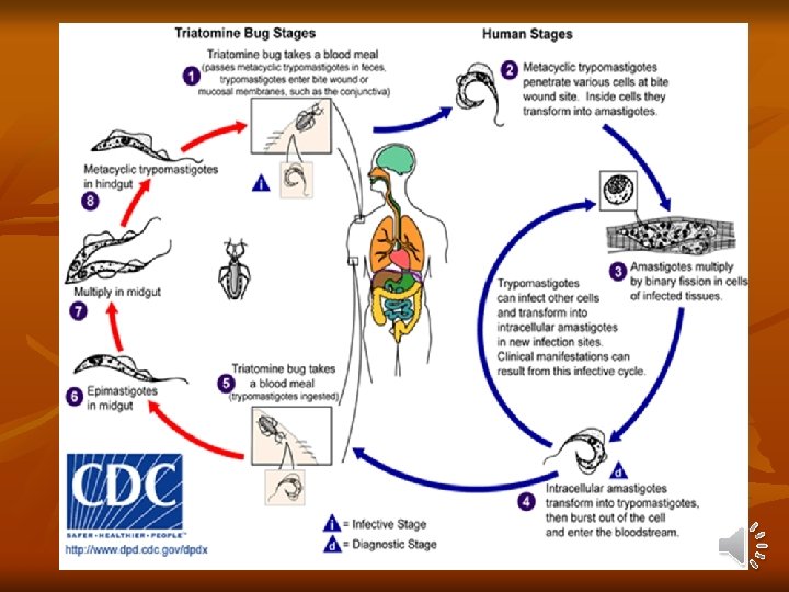
Infective stage: Metacyclic trypomastigotes forms are the infective forms found in feces of reduviid bugs. Mode of transmission: a) Bite of reduviid bug which defecate during the process of feeding. Insect deposits infective feces near or at the bite site and takes place when mucus membranes, conjunctiva, or wound on the surface of the skin is contaminated by feces of the bug containing metacyclic trypomastigotes. b) Blood transfusion, organ transplantation, and congenital. Life Cycle: T. cruzi passes its life cycle in 2 hosts. Development in Man: - Metacyclic trypanosomes do not multiply in this form. They enter the cells of the reticuloendothelial system and striated muscles especially of the heart where they round up and transform into leishmanoid form.
2 -They multiply by binary fission, destroying the host cells and forming cyst-like nests of parasites (pseudocyst). 3. The leishmanoid form turns into trypanosome form, which re-enter the blood to be picked up by the winged bugs and repeat its life cycle. - Trypomastigotes, which are released into the blood stream and are the infective stage for triatomine bug. Development in Reduviid Bugs: - Bugs acquire infection by feeding on an infected mammalian host. - Most triatomine bugs are nocturnal. - The trypomastigotes are transformed into epimastigotes in the midgut, from where they migrate to the hindgut and multiply.
- These, in turn, develop into non-dividing metacyclic trypomastigotes (infective form), which are excreted in feces (stercorarian transmission). - The development of T. cruzi in the vector takes 8– 10 days, the extrinsic incubation period. Pathogenesis and Clinical Features: Incubation period of T. cruzi in man is 1– 2 weeks. The disease manifests in acute and chronic form. Acute Chagas’ disease: - Acute phase occurs soon after infection and may last for 1– 4 months. - It is seen often in children under 2 years of age. - First sign appears within a week after invasion of parasite.
- Chagoma' is the typical subcutaneous lesion occurring at the site of inoculation. Inoculation of the parasite in conjunctiva causes unilateral, painless edema of peri ocular tissues in the eye called as Romana’s sign. This is a classical finding of the acute Chagas' disease. - In few patients, there may be generalized infection with fever, lymphadenopathy, and hepatosplenomegaly. - Patient may die of acute myocarditis and meningoencephalitis. - Usually within 4– 8 weeks, acute signs and symptoms resolve spontaneously and patients then enter the asymptomatic or indeterminate phase of chronic T. cruzi infection
Chronic Chagas’ Disease: - The chronic form is found in adults and older children and becomes apparent years or even decades after the initial infection. - In chronic phase, T. cruzi produces inflammatory response, cellular destruction, and fibrosis of muscles and nerves, that control tone of hollow organs like heart, esophagus, colon, etc. Thus, it can lead to cardiomyopathy, megacolon (dilatation of the colon leading to constipation) or megaoesophagus (dilatation of oesophagus leading to dysphagia). Congenital Infection: - Congenital transmission is possible in both acute and chronic phase of the disease causing myocardial and neurological damage in the fetus
Diagnosis: Microscopy: Microscopic examination of fresh anticoagulated blood (thin blood film) is the simplest way to see motile organisms. Culture: Blood or aspirates from chagoma or enlarged lymph mode cultured on Novy, Neal, and Nicolle (NNN) medium. Xenodiagnoses: In negative blood film and during the early phase of the disease. - Laboratory reared Reduviid bugs are starved for 2 weeks. They are then fed on patients blood. If trypomastigotes are ingested, they will multiply and develop into epimastigotes and trypomastigotes, which can be found in the feces of the bug 2
Serological diagnosis: Indirect immunefluorescence assay (IFA) test and ELISA. Histopathology: Biopsy examination of lymph nodes and skeletal muscles and aspirate from Chagoma may reveal amastigotes of T. cruzi. Treatment : 1 - Nifurtimox: Oral dose of 15 mg/kg daily in 4 divided doses for 3 months. 2 - Benznidazole : Oral dose of 6 mg/kg dialy for about 2 months. Prevention and control: 1 - Eradication of insect intermediate host (winged bug). 2 - Treatment of infected cases to reduce the source of infection. 3 - Health education.