Trilaminar Embryonic Disc The Third Week The significant

Trilaminar Embryonic Disc The Third Week The significant event of third week is Gastrulation

Gastrulation • The process by which the bilaminar disc is converted into a trilaminar disc • It is the beginning of morphogenesis (formation of body form) • Consists of formation of the primitive streak, the three germ layers & the notochord • Embryo is referred to as a Gastrula

Primitive Streak • The primitive streak results from proliferation of the epiblastic cells in the median plane, in the caudal half of the epiblast, and lies along the cranio-caudal axis. • Its cranial end forms primitive node. • A groove, primitive groove, appears in the primitive streak, which continues with a small depression, primitive pit, in the primitive node.

• A circular thickening appears in the hypoblast near the cranial end, in the midline, to form the prechordal plate, that marks the future site of mouth • A circular thickening appears in the hypoblast caudal to primitive streak in the midline to form the cloacal membrane, the future site of the anus

• By this stage of development, it is possible to identify the embryo’s: vcraniocaudal axis vcranial and caudal ends vdorsal and ventral surfaces vright and left sides. Connecting stalk
 proliferate")
Formation of Intraembryonic Mesoderm • The epiblastic cells from the primitive streak (groove) proliferate to form mesenchymal tissue • The newly formed cells invaginate, migrate ventrally, laterally & cranially between the epiblast and hypoblast & organize to form the intraembryonic mesoderm

Formation of Intraembryonic Mesoderm cont. • Intraembryonic mesoderm merges with the extraembryonic mesoderm at the periphery of the embryonic disc • By the end of 3 rd week, mesoderm lies between embryonic ectoderm and endoderm everywhere except in the region of prechordal plate and cloacal membrane, as the embryonic ectoderm & endoderm are fused at these regions

The endoderm, posterior to the region of cloacal membrane, forms a small outgrowth or diverticulum that is called the allantoenteric diverticulum or the allantois.

Formation of Intraembryonic Mesoderm cont. • Some mesenchymal cells displace the hypoblasts forming the embryonic endoderm • Cells remaining in the epiblast form the embryonic ectoderm

Thus the EPIBLAST gives rise to all three germ layers, Ectoderm, Mesoderm, Endoderm in the embryo Each of the three germ layers gives rise to specific tissues and organs

• Cell migration and specification are controlled by fibroblast growth factor 8 (FGF 8), which is synthesized by streak cells themselves. This growth factor controls cell movement by downregulating E-cadherin, a protein that normally binds epiblast cells together. FGF 8 then controls cell specification into the mesoderm by regulating Brachyury (T) expression • Bone morphogenetic protein 4 (BMP 4), is secreted throughout the embryonic disc. In the presence of this protein and fibroblast growth factor (FGF), mesoderm will be ventralized to contribute to kidneys (intermediate mesoderm), blood, and body wall mesoderm (lateral plate mesoderm). In fact, all mesoderm would be ventralized if the activity of BMP 4 were not blocked by other genes expressed in the node

The Fate of Primitive streak • The primitive streak continues to supply new cells until the end of the 4 th week. It regresses gradually and become located at the caudal end of the embryo and then disappears. • Insufficient mesoderm formed in the caudal region may results is congenital defects, involving lumbosacral vertebrae or urogenital system or lower extremities. This is termed Caudal dysgenesis or sirenomelia. • Sacrococcygeal Teratoma.

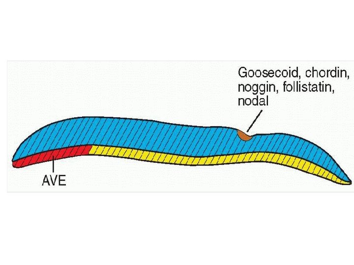
Molecular Determination • Cephalic end is determined by presence of anterior visceral endoderm --- transcription factors OTX 2, LIM 1, HESX 1, Cereberus. • Embryonic disc produces: BMP 4, + FGF (fibroblast growth factor), these regulate formation of mesoderm ---- ventrilization. • Primitive node --- Goosecoid factor --- chordin, noggin, fillostatin and NHE 3 B: antagonist of BMP 4 and important for notochord formation and neural induction.

• In caudal region Brachyury T gene is expressed in the notochordal cells --- DNA-binding proteins, specific for certain several genes (T box) family, important for growth of caudal region of embryo. Deficiency of this factor ---caudal dysgenesis.

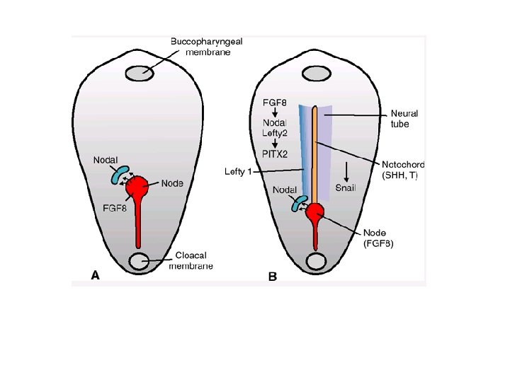
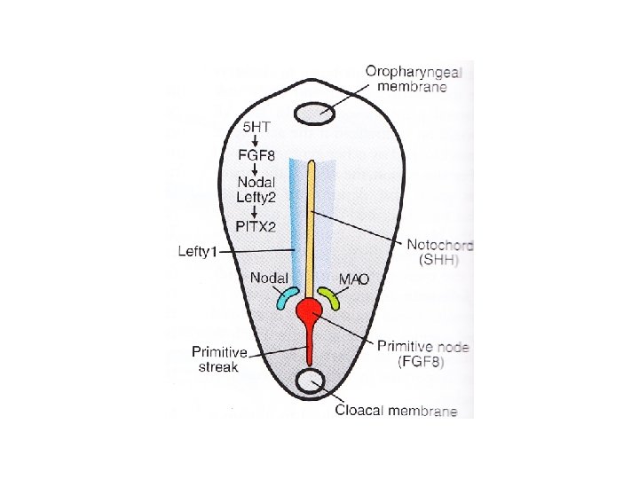
Left and Right sides • Certain genes are expressed in left side but not in the right side: FGF-8, Lefty-2, PITX 2, Serotonin(5 HT) • SHH (sonic hedgehog) gene is expressed in the midline • In right side: snail-transcription factor.

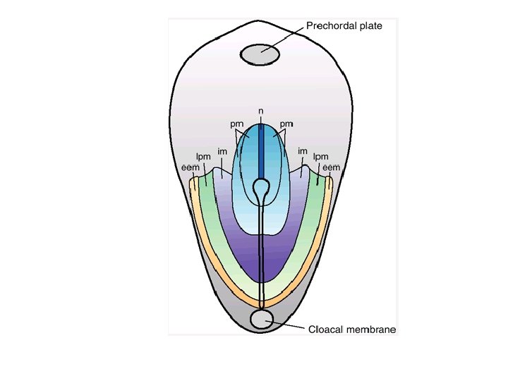
Fate Map • Cells from specific regions of Epiblast , later on form specific part or structure of the embryo • Cells invaginate through the primitive node --notochord • Cells from lateral side of the node and cranial part of primitive streak --- paraxial mesoderm • Cells migrating through mid streak --- intermediate mesoderm • Cells migrating through caudal part of the streak ----lateral mesoderm

Congenital abnormalities • • • Holoprosencephaly ---- alcohol Caudal dysgenesis---sirenomelia Situs inversus Laterality sequences Sacrococcygeal Teratoma

Notochord • A rod of mesenchymal cells located cranially, in the midline, extending between the primitive node and the prechordal plate

Formation of Notochord • Mesenchymal cells migrate cranially from primitive pit toward the prechordal plate, and form a rod like notochordal process • The notochordal process becomes canalized forming a hollow tube, the notochordal canal, communicating with the primitive pit.

Formation of Notochord cont. • The floor of the tube and the underlying endoderm break down, forming a notochordal plate • The notochordal plate becomes continuous with the endodermal layer.

Formation of Notochord cont. • A temporary communication is established between the amniotic cavity and the yolk sac, termed the neurenteric canal.

Dorsal view of a 16 -day embryo

Notochordal plate folds to form the notochord.

Functions of Notochord • Defines primordial axis of the embryo • Provides rigidity to the embryo • Serves as a basis for the development of the axial skeleton • Indicates the future site of the vertebral bodies/column • Regulates differentiation of surrounding structures including the overlying ectoderm (neural plate) and mesoderm (somites).

Fate of Notochord • Degenerates and disappears as the bodies of the vertebrae develop, but it persists as the nucleus pulposus of each intervertebral disc • Remnants of notochordal tissue give rise to tumors called Chordomas

Differentiation of the Intraembryonic Mesoderm • Induced by the notochord • Differentiates (in the region of notochord) into: – Paraxial mesoderm – Intermediate cell mass – Lateral plate mesoderm

Origin of embryonic tissues

- Slides: 32