Trichiasis Conjunctiva Sclera Look at the bulbar the

Trichiasis
 & palpebral (inside of the")
Conjunctiva & Sclera Look at the bulbar (the eye) & palpebral (inside of the lids) conjunctiva Injection & erythema; what is the distribution Discharge; watery, mucous or membranous What do I see?

Scleritis or episcleritis

Scleritis • Red painful eye with decreased vision • Often associated with underlying collagen vascular disease • RA, Lupus • Diffuse, Nodular, Necrotizing forms • Refer!! • Requires systemic immunosuppression • Indocin, Prednisone, Cyclosporin, Cytoxan

Rheumatoid Arthritis

Subconjunctival Hemorrhage • Dramatic but harmless • • Associated with anticoagulation • • Sneezing, coughing, straining, eye rubbing Aspirin If no obvious cause and associated with bruising or repetive than: CBC, Platelet count, Bleeding time, PT/PTT

Subconjunctival Hemorrhage

Pterygium
 • Elastoid degeneration (wrinkle) • Often become")
Pterygium • Benign fibrovascular tumor (UV induced) • Elastoid degeneration (wrinkle) • Often become inflamed • Treatment: • Artificial Tears, Sunglasses, Short term use of vasoconstrictors • Refer if large or conservative fails • Conjunctival Auto graft with Glue

Pingueculum

Bacterial Conjunctivitis

Conjunctivitis: Bacterial • Redness and mucopurulent discharge • • • Minimal discomfort Vision minimally affected Treatment • Will resolve without treatment • Polytrim (polymixin-trimethoprim) q 2 hours the first day then QID for 1 week

Gonoccocal Conjunctivitis

Hyperacute Purulent Conjunctivitis • Sudden onset with rapid progression • Bilateral

Case 4
 • U. resp. Infection • History of contact • • VERY")
Conjunctivits: Viral (EKC) • U. resp. Infection • History of contact • • VERY CONTAGIOUS Sx’s: Photophobia, redness, watery discharge • Bilateral but asymmetric • Preauricalar node • Treatment: None--Avoid Topical Steroids!!

Allergic Conjunctivitis

Conjuntivitis: Allergic • Itching • Seasonal • Bilateral • Mucopurlent discharge, no pre-auricular node • Redness, Chemosis

Allergic Conjunctivitis: Treatment • Avoidance • Associated with Dry Eye • Wash eyes out with tears • Cold Compresses • Ocular antihistamines/mast cell stabilizers • Patenol, Alocril, Zaditor
 Pterygium Epithelium (use fluorescein dye & a")
Cornea Clarity Haze, or scars (including surgical) Pterygium Epithelium (use fluorescein dye & a cobalt blue filter to examine the epithelium for defects including punctate erosions, abrasions, ulcers, dendrites) What do I see?

Case 5

Abrasion • History of Trauma or Contact Lens wear • • Very Painful: More pain nerves per mm than any other location Diagnosis: • Drop of Proparacaine • Flouroscein lights up epithelial defect

Treatment • Relief of Pain and Rapid Visual Rehabilitation • Antibiotic ointment, dilation, patch • Bandage Contact lens • • With Antibiotic Drops • Topical NSAID: Acular or Voltaren Recommend Follow-up (Infection)

Patching

Dry Eye • Postmenopausal women • Sometimes associated with Arthritis • Lupus, RA, Sjorgren’s • Often related to climate/humidity • Exacerbated by systemic medications • Diuretics, antihistamines, and anti-depressant

Dry Eye: Symptoms • Foreign body sensation • Photophobia • May complain of redness • Associated blepharitis or allergic conjunctivitis is common

Dry Eye: Diagnosis • Schirmer’s test • Fluorescein staining • White, quiet eye is common

Flourescein Staining

Rose-Bengal

Schirmer Test Without anesthesia • Measures reflex tear secretion With anesthesia • Eliminates stimulated tearing

Dry Eye: Treatment • Artificial Tears • Watch for preservative toxicity • Saturation therapy • Preservative free drops • • If using more than 4/day Consider punctal occlusion or Restasis (Cyclosporine)
 in lipid vehicle • Treats surface inflammation • Inhibits")
Restasis • Cyclosporine (. 05%) in lipid vehicle • Treats surface inflammation • Inhibits T-cell infiltration of lacricmal gland • Burns on instillation • Administer BID

Dendrite

Treatment of HSV Keratitis • Topical Antivirals Trifluridine • Systemic Acyclovir or Famvir if immunosuppressed or extensive associated skin lesions

Chemical Injuries • Acid or Alkali? • Cation determines speed of penetration • • NH 4+, Na+, K+, Ca++ (OH) Battery Explosions • Chemical plus blunt force trauma • Foreign body

Chemical Injuries • Irrigate, Irrigate and Irrigate • • Topical anesthetic, 7 th nerve block helpful Prognosis determined by: • Type of chemical (acid vs. alkalai) • p. H • Length of exposure • TIME BETWEEN EXPOSURE AND IRRIGATION REFER as soon as possible

Corneal foreign body

Corneal scar

Anterior chamber Clarity; measured by cells & flare Depth

Hypopyon

Hyphema

Cell & Flare

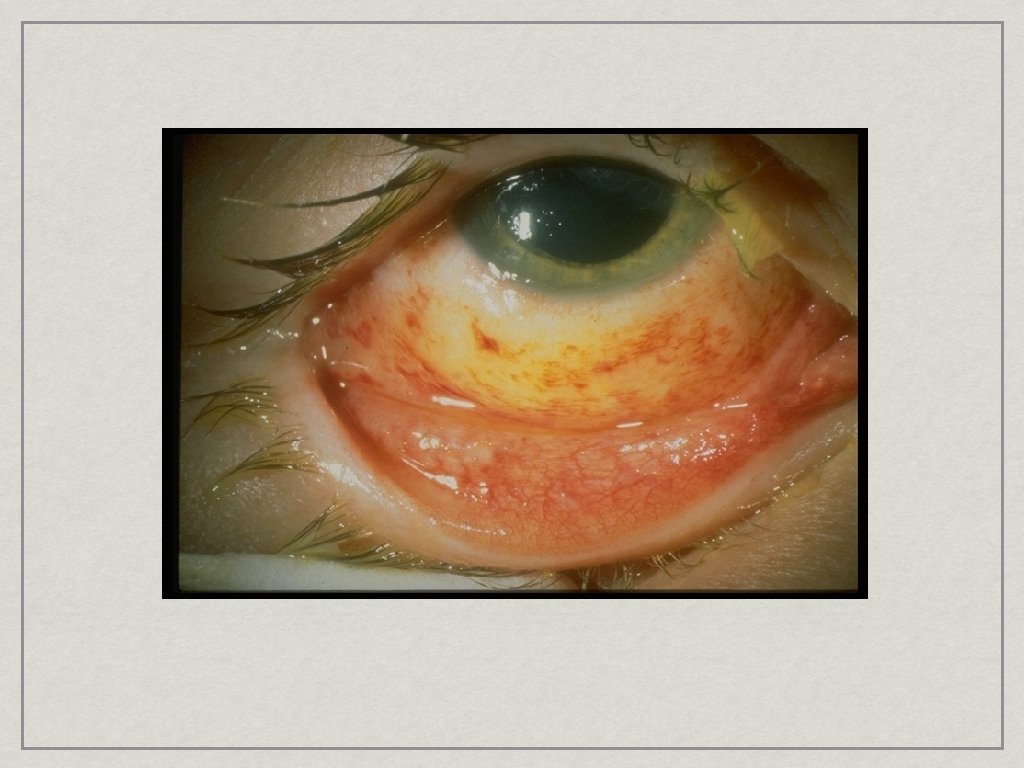
Iritis/Uveitus • “Arthritis of the Eye” • Associated with Collagen Vascular disease • HLA-B 27 associated • Crohn’s disease, RA, Lupus • Sign & Symtom: Photophobia, Floaters, Red Eye, Pain, Decreased vision • Circumlimbal flush

Iritis

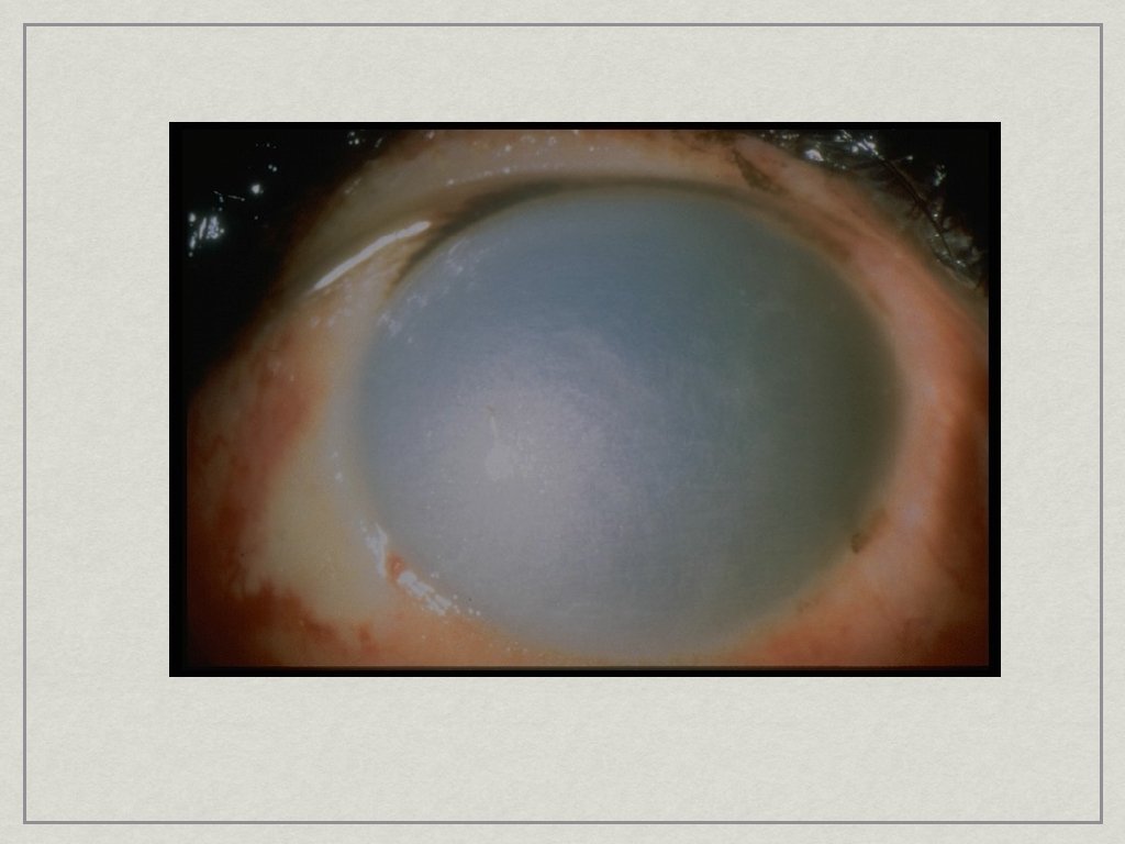
Lens Best examined through a dilated pupil Senile cataracts can appear white or yellow

Cataract

Intraocular lens

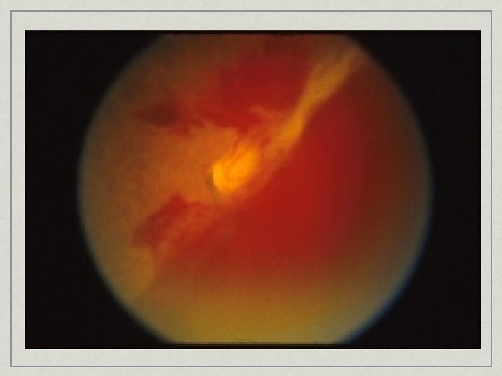
Dilated fundoscopic exam Red reflex with direct ophthalmoscope Dilate with phenylephrine 2. 5% & tropicamide 1% (not used in infants) Get close with the direct ophthalmoscope Vitreous clarity (hemorrhage) Nerve, vessels, macula & periphery with direct ophthalmoscope

Papilledema

Diabetic retinopathy

Vitreous Hemorrhage • Sudden onset of painless decrease in vision • Floaters • Often Diabetic • Dx: No red reflex

Macular degeneration
- Slides: 55