Treatment of osteoporosis Anabolic agents S Kalbasi M

Treatment of osteoporosis Anabolic agents S. Kalbasi, M. D Research Institute for Endocrine Sciences Shahid Beheshti University of Medical Sciences 12 Jun , 2013 Tehran

ANABOLIC AGENTS IN OSTEOPOROSIS TREATMENT

Bone Remodeling Cycle in Normal Bone Osteoclasts digest bone within a sealed resorption vacuole Resorption Bone Reversal Resting Apoptotic osteoclasts Lining cells Mature osteoblasts building osteoid tissue Mineralization Bone Preosteoblasts Bone Formation Illustration Copyright © 2009 Nucleus Medical Art, All rights reserved. www. nucleusinc. com

Many Factors Regulate Bone Remodeling Resorption Formation GM-CSF IL-1 IL-6 RANKL PGE 2 TNF- Bone Reversal Bone Formation Resorption Bone Abbreviations: GM-CSF, granulocyte macrophage colony-stimulating factor; OPG IL, interleukin; OPG, osteoprotegerin; RANKL, RANK-ligand, TGF- PGE 2, prostaglandin E 2; TGF, transforming growth factor; Estrogen TNF, tumor necrosis factor. Illustration Copyright © 2009 Nucleus Medical Art, All rights reserved. www. nucleusinc. com

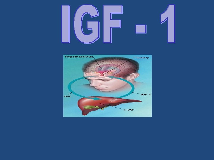
INTRODUCTION : Two important signals that induce the differentiation of osteoblastic lineage cells into mature osteoblasts are bone orphogenetic proteins and Wnt Another important regulator of osteoblast function is insulin-like growth factor I (IGF-I). Anabolic therapeutic agents might be designed to stimulate the actions of these osteoblastic signals directly or to target their extracellular and intracellular regulators. Signals Determining Mesenchymal-Cell Differentiation toward Osteoblasts and Signals Acting on Mature Osteoblasts to Enhance Bone Formation. NEJM 2007

MECHANISMS of ANABOLIC AGENTS Osteoblasts, the cells responsible for bone formation, are rational therapeutic anabolic targets. increase the number of osteoblast precursors. stimulate the differentiation of osteoblasts into mature cells. Enhance osteoblastic function or survival. Finally leads to a net gain of bone tissue. NEJM 2007

The anabolic skeletal agents, in contrast to the antiresorptive drugs, influence processes associated with bone formation. anabolic agents have the potential to positively affect a number of skeletal properties besides bone density. These include bone size and microarchitecture. NEJM 2007
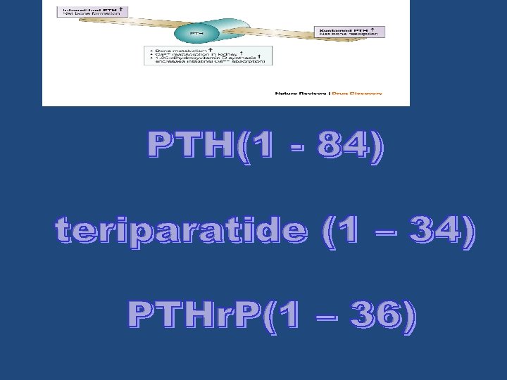

Pharmacokinetics • The peak circulating concentration of teriparatide reached 45– 60 minutes after administration ; complete disappearance of teriparatide occurs within 4– 6 hours. • PTH(1 -84) has a longer half-life than teripara-tide, with serum levels peaking at 120 minutes and disappearing from the circulation after 8 hours. • PTHr. P has a very short half-life, with serum levels peaking at 15 minutes and disappearing from the circulation after only 1– 2 hours. Parathyroid Hormone as an Anabolic Skeletal Therapy. Drugs 2005
 PTH signals through the PTH-1 receptor, a G protein– coupled protein,")
MECHANISMS PTH (1) PTH signals through the PTH-1 receptor, a G protein– coupled protein, which mediates most of the functions of PTH and of its evolutionary relative, PTH-related peptide (PTHr. P). PTH–PTHr. P receptor, it is activated by peptide sequences that include the N-terminal region of either molecule. It is unclear why the intermittent administration of low-dose PTH differs in its effect on bone cells from long-term, sustained PTH exposure in which catabolic effects at cortical sites predominate. NEJM 2007
 The exact signaling pathway responsible for the anabolic effect is not known, but")
(2) The exact signaling pathway responsible for the anabolic effect is not known, but : • The Wnt –β- catenin pathway : of the Wnt antagonist sclerostin is down-regulated by PTH • The anabolic actions of PTH involve direct effects on osteoblast lineage cells and indirect effects through growth factors (e. g. , IGF-I) and growth factor antagonists, such as sclerostin. • Mitogenic properties for osteoblastic cells and decreases osteoblastic apoptosis.

• PTH stimulates preosteoblasts to mature into boneforming osteoblasts that lay down collagen and subsequently mineralize matrix. • Bone formation begins within the first month of PTH treatment and peaks six to nine months after initiation of daily PTH. • Since the remodeling unit is always coupled (ie, bone formation equals bone resorption), once preosteoblasts are stimulated, they release cytokines that can activate osteoclasts resulting in bone resorption.
 • beneficial effects of teriparatide on bone qualities such as bone density, microarchitecture")
(3) • beneficial effects of teriparatide on bone qualities such as bone density, microarchitecture and bone geometry are seen in the cancellous skeleton • on the other hand , PTH stimulates periosteal apposition, which leads to increases in cortical area, cortical thickness, and an overall increase in cross-sectional area
 • PTH likely initially stimulates processes associated with bone formation (bone modeling) and")
(4) • PTH likely initially stimulates processes associated with bone formation (bone modeling) and only later promotes those associated with bone remodeling in which bone resorption predominates. • This sequence of events has led to the concept of the “anabolic window, ” a period of time when the actions of PTH are maximally anabolic

PTH Action on Bone Major action = stimulation of osteoclastic bone resorption However, a classical series of studies elucidated effects on osteoblasts: • PTH extract → increase in skeletal calcium (F. Albright, 1929, H. Seyle, 1932) • PTH receptor primarily located in osteoblast (Rouleau, 1986, 1989) • PTH increases osteoblast number and osteoid formation in mouse radii (Herrmann, 1976) • PTH stimulates osteoblast replication (De. Bartolo, 1982, Canalis, 1989) Anabolic action of PTH is due to stimulation of osteoblast proliferation, and subsequent stimulation of mature osteoblast function

PTH – Mechanism of Action Once daily Osteoblast apoptosis Lining cell differentiation Continuous Rank. L OPG Osteoblast number Osteoclast Bone formation Bone mass Bone strength Bone resorption Serum calcium

FPT study Effect of parathyroid hormone on fractures and BMD in postmenopausal women with osteoporosis Subjects: 1637 post menopausal women with prior vertebral fractures Interventions: 20 mcg PTH , 40 mcg PTH Vs placebo Follow up : 21 mo Primary end points: Vertebral radiographs for fracture , BMD Neer et al, N Engl J Med 2001; 344: 1434

Effect of Teriparatide on Risk of Vertebral Fractures in Postmenopausal Women % of Patients with ≥ 1 Fracture 16 14 12 10 8 65% 6 4 2 0 a RR 0. 35 (95% CI = 0. 22– 0. 55) Placebo Teriparatide 20 µg P <. 001 vs placebo. Neer RM, et al. N Engl J Med. 2001; 344: 1434 -1441. Graphic courtesy of Dr. Paul Miller. a
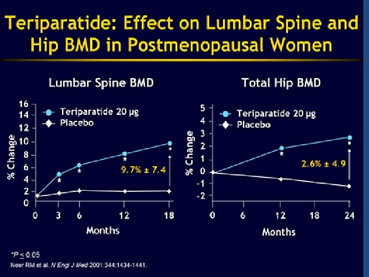

Nonvertebral and Hip Fractures 20 Teriparatide Percent with New Fractures Control Teriparatide 20 µg 15 10 RR = 0. 5 (0. 3, 0. 9) 5 NS 0 Nonvertebral Fractures 5 fragility hip fractures (control + primary treatment group). Nonvertebral fractures = fragility fractures, otherwise not specified. Neer R, et al. N Engl J Med. 2001; 344: 1434– 1441. Graphic courtesy of Dr. Paul Miller. Hip Fractures

New Vertebral fracture rate Non vertebral fractures Number of Hip fractures Placebo 544 20 mcg 541 40 mcg 552 14% 5% 4% 6% 3% 3% 4 2 3

Effect of PTH on Fracture Risk 69% † 54% § RR=0. 35, 95% CI=0. 22 -0. 55. ‡ RR=0. 47, 95% CI=0. 25 -0. 88. † RR=0. 31, 95% CI=0. 19 -0. 50. § RR=0. 46, 95% CI=0. 25 -0. 86. * Data from Neer RM, et al. N Engl J Med. 2001; 344: 1434 -1441.
 IN POSTMENOPAUSAL WOMEN: There is level 1 evidence that")
Beneficial effects of teriparatide (1) IN POSTMENOPAUSAL WOMEN: There is level 1 evidence that h. PTH (1– 34) 20 μg/d and 40 μg/d increase BMD at the lumbar spine and proximal femur and decrease the risk of both vertebral and nonvertebral fractures in postmenopausal women with prior vertebral fractures The decline in BMD at the distal radius seen in h. PTH trials BUT microarchitectural changes due to teriparatide are evident at the cortical sites such as the distal 1/3 radius as well. These geometrical and microarchitectural changes strengthen cortical bone despite the small reduction in bone density Parathyroid hormone for the treatment of osteoporosis : a systematic review ; CMAJ • July 4, 2006.
 There is level 1 evidence that h. PTH(1– 84) increases lumbar spine BMD")
(2) There is level 1 evidence that h. PTH(1– 84) increases lumbar spine BMD in postmenopausal women with osteoporosis incidence of baseline fragility fractures in the phase III PTH(1 -84) study was only 19%. Initial studies in postmenopausal women with osteoporosis suggested that PTHr. P at a daily dose of approximately 400 μg for 3 months increases vertebral BMD by 4. 7%. Serum osteocalcin was increased, but serum calcium and biochemical markers of bone resorption were not affected. Larger and longer trials are required
 IN CORTICOSTEROID – INDUCED OSTEOPROSIS: • There is level 2 evidence that h.")
(3) IN CORTICOSTEROID – INDUCED OSTEOPROSIS: • There is level 2 evidence that h. PTH(1– 34) increases lumbar spine BMD in postmenopausal women with corticosteroid-induced osteoporosis. • Fracture data are lacking in this population. RESULT IN MEN : • There is level 1 evidence that h. PTH(1– 34) increases BMD at the lumbar spine and femoral neck in men with osteoporosis. • Neer etal reported : the risk of vertebral fracture was reduced by 51% (p= 0. 07)
 Health-related quality of life and back pain • There is no evidence of")
(4) Health-related quality of life and back pain • There is no evidence of improved healthrelated quality of life associated with h. PTH (1– 34) or calcium with vitamin D in postmenopausal women with osteoporosis. • There is level 2 evidence that back pain may be reduced in patients given h. PTH (1– 34).

SEQUENTIAL THERAPY WITH TERIPARATIDE AND ANTIRESORPTIVE AGENT WITH ESTROGEN : • Cosman et al. treated postmenopausal women, previously given estrogen for at least 1 year, with teriparatide. Increases in vertebral BMD began with no delay and increased in a linear fashion during the entire 3 -year study. WITH RALOXIFEN : • raloxifene did not impede the effects of teriparatide to increase BMD rapidly and linearly WITH ALENDRONATE: • • alendronate was associated with a 6 - month delay before BMD in the lumbar spine began to increase. some practitioners advocate a 6 -month hiatus between discontinuation of treatment with alendronate and initiation of teriparatide therapy. Others suggest that teriparatide therapy be initiated immediately after the bisphonate. Anabolic Skeletal Therapy for Osteoporosis. (Arq Bras Endocrinol Metab 2006
 • h. PTH(1– 34) results in larger increases in lumbar")
combination therapy: with alendronate(1) • h. PTH(1– 34) results in larger increases in lumbar spine BMD than alendronate • There is level 1 evidence that combination therapy with alendronate and h. PTH(1– 84) may blunt the anabolic effect of h. PTH on BMD.

In previously untreated patients, the effects of PTH are not additive to those of a Bisphonate. These observations appear to result from the ability of alendronate to impair PTH-induced stimulation of new bone formation. Thus, we do not suggest combination PTH and bisphonate therapy for osteoporosis.
 • Combination of teriparatide and raloxifene may have more beneficial effects than")
With raloxifen(2) • Combination of teriparatide and raloxifene may have more beneficial effects than monotherapy with teriparatide in postmenopausal osteoporosis. • Increased to a similar extent in the lumbar spine and femoral neck in both groups, but the increase in total hip BMD was significantly greater in subjects treated with both teriparatide and raloxifene. • Bone formation markers increased similarly in both groups. Bone resorption markers, however, were reduced in the combination group.
 withdrawal (a) when PTH is followed")
Changes in bone density after parathyroid hormone (PTH) withdrawal (a) when PTH is followed with antiresorptive therapy, bone resorption markers fall more rapidly leading to an increase in bone mineral density (BMD); and (b) when PTH is not followed by antiresorptive therapy, bone formation markers fall more rapidly leading to a decrease in bone density. Consequently, it is advisable to administer an antiresorptive agent such as a bisphonate after treatment with teriparatide many experts recommend the continuation of long-term antiresorptive therapy for its own therapeutic benefits.

Indications for Teriparatide q. In the US : postmenopausal women and men with osteoporosis who are at high risk for fracture. The definition of high risk: The T-score very low (i. e. <-3. 0 itself, even without an osteoporotic fracture or a strong family history of osteoporosis. q In many countries in Europe : teriparatide cannot be administered unless a patient has received a previous, unsuccessful course of bisphonate therapy and has had a previous osteoporotic fracture. These restrictive indications are due, in part, to the fact that teriparatide is expensive and is administered by daily subcutaneous injection.
 Teriparatide is available throughout most of the world, but full-length PTH (1– 84)")
) Teriparatide is available throughout most of the world, but full-length PTH (1– 84) is available only in Europe. Duration of teriparatide therapy (2 years in the United States and 18 months in Europe) is relatively short because its safety and efficacy were not evaluated after 2 years in clinical trials. In Europe The cost : $US 7000/year
 • In the European Study of Forsteo (EUROFORS), women who had")
After PTH (1) • In the European Study of Forsteo (EUROFORS), women who had received one year of teriparatide were randomly assigned for a second year to continue teriparatide or to switch to either raloxifene or placebo. • BMD in the women who received teriparatide for a second year continued to increase, whereas BMD was maintained in the women who received raloxifene and decreased in women who received • placebo. • The overall changes in lumbar spine BMD in the three groups from baseline to 24 months were 10. 7, 7. 8, and 3. 8 percent, respectively.
 • Thus, after PTH treatment is discontinued, an antiresorptive, preferably a")
After PTH (2) • Thus, after PTH treatment is discontinued, an antiresorptive, preferably a bisphonate, should be used to preserve or increase gains in BMD acquired with PTH alone. • Raloxifene is an alternative for women who are unable to tolerate oral or intravenous bisphonates. • A persistent reduction in fracture risk with the use of antiresorptive therapy after PTH is discontinued has not yet been firmly established.

Retreatment with PTH • Retreatment with teriparatide does appear to increase spine BMD, but the increase is less than that of initial treatment. • We do not recommend retreatment with teriparatide, particularly in patients who have already received a two-year course, owing to the lack of safety data with longer-term therapy.
 increased risk of osteosarcoma(? ) : There have been no reports of")
Adverse events(1) increased risk of osteosarcoma(? ) : There have been no reports of osteosarcoma in clinical trial subjects Nine h. PTH(1– 34) trials reported post dose hypercalcemia (serum calcium level > 2. 6 mmol/L) that ranged from 3% to 11% among patients taking 20 μg of h. PTH(1– 34) and from 16. 8% to 38% among patients taking 40 μg compared with 0%– 3% among those taking the comparator. episodes were mild: serum calcium levels usually returned to normal within 24 hours, and no clinical sequelae were reported in subjects with transient hypercalcemia. If they persist, they can be managed by decreasing calcium and vitamin D supplements. If these measures fail, a dosage adjustment of teriparatide from daily to everyother- day administration is usually effective. many practitioners check the serum calcium concentration 1 month after initiating therapy.
 Transient hypercalciuria (24 -hour urine collection showing calcium level of 8 mmol/d or")
(2) Transient hypercalciuria (24 -hour urine collection showing calcium level of 8 mmol/d or urine calcium/creatinine ratio > 1. 0) was reported in 6 h. PTH(1– 34) trials and ranged from 0% to 10% difference between h. PTH and comparator There were no reported increases in renal stones A 10% increase in serum creatinine levels was reported in one trial but did not exceed the normal range. Hyperuricemia was reported in 2 h. PTH(1– 34) trials and ranged from 0% to 3% of subjects. Hyperuricemia was associated with gout in 3 subjects taking h. PTH(1– 84).

Teriparatide Contraindications: 1. any hypercalcaemic condition 2. osteosarcoma 3. metastatic bone disease 4. Active Paget’s disease of bone 5. pregnancy 6. x-ray therapy to the skeleton or to soft tissues in which a skeletal port is included(therapeutic radiation, not diagnostic skeletal radiation 0. 7. CHILDREN.

SUMMARY AND RECOMMENDATIONS • PTH is an effective anti-osteoporosis drug that increases bone mineral density and reduces fracture risk. Given its cost, subcutaneous route of administration, long-term safety concerns, and availability of other agents, PTH is generally not used as a first-line drug for treatment or prevention of osteoporosis. • In men and women with severe osteoporosis (low bone mineral density [T score <-2. 5] and at least one fragility fracture) who continue to fracture after one year of bisphonate therapy, we suggest human recombinant PTH therapy (Grade 2 B).

SUMMARY AND RECOMMENDATIONS We also suggest PTH therapy for patients with severe osteoporosis who are unable to tolerate any of the available bisphonates (Grade 2 B). Previous or current bisphonate use, particularly alendronate, "blunts" the response to PTH. However there are no data to suggest that discontinuing long-term bisphonate therapy requires a latency period before starting PTH. Thus, in patients who are switching from bisphonates to PTH, we suggest starting PTH immediately after bisphonates are discontinued (Grade 2 C).

SUMMARY AND RECOMMENDATIONS We suggest stopping PTH treatment after a maximum of 24 months, because of concerns about osteosarcomas which have been observed in rats receiving high doses of PTH (Grade 2 C). For patients at high risk for subsequent fracture after discontinuing PTH, we suggest starting a bisphonate after PTH is discontinued (Grade 2 B). Raloxifene is an alternative for individuals who are intolerant of the available bisphonates.
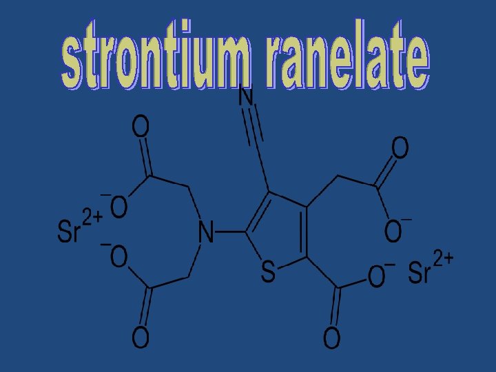
 Strontium was originally detected in lead mines near Strontian, Scotland, in the")
Introduction (1) Strontium was originally detected in lead mines near Strontian, Scotland, in the late 1700 s. It is present in water and food and in trace amounts throughout the skeleton Although absorption is poor when strontium is consumed orally, calcified tissues and areas of active osteogenesis take up 50 to 80 percent of the absorbed dose. excretion is largely renal.
 •")
Strontium • Discovered in 1790 and isolated in 1808 (near Scottish village Strontian) • Divalent cation • Resembles calcium and often mimics action of calcium • up to 1/10 cations incorporated in bone mineral • Anabolic properties • stimulates osteoblast proliferation • inhibits osteoclast activity • Activation of signaling pathways through Ca. SR (? ) • inhibits RANKL expression
 Uptake: Ionic substitution at low rates at the crystal surfaces of the bone")
(2) Uptake: Ionic substitution at low rates at the crystal surfaces of the bone matrix, without affecting mineral structure. An atomic number greater than that of calcium ; will weaken the penetration of x-rays and therefore result in an overestimation of measured BMD and content. Formulas have been derived to correct it. induction of cellular differentiation and the activation of the calcium-sensing receptor and the PTH suppression
 Enhances the replication of preosteoblastic cells")
How does strontium act on bone cells? (1) Enhances the replication of preosteoblastic cells and, consequently, bone matrix synthesis. several studies indicate no alteration of bone mineralization Induces uncoupling in bone remodeling, stimulating bone formation (osteoblast replication) and reducing resorption (osteoclast inhibition) in normal bone. decreases osteoclast activity and number by about 30%.
 Bone ALK P C-telopeptide cross-links At the third month of therapy, the serum")
(2) Bone ALK P C-telopeptide cross-links At the third month of therapy, the serum concentration of bonespecific alkaline phosphatase was higher in the strontium ranelate group than in the placebo group The concentration of serum Ctelopeptide cross-links was lower in the strontium ranelate group than in the placebo group at month 3.

Effects of strontium ranelate on bone mass and strength Increases vertebral bone formation and decreases bone resorption , resulting in increased bone mass. increased trabecular thickness and number, and decreased trabecular separation in the tibial metaphysis , indicating improved bone microarchitecture

Effects on BMD corresponding to differences between the placebo and the treatment groups at three years of 14. 4 percent, 8. 3 percent, and 9. 8 percent, respectively. At three years. the bone mineral density at the lumbar spine, adjusted for the strontium content, showed an increase over the base-line value of 6. 8 percent in the strontium ranelate group and a decrease of 1. 3 percent in the placebo group (P<0. 001).
 +14% Strontium ranelate +8%")
Strontium Ranelate: Effect on Bone Mineral Density BMD LWS (D%) +14% Strontium ranelate +8% (corrected for SR content of bone) Placebo 0 6 12 18 Months Meunier et al. , NEJM 2004; 350: 459 24 30 36
 Effect of Strontium Ranelate on Fractures in Postmenopausal Osteoporosis")
Risk of Vertebral Fractures (%) Effect of Strontium Ranelate on Fractures in Postmenopausal Osteoporosis ↓ RR -41% (0. 48 to 0. 73, p<0. 001) ↓ RR -49% (0. 36 to 0. 74, p<0. 001) 12 Meunier et al. , NEJM 2004; 350: 459 Months 36

Effect of Strontium Ranelate on Hip Fracture Risk Osteoporotic women, n=1977 (age ≥ 74 yrs and T-score FN ≤-3 SD), post-hoc analysis 10 Patients (%) 8 Placebo RRR 36% ARR 2. 1% NNT 48 6 4 Strontium ranelate 2 0 0 6 12 18 Reginster et al, J Clin Endocrinol Metab 2005, 90: 2816 24 30 36 42 Months

Effect of Strontium Ranelate on Non-Vertebral Fracture Risk All non-vertebral fractures 0. 84 (0. 70, 0. 99) Major osteoporotic non-vertebral fractures* 0. 81 (0. 66, 0. 98) Hip fractures, whole population 0. 85 (0. 61, 1. 19) 0. 64 (0. 41, 0. 99) Hip fractures, high risk group 0 0. 5 1 RR (95% CI) after 3 yrs *hip, wrist, pelvis, sacrum, rib, sternum, humerus Reginster et al, J Clin Endocrinol Metab 2005, 90: 2816 1. 5

Anti-fracture efficacy of strontium ranelate • strontium ranelate significantly reduced the incidence of new vertebral and clinical vertebral fractures by 40– 50% after one and three years of treatment. • Reduced by 16% the risk of nonvertebral fractures throughout the three year study compared with placebo. • reduced by 19% the relative risk for major fragility fractures (i. e. hip, wrist, pelvis and sacrum, ribs and sternum, clavicle, humerus). • Accompanied by decreased back pain and body height loss.
 Overally , well tolerated, especially in the upper gastrointestinal area. Like SOTI")
adverse effects(1) Overally , well tolerated, especially in the upper gastrointestinal area. Like SOTI study, the most common adverse effects consisted of nausea and diarrhea, which disappeared after the first three months. There was a lower incidence of gastritis, as diagnosed clinically by the investigators, in the strontium ranelate group than in the placebo group (3. 6 percent vs. 5. 5 percent, P=0. 07).
 Serum calcium concentrations were lower and serum phosphate concentrations higher in the strontium")
(2) Serum calcium concentrations were lower and serum phosphate concentrations higher in the strontium ranelate group than in the control group at month 3, with a plateau thereafter. There was a slight reduction in serum parathyroid hormone from the level at month 6 in both groups (decrease from base-line to six -month values) No changes were observed for serum 25 -hydroxyvitamin D, 1, 25 dihydroxyvitamin D, or calcitonin. Serum creatine kinase concentrations in the skeletal muscle increased in some patients ; at least twice the upper limit of the normal range. No increase in muscular symptoms or other biologic abnormalities were observed. Most of the increases in creatine phosphokinase were transient

Conclusions: Treatment of postmenopausal osteoporosis with strontium ranelate leads to early and sustained reductions in the risk of vertebral fractures.

Changes in bone mass are minimal in both men and women. The lack of a beneficial effect with GH on bone mass could be due to : • The concomitant activation of bone resorption along with formation, so that a net gain does not occur • Another explanation for a lack of effect could be the relatively short, one year, duration of many of these studies.

Recent evidence suggested a delayed and what was ultimately a positive effect of GH on bone • J Bone Miner Res 2006: (A cross-sectional study) patients with growth hormone deficiency who are receiving growth hormone-replacement therapy have a reduced risk of vertebral fractures as compared with untreated patients. • Rev Endocr. Metab. Disord 2005: The use of growth hormone in osteoporosis also is limited by side effects such as weight gain, carpal tunnel syndrome, glucose intolerance, and edema.

• IGF-1 promotes chondrocyte and osteoblast differentiation and growth. • In a prospective clinical studies) Low levels of IGF-1 are associated with a greater risk of spine and hip fractures. • At high doses, IGF-I increases biochemical markers of bone remodeling, whereas at low doses, it increases exclusively markers of bone formation, without an effect on bone resorption. • IGF-I has been studied in patients with anorexia nervosa, a disorder associated with low serum IGF-I levels. In such patients, the administration of IGF-I at doses that normalize serum IGF-I, in combination with estrogenreplacement therapy, increases BMD • A major drawback to the development of IGF-1 as a therapy for osteoporosis is its ubiquitous effect on many organ systems. Similar to GH, potential serious adverse effects could surface with its chronic use.


• Proteasome is a major structure for intracellular protein degradation. • The use of proteasome inhibitors will depend on their skeletal specificity and their safety profile. • such inhibitors can induce cellular toxic effects and the intracellular accumulation of misfolded proteins.

DKK-1 ANTAGONIST A secreted Wnt antagonist that binds LRP 5 and LRP 6

• Dkk-1 neutralization with an antibody increased BMD, trabecular bone volume, and bone formation in rodents. • suggesting that Dkk-1 inhibitors might have potential as an anabolic approach in the treatment of osteoporosis, particularly if they are targeted specifically to bone


• Mutations in SOST, the gene that encodes sclerostin, eliminate the expression of sclerostin; this causes skeletal dysplasias characterized by increased bone mass (sclerosteosis and van Buchem’s syndrome); characterized by hyperostosis, syndactyly, facial palsy, deafness, and the absence of nails. Patients with sclerosteosis, as well as heterozygous carriers, have increased BMD. • Monoclonal antibodies against sclerostin, prevent its binding to Wnt coreceptors, enhancing Wnt signaling and increasing bone mass in rodents and nonhuman primates. • it is possible that enhancement of Wnt signaling through the inhibition of a Wnt antagonist will have unwanted effects in nonskeletal tissues.


• Studies of testosterone in men with osteoporosis are limited, and none have used fractures as a primary end point. • The effects of testosterone on cortical and trabecular bone mass are greatest when it is used in hypogonadal adolescents. • In a 3 -year study of hypogonadal men over 65 years of age, those receiving testosterone-replacement therapy had an 8. 9% greater increase in the bone mineral density of their spine than did those receiving placebo.

The failure of bone mineral density at three sites in the hip to increase during testosterone treatment even in men with relatively low pretreatment serum testosterone concentrations could be at least partly related to the slower response of this bone to changes in gonadal steroids

The effects of testosterone in eugonadal men are more controversial Differences in response in eugonadal men may be related to pretreatment testosterone levels. In a pre specified analysis , men with a testosterone level below 200 ng per deciliter had significant increases in spinal bone mineral density with treatment This analysis showed a minimal effect (0. 9%) of testosterone treatment over that of placebo for a pretreatment serum testosterone concentration of 400 ng/d. L (13. 9 nmol/L), but an increase of 3. 4% for testosterone of 300 ng/d. L (10. 4 nmol/L) and a increase of 5. 9% for a testosterone of 200 ng/d. L (10. 9 nmol/L).

non-aromatizable androgens like nandrolone TESTOSTRON nandrolone placeb • Testosterone seems to be more effective in improving BMD than other non-aromatizable androgens like nandrolone BMD changes in the lumbar spine in men treated with testosterone • nandrolone( • or placeb(o). The dashed line through zero represents no change from baselin

CONCLUSION : • Androgen replacement in hypogonadal AND eugonadal mens remains a controversial issue.
 are a novel class of androgen")
AND FINALLY , Selective androgen receptor modulators (SARMs) are a novel class of androgen receptor (AR) ligands that might change the future of androgen therapy dramatically.
 are present on the osteoblasts, osteoclasts and mesenchymal stromal")
Mechanisms: • Androgen receptors (ARs) are present on the osteoblasts, osteoclasts and mesenchymal stromal cells that differentiate toward the osteoblast lineage. • Androgens increase bone mass through effects on bone cells, enhancing osteoblast activity but inhibiting that of osteoclasts. • responsible for the development of a sexually dimorphic skeleton (i. e. male bones are wider than female) and is a significant determinant of bone strength. AND important for the production of peak bone mass in males.

Testosterone, is also a substrate for the production of estradiol through aromatase activity. Estrogen replacement therapy tends to suppress bone resorption with only modest effects on bone formation; By contrast, androgen replacement data have demonstrated clear effects on bone formation. The skeletal response to androgen administration is characterized as increased cortical and trabecular bone mass, together with increased bone width with surface periosteal expansion but a lack of inner endosteal deposition, in the setting of inhibition of resorption owing to reduced osteoclast activity. Thus maintains trabecular bone and expands cortical bone.

• An unresolved clinical issue is the management of older men with low BMD who have moderately low testosterone levels • A meta-analysis of placebo-controlled trials of testosterone treatment in men with any degree of androgen deficiency (most of them showing low normal or normal testosterone levels at baseline) suggested a beneficial effect on lumbar spine BMD, but equivocal findings at the femoral neck

Agent vetebral Fx Quality of evidence Hip Fx Quality of evidence Alendronate + Good Residronate + Good Etidronate + Good Ibandronate + Good zolendronate + Good + Fair HRT + Good Raloxifen + Good Calcitonin + Fair PTH + Good Ann Intern Med. 2008; 148: 197 -213.
 • Denosumab • Odanacatib • Lasofoxifene • Bazedoxifene")
New and Emerging Treatments Antiresorptive (anticatabolic) • Denosumab • Odanacatib • Lasofoxifene • Bazedoxifene • CE/bazedoxifene • New delivery systems – oral salmon calcitonin Osteo-anabolic (bone-forming) • Sclerostin inhibitor • Variations of PTH • Endogenous PTH stimulation – calcium sensing receptor antagonist (calcilytic) • New delivery systems – transdermal PTH Strontium ranelate Combinations of antiresorptive and anabolic

Conclusion • Monotherapy with calcium or vitamin D has not been shown to reduce fractures. • Combined calcium plus vitamin D , may reduce vertebral and non-vertebral fractures, • Alendronate , risedronate , zolendronate and parathyroid hormone reduce vertebral and non-vertebral fractures compared with placebo • Etidronate , ibandronate , and raloxifene reduce vertebral fractures, but have not been shown to reduce non-vertebral fractures. • Calcitonin may reduce vertebral fractures over 1– 5 years, but has not been shown to reduce non-vertebral fractures. • Hormone replacement therapy may reduce fractures, but it increases the risk of breast cancer and cardiovascular events • Strontium ranelate reduces vertebral and, to some extent, non-vertebral fractures.
 and vitamin D (800 IU) • Oral bisphonates")
Conclusion • Adequate calcium (1200 mg) and vitamin D (800 IU) • Oral bisphonates (alendronate or residronate) as first line therapy • IV bisphonates (zolendronate) for patients who can not tolerate oral bisphonates • • Raloxifen as a second line drug • Calcitonin is not recommended except for pain relief • Teriparatide for patients with severe osteoporosis who are unable to tolerate any of the available bisphonates or who continue to fracture after one year of bisphonates therapy

- Slides: 84