Traumatic Brain Injury C h a p t

Traumatic Brain Injury C h a p t e r 19
 is defined as “an alteration in brain function, or other")
�Traumatic brain injury (TBI) is defined as “an alteration in brain function, or other evidence of brain pathology, caused by an external force. ”

PREVALENCE AND IMPACT �Leading cause of injury related death and disability �Falls 32%, motor vehicle/traffic accidents 19%, struck by/against events 18%, and assaults 10% (USA). �Children, older adolescents/ young adults (less than 25 years old), and older adults are most at risk for experiencing TBI. �The long-term consequences of TBI on the health care system, society, and the individual are high.

MECHANISM OF INJURY AND PATHOPHYSIOLOGY �Primary injury; The brain damage results from external forces that cause brain tissue to make direct contact with an object (bony skull or penetrating object), rapid acceleration or deceleration forces, or blast waves from an explosion. �Secondary injury; that results from a cascade of biochemical, cellular, and molecular events that evolve over time due to the initial injury and injuryrelated hypoxia, edema, and elevated intracranial pressure (ICP).

Primary Injury; Contact injury �Often result in contusions, lacerations, and intracerebral hematomas. �Anterior temporal poles, frontal poles, lateral and inferior temporal cortices, and orbital frontal cortices. �Damage is generally focal in nature as the brain comes into contact with bony protuberances on the inside surface of the skull or damage from the penetrating object

Primary Injury; rapid Acceleration/ deceleration �Acceleration and deceleration cause shear, tensile, and compression forces within the brain, which causes diffuse axonal injury (DAI), tissue tearing, and intracerebral hemorrhages.

Diffuse Axonal Injury �Diffuse axonal injury is the predominant mechanism of injury in most individuals with severe to moderate TBI. �It is common in high-speed motor vehicle accidents (MVAs) and can be seen in some sports related TBIs. �The term diffuse is somewhat misleading because DAI most often occurs in discrete areas: the parasagittal white matter of the cerebral cortex, the corpus callosum, and the pontinemesencephalic junction adjacent to the superior cerebellar peduncles. �The mechanism of DAI is microscopic, so often there are minimal initial findings on computed tomography (CT) and magnetic resonance imaging (MRI). �The acceleration/deceleration forces cause disruption of neurofilaments within the axon leading to Wallerian-type axonal degeneration. 13

Blast Injury �Blast injury is considered a signature injury of the U. S. military conflicts in the Middle East. �When an explosive device detonates a transient shock wave is produced, which can cause brain damage. �Primary blast injury results from the direct effect of blast overpressure on organs (in this case the brain), secondary injury results from shrapnel and other objects being hurled at the individual, and tertiary injury occurs when the victim is flung backward and strikes an object.

Secondary Injury �Secondary cell death occurs as a result of a chain of cellular events that follow tissue damage in addition to the secondary effects of hypoxemia, hypotension, ischemia, edema, and elevated ICP.

SEQUELAE OF TRAUMATIC BRAIN INJURY �Traumatic brain injury is associated with a wide spectrum of neuromuscular, cognitive, and behavioral impairments that can lead to limitations in activity, restrictions in social participation, and diminished quality of life. �Although physical therapy interventions primarily address physical limitations related to mobility, the cognitive and behavioral changes associated with TBI are often more disabling.
 and")
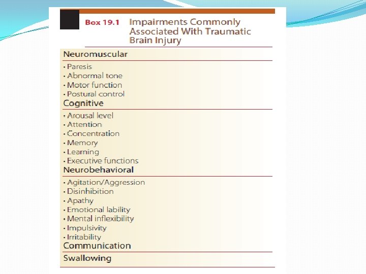
Neuromuscular Impairments �Individuals with TBI commonly exhibit impaired motor function. �Upper extremity (UE) and lower extremity (LE) paresis, impaired coordination, impaired postural control, abnormal tone, and abnormal gait may be present as lifelong impairments. �Abnormal, involuntary movements such as tremor and chorea form and dystonic movements are less common. �Patients may also present with impaired somatosensory function, depending on the location of the lesion.

Cognitive Impairments �Cognition includes arousal, attention, concentration, memory, learning, and executive functions. �Executive functions include planning, cognitive flexibility, initiation and selfgeneration, response inhibition, and serial ordering and sequencing.

Neurobehavioral Impairments �Patients can exhibit profound behavioral changes as they progress through recovery. �These impairments can be closely linked to cognitive impairments and are often more debilitating in the long run than physical disability. �Common behavioral sequelae include low frustration tolerance, agitation, disinhibition, apathy, emotional lability, mental inflexibility, aggression, impulsivity, and irritability.

Communication �Language and communication deficits after brain injury are generally nonaphasic in nature and are related to cognitive impairment. �Disorganized and tangential or written communication, imprecise language, word retrieval difficulties, and disinhibited and socially inappropriate language. �Patients may also exhibit difficulties communicating in distracting environments, reading social cues, and adjusting communication to meet the demands of the situation. �These communication deficits can affect employability, social integration, and quality of life.

Dysautonomia �Elevated sympathetic nervous system activity occurs as a normal response to trauma; following TBI this response may become overactive. Increased sympathetic activity results in increased heart rate, respiratory rate, and blood pressure; diaphoresis; and hyperthermia. �Other symptoms of dysautonomia include decerebrate and decorticate posturing, hypertonia, and teeth grinding. �The term paroxysmal sympathetic hyperactivity accurately describes this phenomenon. �The incidence of paroxysmal sympathetic hyperactivity ranges from 8% to 33% in patients with TBI in the intensive care unit.

Post-traumatic Seizures �Between 12% and 50% of people with severe TBI develop post-traumatic seizures. �For adults with severe injury, phenytoin (an anticonvulsant) is effective in decreasing the risk of early post-traumatic seizures.

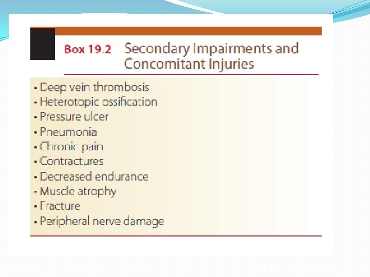
Secondary Impairments and Medical Complications �Due to the high potential of prolonged immobility and concomitant injury, patients with TBI are at risk of developing a number of secondary impairments and other medical issues. �Up to 50% of patients with severe brain injury develop gastrointestinal difficulties, 45% develop genitourinary problems, 34% develop respiratory problems, 32% develop cardiovascular problems, and 21% develop dermatological complications. �Urinary and bowel incontinence, deep vein thrombosis (DVT), heterotopic ossification, pressure ulcer, pneumonia, and chronic pain.

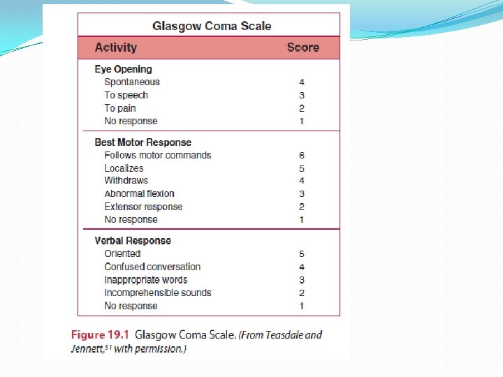
DIAGNOSIS AND PROGNOSIS �Traumatic brain injury is generally categorized as severe, moderate, or mild using the Glasgow Coma Scale (GCS) developed by Teasdale and Jennett, �Scores of 8 or less are classified as severe, scores between 9 and 12 are defined as moderate, and scores of 13 to 15 are classified as mild brain injury.

Interdisciplinary Team members �Patient and family �Physician �Speech and language Pathologist �Rehabilitation Nurse �Occupational therapist �Physical therapist �Neuropsychologist �Social worker � case manger / team coordinator

EARLY MEDICAL MANAGEMENT �Starts at the scene of the accident. �Early resuscitation to stabilizing the cardiovascular and respiratory systems is important to maintain sufficient blood flow and oxygen to the brain. �Minimize secondary brain injury by optimizing cerebral blood flow and oxygenation �stabilize vital signs, perform a complete examination, identify and treat any nonneurological injuries, and continuously monitor the patient.

�Systolic blood pressure should be kept above 90 mm Hg and oxygen saturation above 90%. �Intubation �The patient’s neck should be stabilized with a collar and the head elevated 30 degrees to protect the spine in case of instability, as well as avoid an increase in ICP. �The GCS is used to determine the severity of the brain injury. �A complete neurological examination is also done. �x-ray films, CT and MRI. �Surgical evacuation for heamatoma �The patient is monitored continuously. For patients with a GCS of 8 or less, any acute abnormality on CT, a systolic pressure of less than 90 mm Hg, or age greater than 40 years, ICP monitoring is recommended.

�An external ventricular drain provides the most accurate and reliable data and also provides a means to control ICP by cerebrospinal fluid removal. �Other monitoring options that are less invasive include the subdural bolt and a fiberoptic catheter. �Elevated ICP can be treated with the use of sedating medications, moderate head-up positioning (head elevated to 30°), osmotherapy, hypothermia, surgical decompression, and barbiturates. �Intracranial pressure should be less than 20 mm Hg and the cerebral perfusion pressure (CPP) greater than 60 mm Hg. �If ICP cannot be treated successfully, inducing a pharmacologic coma or surgical decompression may be necessary.

PHYSICAL THERAPY MANAGEMENT OF MODERATE TO SEVERE TRAUMATIC BRAIN INJURY IN THE ACUTE STAGE �Physical therapy management of patients with severe to moderate TBI during the early stages of recovery �Physical therapy management of patients with severe to moderate TBI during active rehabilitation �Physical therapy management of patients with mild TBI.

�Patients in the early stage of recovery after severe to moderate TBI often demonstrate low levels of arousal. The primary goal of physical therapy is to prevent secondary complications due to the TBI and prolonged bed rest/immobilization, begin early mobilization when medical clearance is received, and initiate patient and family education. �Depending on the patient’s presentation, some of the examination and intervention procedures described in the section on physical therapy management during active rehabilitation can be used in the early stage of recovery as well.

Examination �Complete medical record review. �The patient may be on a ventilator with ongoing monitoring of ICP. �He or she may have weightbearing precautions and range of motion (ROM) restrictions owing to musculoskeletal injuries and/or interventions, and open wounds. �A thorough medical record review provides a comprehensive perspective about the patient’s condition, as well as a complete understanding of the precautions and contraindications that must be observed during the examination and subsequent treatment.

�Because the patient’s medical status may be dynamic at these stages, it is important to check with the patient’s primary nurse before beginning any session. �Team members should always observe universal precautions and may need to wear gowns, gloves, and/or masks or other personal protective equipment when treating the patient. �Examine; Arousal, attention, and cognition, Integument integrity, Sensory integrity, Motor function, Range of motion, Reflex integrity, Ventilation and respiration/gas exchange �Patients with severe TBI who are in low arousal state (coma, vegetative, or minimally conscious) may present with abnormal tone and posturing. �Primitive postures may include those associated decorticate or decerebrate rigidity. �Decorticate rigidity; the upper extremities are in a flexed posture and the lower extremities are extended. �Decerebrate rigidity; both the upper and lower extremities are positioned in extension.

� Abnormal tone; spastic hypertonia. �Spastic hypertonia may range from spasticity that severely affects the entire body and greatly inhibits normal, functional movement, to lesser levels of tone that affect individual muscle groups. �If not medically contraindicated, the examination should include sitting on the side of the bed with assistance. �The therapist should monitor vital signs and document any changes in tone or head and trunk control. �When appropriate, the patient should be transferred into a wheelchair. �The patient may require the assistance of two to three people to transfer at this stage. �In most cases, a reclining or tilt-in-space wheelchair is the best option for positioning, with a specialized pressure reducing cushion. �Often it may require several treatment sessions to complete the entire examination. Because early patient status is often dynamic, any signs of progress or regression
 is recommended to assess patients with disordered")
Outcome Measures �The Coma Recovery Scale–Revised (CRS-R) is recommended to assess patients with disordered consciousness. The CRS-R is a valid and reliable 23 item measure with six subscales: auditory, visual, motor, oromotor, communication, and arousal. Scores range from 0 to 23. Data are useful in distinguishing between different states of consciousness (vegetative state, minimally conscious state, and emerging), determining the prognosis, and informing treatment planning.
 scale is a descriptive scale used")
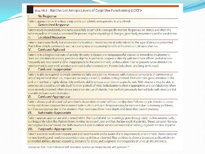
�Rancho Los Amigos Levels of Cognitive Functioning (LOCF) scale is a descriptive scale used to examine cognitive and behavioral recovery in individuals with TBI (Box 19. 3) as they emerge from coma. 71 his scale does not address specific cognitive deficits, but is useful for communicating general cognitive and/or behavioral status and for treatment planning. he eight categories describe typical cognitive and behavioral progress after a brain injury. Patients may plateau at any level. The LOCF has been shown to be a reliable and valid measure of cognitive and behavioral function for individuals with brain injury. 72

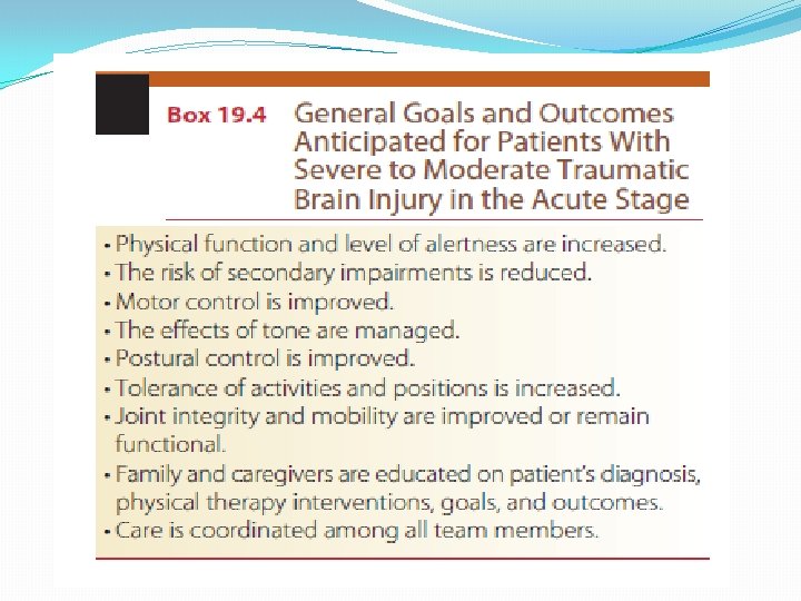
Plan of Care; Outcomes/Goals �A list of general goals and outcomes anticipated for patients in Levels I, II, and III (LOCF) adapted from the American Physical Therapy Association’s Guide to Physical Therapist Practice 73 are presented in Box 19. 4. �They can be used to guide the development of specific anticipated goals and expected outcomes for an individual patient.

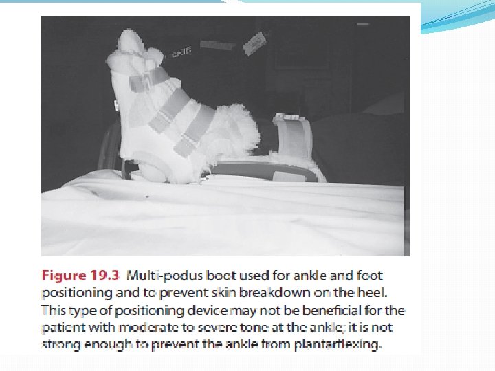
Plan of Care; Interventions �Preventing Secondary Impairments �Because of the patient’s inability to move at these levels, he or she is susceptible to indirect impairments such as contractures, decubiti, pneumonia, and DVT. �If prevention is not addressed early in rehabilitation, these impairments are likely to impede future progress, and can be life threatening. �Proper positioning both in bed and in a wheelchair is essential.

Early Mobility �Upright sitting is extremely important because it addresses elements of treatment goals for the early levels of recovery. �As soon as medically stable, the patient should be transferred to a sitting position and out of bed to a wheelchair. �All precautions should be observed. �The head should be properly supported, because the patient is not likely to have adequate neck and head control to maintain an upright posture without support.

�Co-treatment with an occupational therapist provides two skilled professionals to assist the patient, because maximal assistance is often required. �Use of a tilt table is also advantageous because it allows early weight-bearing through the LEs. �The upright position, both on a tilt table and in a wheelchair, may improve overall level of alertness.

Sensory Stimulation �Sensory stimulation is an intervention used to increase the level of arousal and elicit movement in individuals in a coma or persistent vegetative state. he theory is that by providing stimulation in a controlled, multisensory manner, with a balance of stimulation and rest, the reticular activating system may be stimulated causing a general increase in arousal.

PHYSICAL THERAPY MANAGEMENT OF MODERATE TO SEVERE TRAUMATIC BRAIN INJURY DURING ACTIVE REHABILITATION �Require extensive and protracted rehabilitation across a variety of settings throughout the continuum of care. �This may include acute and subacute inpatient rehabilitation, postacute rehabilitation, day treatment program, and outpatient or home care. �The many cognitive, physical, and/or behavioral impairments that affect activity levels and social participation often necessitate multiple episodes of physical therapy care over the patient’s lifetime. �Goals and interventions should be focused on the patient’s abilities and personal goals regardless of the setting.

Examination �Irrespective of injury chronicity, some patients with TBI will have cognitive and behavioral impairments that pose barriers to the examination process. �These barriers may include disorientation, confusion, physical aggression, memory deficits, and limited attention span. �It may be difficult to gather data using standardized tests and measures, such as goniometry or manual muscle testing, because the patient may be unable to cooperate.

�The physical therapist should determine the patient’s cognitive abilities, because these will affect the ability to relearn motor skills. his will include orientation, attention span, memory, insight, safety awareness, and alertness. Key initial questions that warrant consideration include the following: �Is the patient able to follow commands: one-step, two-step, or multistep commands? �Is the patient oriented to person, place, and/or time? �Does the patient recognize family members? �Does the patient demonstrate any insight into what has happened?

�Determination of the patient’s functional abilities should be done in a variety of environments, because some patients may perform well in the closed environment of a private room, but performance may deteriorate in an open environment with multiple distractions.
 �The")
Outcome Measures: Body/Structure Function Balance �The Berg Balance Scale �Functional Independence Measure (FIM) �The Community Balance and Mobility Scale (CB&M) �High-Level Mobility Assessment Tool (Hi. MAT)
 is an observational rating scale")
Attention and Cognition �The Moss Attention Rating Scale (MARS) is an observational rating scale that provides a reliable and valid measure of attention-related behavior after TBI. �Test of Everyday Attention �Trail Making Test Part B. �The Galveston Orientation and Amnesia Test (GOAT)

Outcome Measures: Activity and Participation Global Functioning �The Functional Independence Measure is a commonly used measure of functional mobility, ADL function, cognition, and communication. �The FIM was designed to measure level of disability and burden of care in individuals undergoing inpatient rehabilitation, and is useful for monitoring patient progress and evaluating outcomes
 � 6 -Minute Walk Test")
Locomotion �OGA � 10 -Meter Walk Test (10 MWT) � 6 -Minute Walk Test (6 MWT) �Modified Walking and Remembering Test (WART)

Plan of Care; Goals/Outcomes �The American Physical Therapy Association’s Guide to Physical Therapist Practice offers examples of general anticipated goals and expected outcomes pertinent to patients with TBI. Examples include the following: �The risk of secondary impairments is reduced. �Performance of functional mobility and ADL skills is increased. �Ability to assume or resume self-care and home management roles is improved.
Learning Strategies �Restorative Versus Compensatory-Based Interventions �Assistive technology �Physical")
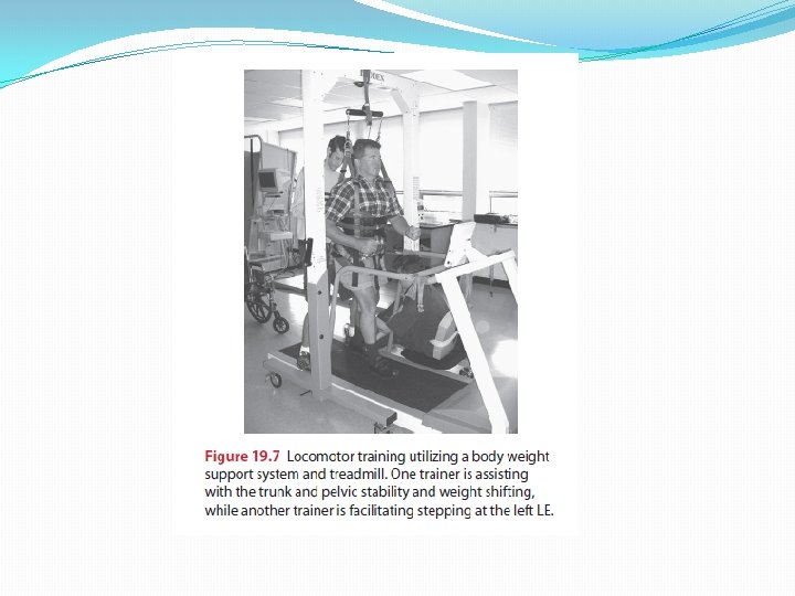
Plan of Care; Interventions �Motor (Re)Learning Strategies �Restorative Versus Compensatory-Based Interventions �Assistive technology �Physical barriers �Restorative Interventions and Neural Plasticity �Task-Oriented Approach �Loco motor Training with Body Weight Support �Constraint-Induced Therapy �Aerobic and Endurance Conditioning �Resistance Training �Electrical Stimulation �Dual-Task Performance

Patient/Family/Caregiver Education �Patient/family/caregiver education and training are important goals across each level of rehabilitation. �The goals of this education and training will vary based on the cognitive and behavioral abilities of the patient. �Clients in the early phase of recovery may go through a period where they are significantly confused and agitated. �It is difficult to provide education for the patient at this level; the patient has very little, if any, ability for new learning. �However, it is extremely important to provide education for the patient’s family.

Behavioral Factors � Therapists may encounter a variety of behavioral barriers to examining and treating patients with moderate to severe TBI. � As the patient begins to emerge from coma, he or she often experiences a period of acute post-traumatic agitation. � The confusion, amnesia, and disorientation during this phase of recovery often result in agitation, aggression, noncompliance, and combative behavior. � The therapist should incorporate creativity and flexibility when designing and providing interventions. his is particularly true with individuals who are in the confused and agitated stage of recovery. � using familiar activities, rather than progress to more � At this stage, therapist should work near the patient’s physical level of function challenging skills that require new learning, because the patient does not have the capacity for new learning at this level of recovery.

�The neuropsychologist can assist the team by providing insight into different ways to manage the patient’s agitated behavior and may set up a behavioral modification program. �Behavioral modification techniques such as positive reinforcement using a point or reward system, redirection, and compliance training are useful in managing these inappropriate behaviors and improving participation in therapy.

Community Reentry Programs �Many clients will make significant progress in the early phase of rehabilitation. Before discharge, it is crucial to begin to wean the patient from the external structure provided by the hospital setting that was so important in the early stages of recovery. �As the patient becomes better able to control himself or herself, external control provided by the environment should be lessened. �Doing so will prepare the patient for the challenges of the next level of rehabilitation, postacute, community reentry programming.
- Slides: 60