
Trauma in Pregnancy Dr Zohair Alaseri MD FRCPc




- Slides: 96
Trauma in Pregnancy Dr. Zohair Alaseri, MD FRCPc, Emergency Medicine FRCPc, Critical Care Medicine Intensivest and Emergency Medicine Consultant Chairman, Department of Emergency Medicine King Khalid University Hospital King Saud University Hospitals, Riyadh, KSA
Trauma in Pregnancy Facts • 6% to 7% of all pregnancies. • Leading cause of maternal death
Trauma in Pregnancy Objectives • • • Physiological and anatomical changes in pregnancy Some Alterations Mimic Shock Clinical features of trauma in pregnancy Diagnostic challenges Management
Physiological and anatomical changes in pregnancy Pregnancy produces changes that alter the usual course of trauma management.
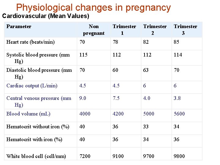
Physiological changes in pregnancy Cardiovascular (Mean Values) Parameter Non pregnant Trimester 1 Trimester 2 Trimester 3 Heart rate (beats/min) 70 78 82 85 Systolic blood pressure (mm Hg) 115 112 114 Diastolic blood pressure (mm Hg) 70 60 63 70 Cardiac output (L/min) 4. 5 6 6 Central venous pressure (mm Hg) 9. 0 7. 5 4. 0 3. 8 Blood volume (m. L) 4000 4200 5000 5600 Hematocrit without iron (%) 40 36 33 34 Hematocrit with iron (%) 40 36 34 36 White blood cell (cell/mm) 7200 9100 9700 9800
Physiological changes in pregnancy Some Alterations Mimic Shock • Blood pressure declines in the first trimester • More problematic is the supine hypotensive syndrome.
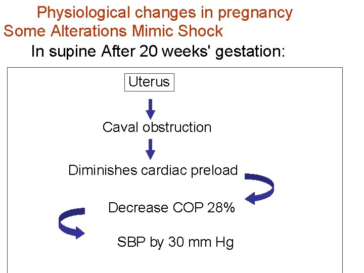
Physiological changes in pregnancy Some Alterations Mimic Shock In supine After 20 weeks' gestation: Uterus Caval obstruction Diminishes cardiac preload Decrease COP 28% SBP by 30 mm Hg
Physiological changes in pregnancy Some Alterations Mimic Shock • central venous pressure (CVP) measurements can be lowered in the last two trimesters
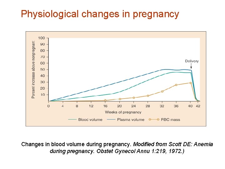
Physiological changes in pregnancy Some Alterations Hide Shock • Blood volume increases 48% to 58% above normal, peaking at 32 to 34 weeks‘. so clinical signs of maternal hypotension from acute traumatic bleeding may be delayed.
Physiological changes in pregnancy Changes in blood volume during pregnancy. Modified from Scott DE: Anemia during pregnancy. Obstet Gynecol Annu 1: 219, 1972. )
Some Alterations Can Exacerbate Traumatic Bleeding. Blood flow to the uterus increases from 60 m. L/min 600 m. L/min can be a major source of blood loss
Some Alterations Can Exacerbate Traumatic Bleeding Marked venous congestion in the pelvis in the third trimester increasing the potential for hemorrhage from both bony and soft tissue pelvic injuries.
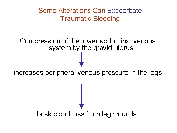
Some Alterations Can Exacerbate Traumatic Bleeding Compression of the lower abdominal venous system by the gravid uterus increases peripheral venous pressure in the legs brisk blood loss from leg wounds.
Physiological changes in pregnancy Pregnancy Is Associated with a Greater Risk of Thromboembolism Stasis is potentiated by • increased venous capacity • inferior vena cava compression • posttrauma bed rest.
Physiological changes in pregnancy Pregnancy Is Associated with a Greater Risk of Thromboembolism By the third trimester increases in coagulation factors V, VIII, IX, X, and XII and fibrinogen exceed fibrinolytic activity.
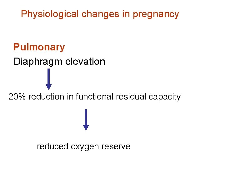
Physiological changes in pregnancy Pulmonary Diaphragm elevation 20% reduction in functional residual capacity reduced oxygen reserve
Physiological changes in pregnancy Gastrointestinal Increase the possibility of aspiration: • Reduced Gastroesophageal sphincter • Decrease gastrointestinal motility • The stomach's increased acid production aspiration more ominous than usual.
Anatomic Changes in Pregnancy The diaphragm rises, flaring of the ribs & hyperventilation Tension pneumothorax may develop more quickly chest tube should be placed one or two interspaces higher
Anatomic Changes in Pregnancy • Altered pain location Abdominal viscera are pushed upward by the enlarging uterus, resulting in • The gravid uterus increases the likelihood of bowel injury with penetrating trauma to the upper abdomen. • The bladder is also displaced into the abdominal cavity beyond 12 • bladder becomes hyperemic, and injury may lead to a marked increase in blood loss
Changes in Laboratory Values with Pregnancy • The physiologic anemia • The leukocyte count averages 9000 to 10, 000 in the last two trimesters • ESR rises to a mean of 78 mm per hour at term. • Increased coagulation factors shorten prothrombin times & Partial thromboplastin times • Fibrinogen levels begin to rise in the third month of pregnancy.
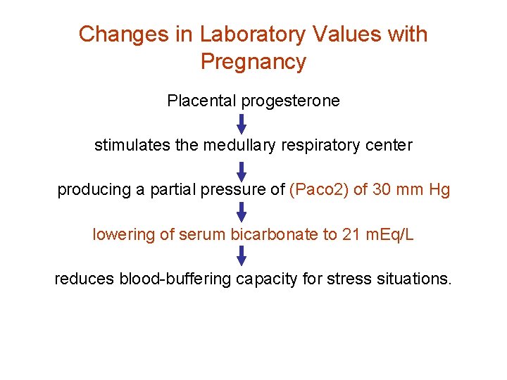
Changes in Laboratory Values with Pregnancy Placental progesterone stimulates the medullary respiratory center producing a partial pressure of (Paco 2) of 30 mm Hg lowering of serum bicarbonate to 21 m. Eq/L reduces blood-buffering capacity for stress situations.
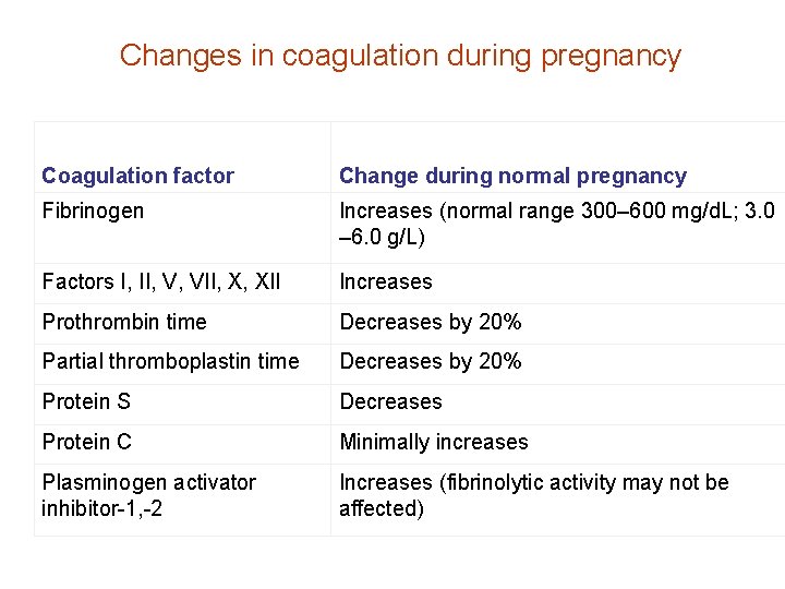
Changes in coagulation during pregnancy Coagulation factor Change during normal pregnancy Fibrinogen Increases (normal range 300– 600 mg/d. L; 3. 0 – 6. 0 g/L) Factors I, II, V, VII, X, XII Increases Prothrombin time Decreases by 20% Partial thromboplastin time Decreases by 20% Protein S Decreases Protein C Minimally increases Plasminogen activator inhibitor-1, -2 Increases (fibrinolytic activity may not be affected)
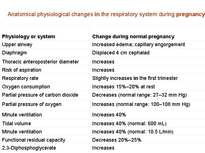
Anatomical physiological changes in the respiratory system during pregnancy Physiology or system Change during normal pregnancy Upper airway Increased edema; capillary engorgement Diaphragm Displaced 4 cm cephalad Thoracic anteroposterior diameter Increases Risk of aspiration Increases Respiratory rate Slightly increases in the first trimester Oxygen consumption Increases 15%– 20% at rest Partial pressure of carbon dioxide Decreases (normal range: 27– 32 mm Hg) Partial pressure of oxygen Increases (normal range: 100– 108 mm Hg) Minute ventilation Increases 40% Tidal volume Increases 40% (normal: 600 m. L) Minute ventilation Increases 40% (normal: 10. 5 L/min) Functional residual capacity Decreases 20%– 25% 2, 3 -Diphosphoglycerate Increases
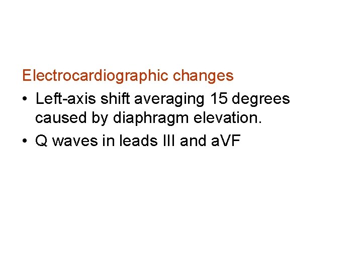
Electrocardiographic changes • Left-axis shift averaging 15 degrees caused by diaphragm elevation. • Q waves in leads III and a. VF
CLINICAL FEATURES OF TRAUMA IN PREGNANCY Blunt Trauma Motor Vehicle Crashes • Higher risk of adverse fetal outcome if not properly restrained. • Dummy trials show that improper placement of the lap belt over the pregnant abdomen causes a three- to fourfold increase in force transmission through the uterus.
CLINICAL FEATURES OF TRAUMA IN PREGNANCY Blunt Trauma Motor Vehicle Crashes Correct positioning seat belt • the lap belt underneath the pregnant abdomen against the pelvis • the shoulder belt to the side of the pregnant abdomen, between the breasts, and over the midportion of the clavicle.
Expectant mom with seat on © Mark Pearlman MD
CLINICAL FEATURES OF TRAUMA IN PREGNANCY Blunt Trauma Physical Abuse • The prevalence of physical abuse during pregnancy ranges from 4% to 17%.
CLINICAL FEATURES OF TRAUMA IN PREGNANCY Falls • more prevalent after the 20 th week of pregnancy.
CLINICAL FEATURES OF TRAUMA IN PREGNANCY Mechanisms of Fetal Loss: Clinical Data g g g Abruptio placentae (50 -80%) loss of maternal life (~10%) unrecognized cause (>10%) direct fetal injury (<10%) maternal hypovolemic shock (<5%)
CLINICAL FEATURES OF TRAUMA IN PREGNANCY Penetrating Trauma • in the upper abdomen increased probability of harm (approaching 100%) to the bowel, liver, or spleen • below the uterine fundus visceral injuries are less likely (0% in some studies). But it places the fetus in a position more susceptible to injury
CLINICAL FEATURES OF TRAUMA IN PREGNANCY Fetal Injury • Trauma is associated with a high risk for fetal loss. Poor fetal outcome is predicted by • Maternal hypotension • Sever acidosis
CLINICAL FEATURES OF TRAUMA IN PREGNANCY Fetal Injury • Only cardiotocographic monitoring for a minimum of 4 hours is useful to predict fetal outcome • Fetal injuries from blunt trauma usually involve intracranial hemorrhage and skull fractures
CLINICAL FEATURES OF TRAUMA IN PREGNANCY Placental Injury In blunt trauma • 50% to 70% of all fetal losses result from placental abruption. • abruption can occur with little or no external sign of injury to the abdominal wall.
CLINICAL FEATURES OF TRAUMA IN PREGNANCY Placental Injury Placental abruption Classical findings • vaginal bleeding, 37% of cases • abdominal cramps • uterine tenderness • maternal hypovolemia • fetal heart rate changes.
CLINICAL FEATURES OF TRAUMA IN PREGNANCY Placental Injury Placental abruption 1 --Fetal distress sensitive indicator of placental abruption 2 -- uterine activity. One study reported that if 12 or more contractions occurred in any hour of a 4 -hour cardiotocographic monitoring period, the risk of abruption was 14%; abruption did not occur in this study if contractions occurred less than once every 10 minutes.
CLINICAL FEATURES OF TRAUMA IN PREGNANCY Placental Injury Placental abruption • US is less than 50% accurate • If the abruption bleeds externally, not enough blood collects to be seen sonographically. • The risk of preterm delivery is increased with even mild abruptions
CLINICAL FEATURES OF TRAUMA IN PREGNANCY Placental Injury Placental abruption If fetus less than 32 weeks' Where mother and fetus are stable, expectant management can be tried for partial placental abruptions.
CLINICAL FEATURES OF TRAUMA IN PREGNANCY Placental Injury Placental abruption • 54 times more likely to have coagulopathies and DIC Thromboplastin
CLINICAL FEATURES OF TRAUMA IN PREGNANCY Uterine Injury Uterine contractions The most common obstetric problem caused by maternal trauma
CLINICAL FEATURES OF TRAUMA IN PREGNANCY Uterine Injury Uterine contractions • The routine use of tocolytics for premature labor has come under question • 90% of contractions stop spontaneously • The option to use tocolytics ends when cervical dilation reaches 4 cm.
CLINICAL FEATURES OF TRAUMA IN PREGNANCY Uterine Injury Uterine rupture • rare • pelvic fractures strike directly against the uterus.
CLINICAL FEATURES OF TRAUMA IN PREGNANCY Uterine Injury Uterine rupture Typical findings • Maternal shock • abdominal pain • easily palpable fetal anatomy caused by extrusion into the abdomen • fetal demise
CLINICAL FEATURES OF TRAUMA IN PREGNANCY Uterine Injury Uterine rupture Diagnosing difficult. Optimal treatment • between suturing the tear or performing a hysterectomy, depends on the extent of uterus and uterine vessel tears and the importance of future childbearing.
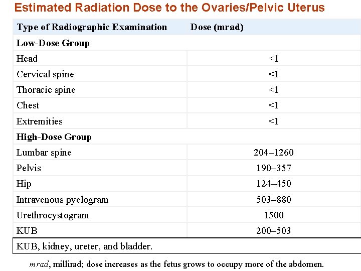
DIAGNOSTIC STRATEGIES RADIOGRAPHY Plain Films • Sensitivity to radiation is greater during intrauterine development • the risk to the fetus of a 1 rad (1000 mrad) exposure, approximately 0. 003%, is thousands of times smaller than the spontaneous risks of malformations, abortions, or genetic disease. • At 15 rad there is approximately a 6% chance that the fetus could experience severe mental retardation
DIAGNOSTIC STRATEGIES RADIOGRAPHY Plain Films • Coned x-ray beams aimed more than 10 cm away from the fetus are not harmful • diagnostic studies should not be withheld out of concern for fetal radiation exposure. • When appropriate, shielding and collimation should be used.
Estimated Radiation Dose to the Ovaries/Pelvic Uterus Type of Radiographic Examination Dose (mrad) Low-Dose Group Head <1 Cervical spine <1 Thoracic spine <1 Chest <1 Extremities <1 High-Dose Group Lumbar spine 204– 1260 Pelvis 190– 357 Hip 124– 450 Intravenous pyelogram 503– 880 Urethrocystogram KUB 1500 200– 503 KUB, kidney, ureter, and bladder. mrad, millirad; dose increases as the fetus grows to occupy more of the abdomen.
DIAGNOSTIC STRATEGIES Ultrasonography • safe procedure. • the best modality for simultaneous assessment of both the mother and the fetus.
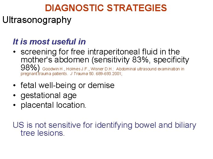
DIAGNOSTIC STRATEGIES Ultrasonography It is most useful in • screening for free intraperitoneal fluid in the mother's abdomen (sensitivity 83%, specificity 98%) Goodwin H. , Holmes J. F. , Wisner D. H. : Abdominal ultrasound examination in pregnant trauma patients. J Trauma 50. 689 -693. 2001; • fetal well-being or demise • gestational age • placental location. US is not sensitive for identifying bowel and biliary tree lesions.
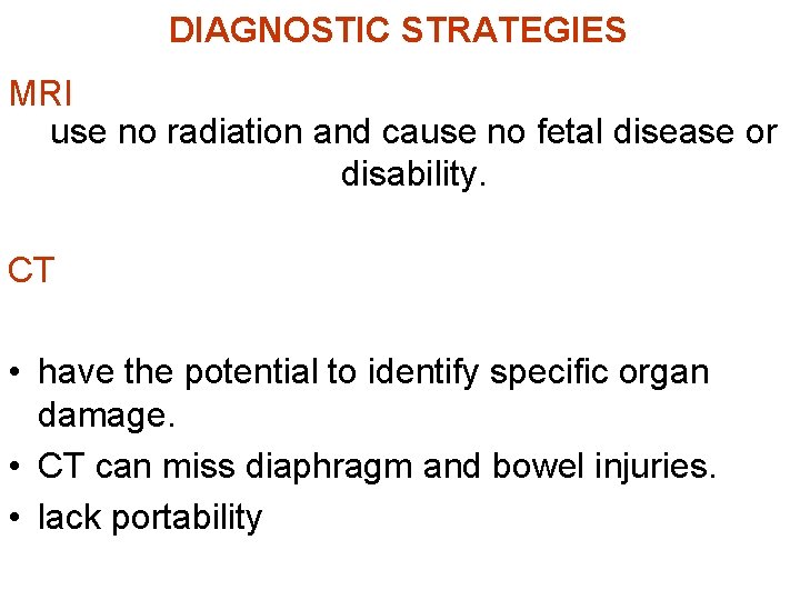
DIAGNOSTIC STRATEGIES MRI use no radiation and cause no fetal disease or disability. CT • have the potential to identify specific organ damage. • CT can miss diaphragm and bowel injuries. • lack portability
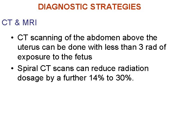
CT & MRI DIAGNOSTIC STRATEGIES • Radiation from CT scanning is a concern in the pregnant trauma patient. • with shielding, fetal exposure from head and chest CT scans can be kept below an acceptable 1 rad limit.
CT & MRI DIAGNOSTIC STRATEGIES • CT scanning of the abdomen above the uterus can be done with less than 3 rad of exposure to the fetus • Spiral CT scans can reduce radiation dosage by a further 14% to 30%.
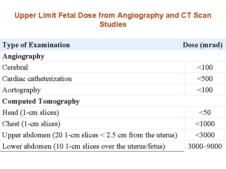
Upper Limit Fetal Dose from Angiography and CT Scan Studies Type of Examination Angiography Cerebral Cardiac catheterization Aortography Computed Tomography Head (1 -cm slices) Chest (1 -cm slices) Upper abdomen (20 1 -cm slices < 2. 5 cm from the uterus) Lower abdomen (10 1 -cm slices over the uterus/fetus) Dose (mrad) <100 <500 <100 <50 <1000 <3000– 9000
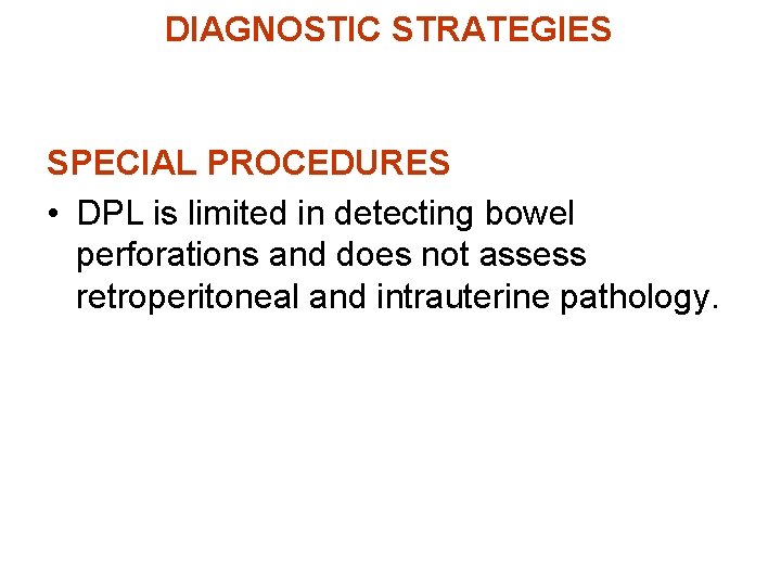
DIAGNOSTIC STRATEGIES SPECIAL PROCEDURES • DPL is limited in detecting bowel perforations and does not assess retroperitoneal and intrauterine pathology.
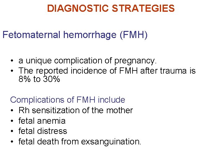
DIAGNOSTIC STRATEGIES Fetomaternal hemorrhage (FMH) • a unique complication of pregnancy. • The reported incidence of FMH after trauma is 8% to 30% Complications of FMH include • Rh sensitization of the mother • fetal anemia • fetal distress • fetal death from exsanguination.
DIAGNOSTIC STRATEGIES Fetomaternal hemorrhage (FMH) • more concern after 12 weeks' gestation when the uterus rises above the pelvis and becomes susceptible to direct trauma.
DIAGNOSTIC STRATEGIES Fetomaternal hemorrhage (FMH) Trauma patients at risk for massive FMH present with • major injuries • abnormal obstetric findings such as – Uterine tenderness – Contractions – Vaginal bleeding
DIAGNOSTIC STRATEGIES Kleihauer-Betke Test • identifies fetal cells in a maternal blood sample. • Most laboratories screen for FMH of 5 m. L or more. • Unfortunately, the amount of FMH sufficient to sensitize most Rh-negative women is well below this 5 -m. L sensitivity level • Therefore, all Rh-negative mothers who have a history of abdominal trauma should receive one prophylactic dose of Rh immune globulin (Rh. IG).
DIAGNOSTIC STRATEGIES Kleihauer-Betke Test • In the first trimester, one 50 -μg dose is used because total fetal blood volume is only 4. 2 m. L by 12 weeks' gestation and a 50 -μg dose covers 5 m. L of bleeding. • During the second and third trimesters, a 300 -μg dose of Rh. IG is given, which protects against 30 m. L of FMH.
DIAGNOSTIC STRATEGIES Kleihauer-Betke Test • useful in screening FMH that exceeds the efficacy of one 300 -μg dose of Rh. IG at 16 weeks' gestation, when total fetal blood volume reaches 30 m. L. • Because Rh. IG can effectively prevent Rh isoimmunization when administered within 72 hours of antigenic exposure, the results of the KB test are not immediately needed in the emergency department.
MANAGEMENT • Advance notification by out-of-hospital personnel • An obstetrician can be called • if the patient is in her last trimester, a pediatrician's presence is desirable. • A fetal monitor, portable US, and neonatal resuscitation equipment should be available in the emergency department.
MANAGEMENT Maternal Resuscitation Primary Survey • it is reasonable also to gather preliminary information about the fetus in the primary survey
MANAGEMENT Pregnant patients are a special challenge for several reasons • (1) the incidence of difficult intubation is 17 fold higher in advanced pregnancy • (2) aspiration risk is higher • (3) risk of hypoxia is increased during intubation because of reduced functional residual capacity and increased oxygen consumption.
MANAGEMENT Airway and Breathing • Rapid sequence intubation is recommended when intubation is performed. • Mechanical respirators need to be adjusted for increased tidal volumes and respiratory alkalosis consistent with the physiologic Paco 2 of 30 mm Hg in the last stage of pregnancy.
MANAGEMENT Airway and Breathing Fetus is very vulnerable to any reduction in oxygen delivery • Oxygen therapy should be instituted early.
MANAGEMENT Circulation Early intravenous access for volume resuscitation is indicated. Maternal blood pressure and heart rate are not consistently reliable predictors of fetal and maternal well-being. The mother can be bleeding but not show early signs of hypotension. vasopressors do not have a role in treating hypovolemic shock, and the pregnant patient is no exception.
MANAGEMENT Circulation • When traditional signs of shock appear, fetal mortality rates as high as 85%. • Lactated Ringer's solution is the preferred intravenous fluid for initial resuscitation.
MANAGEMENT Circulation • Beyond 20 weeks' gestation, the patient should be tilted 15 to 30 degrees to the left when on a backboard or should have the right hip elevated. • Tilting to the right is less effective in removing the uterus from the inferior vena cava.
MANAGEMENT Circulation • For severe injuries, a CVP line is helpful in assessing cardiac preload.
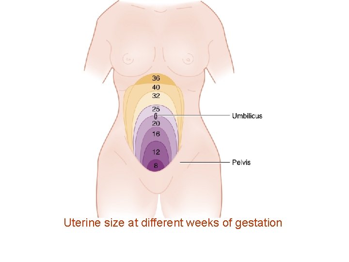
MANAGEMENT Circulation Estimating gestational age • Uterine size Usually, 24 to 25 weeks is used as the cutoff point for fetal viability
Uterine size at different weeks of gestation
MANAGEMENT Circulation As a rough guide, the fetus is potentially viable when • Dome of the uterus extends beyond the umbilicus • Fetal heart tones can be detected by auscultation at 20 weeks' gestation or by Doppler probe at 10 to 14 weeks.
MANAGEMENT Circulation treatment is directed solely at the mother If either • the uterus is less than 24 cm in size • or fetal heart tones are absent
MANAGEMENT Tocolytic Intravenous magnesium sulfate and the betamimetics • potent tocolytic agents • may exacerbate maternal hypotension by inducing maternal vasodilatation • invasive blood pressure monitoring is recommended • these drugs are usually inappropriate in trauma management.
MANAGEMENT Secondary Survey • Physical examination of the abdomen is unreliable • Information can be gathered about uterine tenderness, contraction frequency, and vaginal bleeding.
MANAGEMENT Secondary Survey Speculum Pelvic examination • Vaginal bleeding suggests placental abruption • watery discharge suggests rupture of the membranes. • If a vaginal fluid sample placed on a slide dries and crystallizes in a ferning pattern, it is amniotic fluid and not urine. • Cervical cultures for group B streptococci, Neisseria gonorrhoeae, and Chlamydia should be obtained if there is evidence of amniotic fluid leak. Bimanual examination • should be limited to assessing for pelvic bone injury or progression of advanced labor.
MANAGEMENT Fetal Evaluation • Fetal evaluation focuses on the fetal heart rate and detection of fetal movement. • If the fetus is viable (24 weeks or more) continuous external monitoring should be initiated quickly and maintained throughout all diagnostic and therapeutic procedures.
MANAGEMENT Fetal Evaluation Fetal Distress can be a sign of occult maternal distress. Fetal Distress Signs • abnormal baseline rate • decreased variability of heart rate • fetal decelerations after contractions. • The normal fetal heart rate ranges between 120 and 160 beats/min. Rates outside or trending toward these limits are ominous.
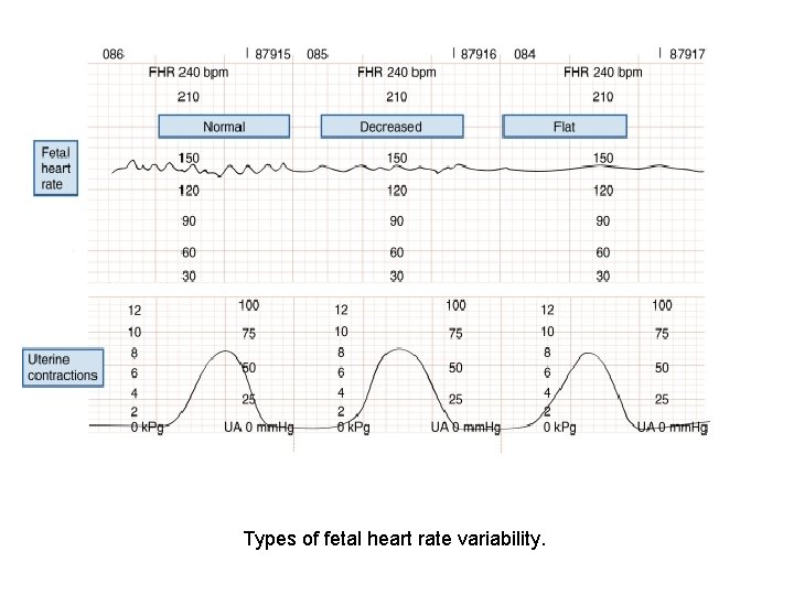
MANAGEMENT Fetal Evaluation Heart rate variability Two components • Beat-to-beat variability measures autonomic nervous function • Long-term variability indicates fetal activity. Heart rate variability increases with gestational age. • The loss of beat-to-beat and long-term variability warns of fetal central nervous system depression and reduced fetal movement caused by fetal distress
MANAGEMENT Heart rate variability Fetal Evaluation Late decelerations • indication of fetal hypoxia. • relatively small in amplitude and occur after the peak or conclusion of a uterine contraction.
MANAGEMENT Fetal Evaluation Heart rate variability Early decelerations are • larger • occur with the contraction • recover to baseline immediately after the contraction. • Early decelerations may be vagally mediated when uterine contractions squeeze the fetal head, stretch the neck, or compress the umbilical cord. Variable decelerations are large, occur at any time, and are possibly caused by umbilical cord compression
Types of fetal heart rate variability.
MANAGEMENT 3 Categories 1)Mother Stable, Fetus Stable • focus of care is directed toward the fetus. • For the viable fetus, monitoring is the next step. • The recommended 4 hours of cardiotocographic observation of the viable fetus should be extended to 24 hours if, at any time during the first 4 hours, there are – – – more than three uterine contractions per hour persistent uterine tenderness a worrisome fetal monitor strip vaginal bleeding rupture of the membranes any serious maternal injury is present.
MANAGEMENT 1)Mother Stable, Fetus Stable Mother should be instructed to record fetal movements for 1 week. • If fewer than four movements per monitored hour are noted, the patient should see her obstetrician immediately and a nonstress test should be performed. • Serial US and fetal heart rate tests should be performed on viable fetuses a few days after all trauma episodes
MANAGEMENT 2)Mother Stable, Fetus Unstable • If a viable fetus remains in distress despite optimization of maternal physiology, cesarean section should be performed. • Emergency decisions on fetal viability made on the basis of the best gestational age information available.
MANAGEMENT 2)Mother Stable, Fetus Unstable • Fetal heart tones is an important survival marker. • No infant survives if there is no fetal heart tone before emergency cesarean section commences. • If fetal heart tones are present and the gestational age is 26 weeks or more, then infant survival is 75%.
MANAGEMENT 2)Mother Stable, Fetus Unstable Reasons for a cesarean section include • besides fetal distress • uterine rupture • fetal malpresentation during premature labor • uterus mechanically limits maternal repair. Fetal death is not an indication for cesarean section because spontaneous labor usually occurs within 1 week.
MANAGEMENT 3)Mother Unstable, Fetus Unstable • The best initial action on behalf of the fetus is early restoration of normal maternal physiology. • If it is felt that the unstable mother can tolerate an emergency cesarean section, it can be attempted for the distressed, viable fetus. • Any above-the-uterus intraperitoneal gunshot wounds should be explored.
Decision-making algorithm in emergency obstetric care. C-section, cesarean section; FHTs, fetal heart tones; U/S, ultrasonography.
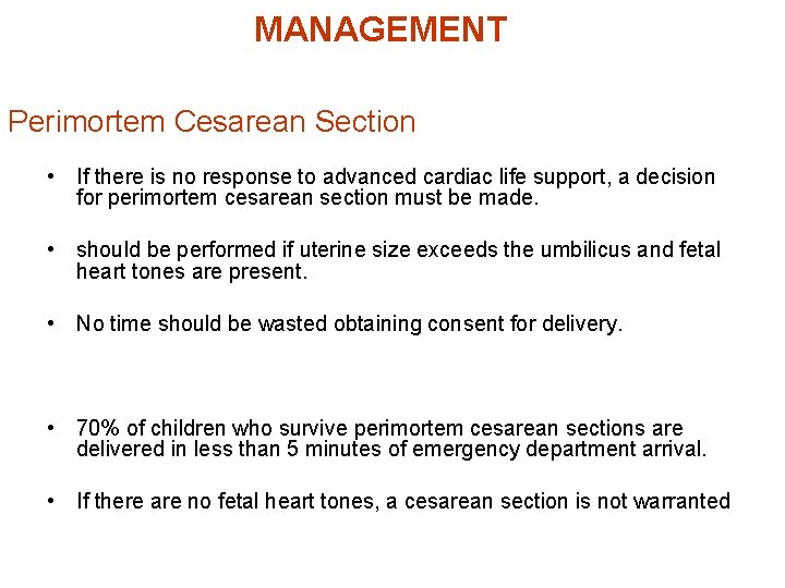
MANAGEMENT Perimortem Cesarean Section • If there is no response to advanced cardiac life support, a decision for perimortem cesarean section must be made. • should be performed if uterine size exceeds the umbilicus and fetal heart tones are present. • No time should be wasted obtaining consent for delivery. • 70% of children who survive perimortem cesarean sections are delivered in less than 5 minutes of emergency department arrival. • If there are no fetal heart tones, a cesarean section is not warranted
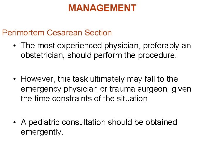
MANAGEMENT Perimortem Cesarean Section • The most experienced physician, preferably an obstetrician, should perform the procedure. • However, this task ultimately may fall to the emergency physician or trauma surgeon, given the time constraints of the situation. • A pediatric consultation should be obtained emergently.
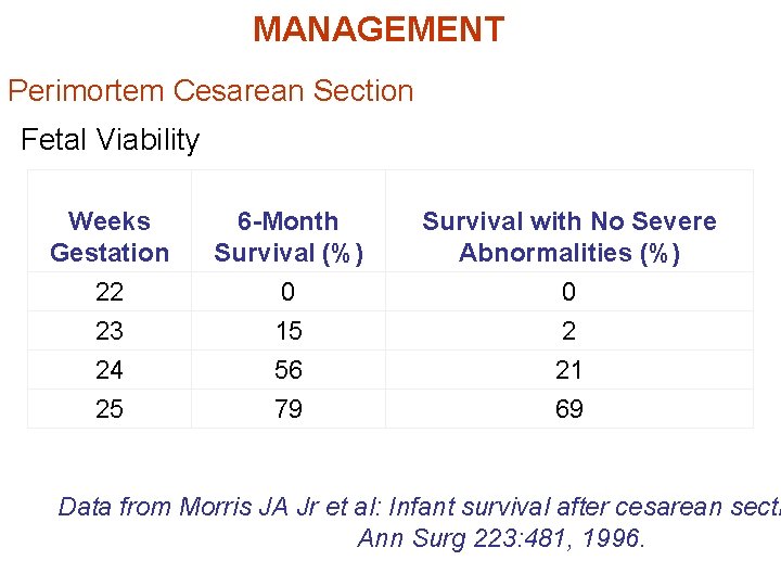
MANAGEMENT Perimortem Cesarean Section Fetal Viability Weeks Gestation 6 -Month Survival (%) Survival with No Severe Abnormalities (%) 22 0 0 23 15 2 24 56 21 25 79 69 Data from Morris JA Jr et al: Infant survival after cesarean secti Ann Surg 223: 481, 1996.
MANAGEMENT Perimortem Cesarean Section • Of 441 pregnant trauma victims, 32 underwent emergency cesarean section because of either fetal or maternal distress • Survival rates were 45% for the infants and 72% for the mothers. • Their conclusion was that a fetus with an estimated gestational age of 26 weeks that has cardiac activity is salvageable, irrespective of maternal injury Morris J. A. , Rosenbower T. J. , Jurkovich G. J. , et al: Infant
Trauma in Pregnancy Tetanus toxoid and immune globulin • No detrimental effect on the fetus. • Proper immunization of pregnant women decreases the incidence of neonatal tetanus because the tetanus antibody crosses the placenta. Electrical flow • little effect on the pregnancy. • Maternal elective and emergent cardioversion has been performed safely for cardiac dysrhythmias in all three stages of pregnancy. • the fetal heart should be monitored during cardioversion.
Trauma in Pregnancy DISPOSITION • Any pregnant woman at 24 or more weeks of gestation who has suffered blunt body trauma should be admitted for fetal monitoring even if she looks well.
Trauma in Pregnancy References 1. Trauma in Pregnancy Emergency Medicine Clinics of North America - Volume 25, Issue 3 (August 2007) Seric S. Cusick 2. Trauma in Pregnancy Obstetrics and Gynecology Clinics - Volume 34, Issue 3 (September 2007) Michael V. Muench 3. Initial trauma management in advanced pregnancy. Review Full Text Abstract PDF Find Similar Articles Meroz Y - Anesthesiol Clin - 01 -MAR-2007; 25(1): 117 -29, x 4. Rosen 2006