Transportation in plants and animals Transportation is a


 Blood circulatory system")


")


 The heart in mammals")


- Slides: 44
Transportation in plants and animals Transportation is a life process in which a substance absorbed or made in one part of the body of an organism , is carried to other parts of its body.
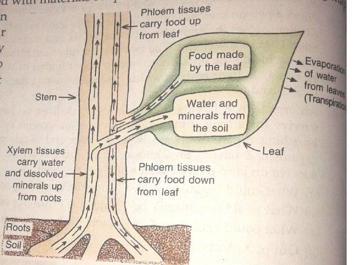
Transportation in plants • Transportation of materials in plants is done by two types of vascular tissues-xylem and phloem. • Xylem helps in transportation of water and minerals. • Phloem helps in transportation of food( from leaves to other parts of plants) and hormones( from root and shoot tip)
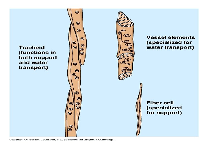
Types of xylem and phloem Xylem parenchyma Xylem Sclerenchyma Xylem vessels Phloem Parenchyma Phloem Sclerenchyma Sieve Tube Xylem tracheids Companion cells In Case of xylem only xylem parenchyma is the living tissue others are dead. While in case of Phloem except phloem Sclerenchyma all other types are living.
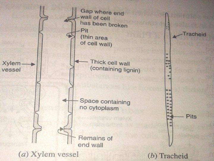
Transportation of water and minerals • Plant require water for the process of photosynthesis. They also need minerals for various purpose like making proteins. • Water and minerals present in the soil is absorbed by the roots and transported to various parts of the plant like stem, leaves and flowers via conducting tissue called xylem. • Two types of elements of xylem tissue called xylem vessels and tracheids helps in transportation process. Both are non living tissue having thick. wall.
Description of xylem vessels & tracheids Xylem vessels Xylem traheids It is a non living , long tube which runs like Tracheids are long, thin, spindle shaped a drainpipe throughout the plant. cells with pits in their thick cell wall. A xylem vessel is made of many hollow, dead cells, joined end to end. The end walls of the cells have broken down so a long, open tube is formed They are elongated cells with tapering ends. They do not have open ends, so they do not form vessels. Dead cells-do not contain the cytoplasm or nuclei. The walls of xylem vessels are made of cellulose and lignin. Dead cells with lignified walls. Xylem vessels run from the roots of the Tracheids have pits in their walls, so water plant right up through the stem and reach can pass from one tracheids to another the leaves. through these pits.
Few terms Epidermis The outer layer of cells in the root. It is only one cell thick. Endodermis The layer of cells around the vascular tissues ( xylem and phloem). It is the inner most layer of the cortex. Root cortex The part of root between the epidermis and the endodermis. Root xylem Xylem tissue present in roots. Xylem sap Water and minerals carried by xylem tissue
Important points • Water absorbed by root hairs----pass through epidermis----root cortex----endodermis, then it reaches to root xylem. • The minerals needed by the plants are taken up by the plants in inorganic form such as nitrates and phosphates. These minerals are present in soil. • When water is transported by the root of the plant to its leaves, then the minerals dissolved in it also get transported along with it.
Mechanism of transport of water and minerals in a plant
• Water and minerals present in the soil • Root hairs absorb these materials form the soil By diffusion process • These materials pass from cell to cell by osmosis through the epidermis, root cortex, endodermis and reach the root xylem • Water + dissolved materials enters from the root xylem vessels to stem xylem vessels and finally branch into leaves of the plant. Only 1 -2 % of the water absorbed by the plant is used up by it in photosynthesis and other metabolic activities. The rest of water is lost as water vapour through transpiration process.
Importance of transpiration in transportation of water High pressure • A lot of water from the leaves keeps on evaporating into the air Low through the stomata present on pressure the surface of the leaves. • So water from the xylem vessels in the leaf will travel to the cells by osmosis to make up this loss of water. • This reduces the effective pressure at the top of the xylem vessels. • So water flows into them from the soil. • Thus, the continuous evaporation of water/transpiration from the cells of a leaf create a kind of Upward suction which pulls up water movement through the xylem vessels. of water from roots to leaves
Transportation of food and other substances • The transportation of food from the leaves to other parts of the plant is called translocation. • Phloem tanslocates the food made in the leaves to other parts of plant. • The movement of food materials and other substances like hormones through phloem depends on the action of living cells called sieve tube ( a kind of phloem)
Phloem
Phloem • Phloem is made of tubes many cells joined end to form long tube. • However the end walls of the cells which form phloem arenot completely broken down. • The end walls of cells in the phloem form sieve plates , which have small holes in them. • These holes in the sieve plates allow the food to pass along the phloem. The cells of phloem are called sieve tubes. • Sieve tubes are living cells which contain cytoplasm but no nucleus. They donot have lignin in their walls. • Each sieve tube has a companion cells next to it. • Companion cell has a nucleus and may other organelles. These cells supply the sieve tubes with some of their requirements.
Mechanisms of transport of food in plants • The food is prepared by the mesophyll cells of leaves • The food is loaded into the sieve tubes of phloem tissue by utilizing energy from ATP. • Water now enters into sieve tubes containing sugar by the process of osmosis. • Pressure in the phloem tissue rises. • Food moves to all the parts of the plant having less pressure in their tissues. In this way phloem transport food according to needs of the plant. For e. g. - in Spring, even the sugar stored in the root or stem tissue of a plant would be transported through phloem to the buds which need energy to grow.
Difference Xylem Phloem Conducts water and minerals Conducts food materials and hormones. Conduction is mostly unidirectional i. e from roots to apical parts of plant. Conduction may be bi directional i. e from leaves to roots or storage organs or growing parts depending upon the requirement. ATP is not required for conduction. Transpiration causes a kind of suction pump to transport water and minerals. ATP is required for translocation of food. Conducting elements are tracheids and vessels. Conducting elements are sieve tubes. In addition to conduction , it also provides mechanical strength to plant. Performs no mechanical function for the plant.
Transportation in animals
Human circulatory system • Human circulatory system consists of : (a) Blood circulatory system (b) Lymphatic system Blood circulatory system consists of : - 1. Blood 2. Blood vessels 3. Heart
Blood- It is a connective tissue, consisting of blood plasma&blood cells Blood plasma Blood cells It is a colurless liquid which consists 1. RBC-Red blood cells- Donot mainly of water with many contain nucleus, Contain substances dissolved in it like Hemoglobin pigment. Normal life proteins, digested food, common span is 120 days. Helps in salts, waste products ( CO 2, urea) and transportation of oxygen hormones. It contains 90% water. throughout the body. Plasma carries all these dissolved 2. WBC- White blood cellssubstances from one part to another Nucleated, less in no. as compared part in the body. Blood to RBC. Helps in fighting against cells/corpuscles are immersed in this infection by engulfing the germs fluid. or producing antibodies. 3. Platelets- Donot contain nucleus. Help in clotting of blood and thus prevent excessive loss of blood during an inquiry.
Functions of blood • Regulates the body temperature- the blood capillaries in our skin help to keep our body temperature constant at about 37 degree Celsius. • Carries a waste product called urea from the liver to the kidneys for excretion in urine. • Carries hormones from the endocrine glands to different organs of the body. • Carries digested food from the small intestine to all the parts of the body. • Protects the body from diseases (By WBC) • Carries oxygen from lungs to different parts of the body. • Carries CO 2 from the body cells to the lungs for breathing out.
Blood vessels- these are tube kind of structure through which blood flows in the body. • Types of blood vessels: (a) Arteries (b) Veins (c) Capillaries
Comparison Arteries Thick walled Veins Thin walled Carry blood from heart to all other part of Carry blood from all the parts of the body back to the heart. Blood flowing through them under high pressure. Blood flowing thorough them under low pressure. Donot have valves. Have valves to prevent back flow of blood. The Main artery is called the aorta which is connected left ventricle of heart through a valve called semilunar valve. Aorta carries oxygenated blood from left ventricle to all other parts of the body ( except lungs) The main vein is called Vena cava which is connected to right atrium of heart. Vena cava carries deoxygenated blood from all the parts of the body ( except lungs) back to the heart. Generally arteries carry oxygenated blood but pulmonary artery which is connected to right ventricle ( Through semilumar valve) carries deoxygenated blood and sent it to lungs for oxygenation. Generally veins carry deoxygenated blood, but pulmonary vein carries oxygenated blood from lungs to left atrium.
Capillaries • Thin walled • Extremely narrow tubes which connects arteries to veins. • Food and oxygen go from blood into body cells through capillaries. • Waste materials like CO 2 go from body cells into the blood through capillaries. • So we can say tat exchange of varies materials like oxygen, food, carbon dioxide etc between the blood and body cells takes place through capillaries.
Heart ( draw any one diagram)
Structure of heart • • • Heart is a muscular organ which pumps blood to all parts of the body. Made up of cardiac muscles. Protected by pericardium layer. No. of chambers- 4, upper two chambers are left and right atrium and lower two chambers are left and right ventricles. Atria are smaller chambers than ventricles. Walls of ventricles are much thicker than atria because ventricles supply blood to the different organs. Septa- partition to separate 4 chambers. (a)Inter atrial septa separates right and left atria, (b) inter ventricular septa separates right and left ventricles, these septas separates left and right chambers and prevent mixing of oxygenated and deoxygenated blood. ©atrial ventricular septa separates atrium and ventricles of same side. Valve- prevent any back flow of blood i. e allows flow of blood only in one direction. (a)Tricuspid valve. -Guards the opening between right atrium and right ventricle. (b) Bicuspid valve ( mitral valve) guards the opening between left atrium and left ventricle. (c) Semilunar valve ( of right side)regulates opening between right ventricles into pulmonary artery, (d) another semilunar valve ( of left side) regulates opening between left ventricle and aorta.
Continue • • The left side of the heart carries oxygenated blood while the right side of the heart carries deoxygenated blood. The left atrium receives oxygenated blood though pulmonary vein ( from lungs). The main vein i. e vena cava collects deoxygenated blood from all parts of the body and pours into right atrium. The left ventricles transports oxygenated blood to all parts of the body through main artery i. e aorta. Aorta is connected to left ventricle through a valve V 3( semi lunar valve). The right ventricle transports deoxygenated blood to lungs through pulmonary artery. Pulmonary artery is connected to the right ventricle through a valve V 4 ( semi lunar valve) The left atrium is connected to left ventricle through (bicuspid) V 1 valve, while the right atrium is connected to right ventricle through V 2 valve ( tricuspid) valve. These valves prevent back flow of blood intro atrium when the ventricles contract (to pump blood out of the heart to the rest of the body). When the ventricles contract, the valves V 1 and V 2 close automatically. All the atria and ventricles of the heart contract and relax at approximate times and make the heart behave like a pump.
Blood circulation 1. The heart beats non stop all the time. It is due to the rhythmic contraction and relaxation of the heart muscles (which make up the atria and the ventricles. ) 2. The two atria contract together and relax together. Same is applicable for ventricles. 3. The contraction of two atria is immediately followed by contraction of two ventricles. 4. The heart beat circulates the blood in the human body
ONLY FOR INFORMATION
1. 2. 3. 4. 5. 6. When the muscles of all the four chambers of the heart are relaxed, the pulmonary vein brings the oxygenated blood from the lungs into the left atrium(LA) of the heart. When the LA contracts, the oxygenated blood is pushed into the left ventricle (LV) through the valve V 1( Bicuspid valve). When the LV contracts, the oxygenated blood is forced into the main artery called aorta. Aorta branches intro smaller arteries which go into different body organs ( except lungs. The smaller arteries ( arterioles) further branch into capillaries. Aorta carries blood to all the organs. When the oxygenated blood passes through the capillaries of the body organs, then it gives oxygen to the body cells. It also gives digested food and other dissolved materials to the body cells. At the same time CO 2 is produced as waste material during respiration process. The blood now becomes deoxygenated. Vena cava ( main vein) carries deoxygenated blood from all parts of the body and pours into RA. When the RA contracts, deoxygenated blood is pushed into the RV through valve V 2 ( tricuspid valve) When the RV contracts, the deoxygenated blood is pumped into the lungs through the pulmonary artery. In the lungs the blood becomes oxygenated , which is again sent to the LA of the heart by pulmonary vein for circulation in the body.
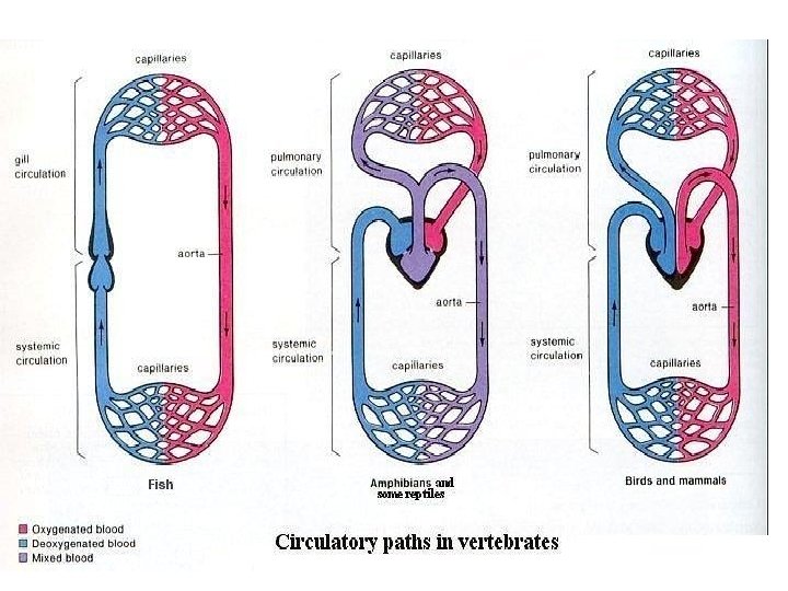
Double circulation in human beings • A circulatory system in which the blood travels twice through the heart in one complete cycle of the body is called double circulation. • Double circulation= pulmonary+ Systemic circulation. 1. Pulmonary circulation- in human circulatory system the pathway of blood from the heart to the lungs ( deoxygenated blood) and back to the heart ( oxygenated blood) is called pulmonary circulation. 2. Systematic circulation- the pathway of blood from heart to the rest of the body ( oxygenated blood) and back to the heart ( deoxygenated blood) is called the systemic circulation
Hearts of mammals, birds, amphibians, reptiles, and fishes : ●i) The heart in mammals : - and birds have four chambers and the right and left sides of the heart is separated by a septum. This prevents mixing of oxygenated and deoxygenated blood and provides efficient supply of oxygen. This is necessary because they need more energy to maintain their body temperature. All the animals having four chambered heart have double circulation. ●ii) The heart in amphibians and reptiles : - have three chambers ( two atria and one ventricle) and allows some mixing of oxygenated and deoxygenated blood because the do not use energy to maintain their body temperature. Their body temperature is the same as the temperature of the surroundings. They are cold blooded animals. Due to incomplete division within their heart, the oxygenated and deoxygenated bloods mix to some extent. This reduces the production of energy. They have however double circulation that delivers blood to the lungs and the rest of the body respectively. ●iii) The heart in fishes : - have only two chambers ( one atrium and one ventricle) and blood is oxygenated in the gills. The heart pumps deoxygenated blood to the gills. Oxygenation of blood takes place in the gills. The oxygenated blood from the gills is supplied to the body parts. The deoxygenated blood returns to the heart to be pumped into the gills again. This is a case of single circulation because the blood passes through the heart of fish only once in one complete cycle of the body.
Heart beat • The heart pumps blood into our arteries by contracting. • When the heart contracts ( systole), it becomes smaller in size and pushes the blood into the main artery ( aorta) with a great force. • When the heart relaxes(diastole), it gets filled up with blood from pulmonary vein. • In this way the heart keeps on contracting and relaxing again and again to pump blood into the body continuously. • One complete contraction and relaxation of the heart is called a heart beat. • Normal heart beat is 70 -72 times a minute. • Heart beats faster during and after an exercise because the body needs more energy under these conditions Faster heart beat---more supply of blood----more supply of O 2 ---rapid respiration-----more energy
Cardiac cycle Only for information
AV valves* Semilunar valves† Status of ventricles and atria • Atria contract and pump blood 1. Atrial Systole 2. Isovolumetric Contraction 3. Ventricular Ejection 4. Isovolumetric Relaxation open closed Closed • Ventricles, already partially filled from phase 5, receive last ~30% of blood, for a final resting volume of approximately 130 m. L. • Ventricles begin to contract. Ventricular muscle initially shortens only a little, but intraventricular pressure rises sharply • Ventricular volume unchanged closed Open Closed • Pressures in left and right Ventricle exceed pressures in Aorta (80 mm. Hg) and Pulmonary Artery (10 mm. Hg). Ejection is rapid at first, slowing down as systole progresses. • Amount ejected each ventricle per stroke at rest is 70 -90 m. L. Approximately 50 m. L of blood remains in each ventricle at the end of systole • Valves close as Ventricles relax and pressure within Ventricles drops below 120 mm. Hg. This ends once Ventricular Pressure falls below Atrial pressure and AV valves open • pump blood to rest of body • ventricles relaxed 5. Ventricular Filling open Closed • ventricles passively fill with approximately 70% of their final volume. As the ventricles fill, rate of filling decreases and the AV valves drift towards closing • atria expand are filling * AV (atrioventricular) valves: 1) mitral valve – between the left atrium and the left ventricle 2) tricuspid valve – between the right atrium and the right ventricle † Semilunar valves: 1) aortic valve – between the left ventricle and the aorta 2) pulmonic valve – between the right ventricle and
Pulse • Every time the heart beats, blood is forced into arteries. • This blood makes the arteries expand a little. • The expansion of an artery each time the blood is forces into it, is called pulse. • Each heartbeat generates one pulse in the arteries, so the pulse rate of a person is equal to the no. of heartbeats per minute. • The pulse rate of an adult person while resting is 70 -72 per minute, which increases after a physical exercise or when a person is excited.
Blood pressure • The pressure at which blood is pumped around the body by the heart is called blood pressure. • The phase of the heart beat when the heart contracts and pump blood into arteries is called systole, and the phase of heart beat when the heart relaxes and allows the chambers to fill with blood is called diastole. • Blood pressure of a person is always expressed in the form of two values- 1. systolic pressure & 2. Diastolic pressure. • The maximum pressure at which the blood leaves the heart through the main artery ( aorta) during contraction phase , is called systolic pressure. this maintains steady flow of blood in all the arteries towards the capillaries. • The minimum pressure in the arteries during relaxation phase of heat is called the diastolic pressure. Normal blood pressure – 120/80, that means • Systolic pressure: 120 mm Hg, • diastolic pressure- 80 mm Hg. • Blood pressure values vary from person to person and from time to time. • High blood pressure is called hypertension. It is caused by the constriction( narrowing) of arterioles ( very small arteries) which results in increased resistance to blood flow. Very high blood pressure can lead to rupture of an artery and internal bleeding.
How to measure blood pressure by using sphygmomanometer Pressure applied=200 mm Hg to the brachial artery. Brachial artery closed, no blood flows trough it, hence no tapping sound is heard. Cuff pressure is reduced gradually by deflating it. Cuff pressure =systolic pressure, brachial artery opens up slightly, intermittent blood flow. Cuff pressure is reduced further. Brachial artery opens up fully, blood flow in it is fully restored.
How do food and oxygen reach body cells • Tissue fluid- the liquid from the blood which is forced through the capillary walls and moves between all the body cells, providing them with food and O 2 and removing CO 2 is called tissue fluid. • The walls of blood capillaries are very thin. So when blood flows through them, tissue fluid leaks from the blood capillaries and goes into tiny spaces between the various body cells in the tissue. • The tissue fluid carries food and oxygen from the blood to the cells and picks up their waste products like CO 2. • After this most of the tissue fluid seeps back into blood capillaries. • The remaining tissue fluid carrying large protein, digested fat, germs from the cells and fragments of dead cells, enters into lymph capillaries and becomes lymph. • This lymph is returned to the blood by another type of transport system in the human body called lymphatic system.
Lymphatic system • A system of tiny tubes called lymph vessels and lymph nodes in the human body which transports the liquid called lymph from the body tissues to the blood circulatory system is called lymphatic system. • The lymphatic system consists of : - Lymph capillaries, larger lymph vessels. Lymph nodes/glands and lymph
components of lymphatic system Lymph capillaries Smaller tubes Tiny tubes , present in the whole body. Lymph capillaries are close ended, so the tissue fluid can only seep into the walls of the lymph capillaries. Pores in the walls of lymph capillaries are bigger, so even large protein molecules present in the tissue fluid can enter into it. Lymph vessels The lymph capillaries join to form larger lymph vessels Lymph nodes . The lymph vessels have lymph nodes at intervals. The lymph vessels are connected to large veins of the blood circulatory system ( subclavian veins) Lymph Light yellow liquid which is somewhat similar in composition to blood plasma( does not contain RBCs). It contains large protein molecules , digested fats. , germs from the cells and fragments of dead cells . It is another medium of circulation in the human body. It flows only in one direction i. e from body fluid to heart. Lymph is also called as extra cellular fluid as it remains outside the c ells of the body. Lymph contain lymphocytes( a type of WBC) which helps in fighting infection and diseases. The lymph nodes contain special type of cells called lymphocytes, which is involved in the cleaning of lymph and protecting the body from diseases.
How lymphatic system works? ? 1. Lymph containing large protein molecules, digested fat, germs and fragments of dead cells from the tissue fluid seeps into the lymph capillaries. 2. From lymph capillaries lymph passes into larger lymph vessels containing lymph nodes. 3. In the lymph nodes lymph is cleaned by lymphocytes. Lymphocytes eat the germs, dead cells and make antibodies for protecting body against diseases. 4. The cleaned lymph containing large protein, digested fat and other useful material is transported by lymph vessels to the large veins called subclavian veins. 5. Subclavian veins carry the lymph to the heart. 6. Circulation of lymph from the body tissues to the heart is completed.