Transport Whats wrong with this picture Transport Process


 • Blood")


= Vessel narrowed by fatty plaques, blood")

 Atrioventricular Valve (Bicuspid, Mitral) Rt")
 Inferior Vena Cava (Vein) Tricuspid Valve Rt Atrium")


 • • • Atria fill with blood Atrioventricular")


")


")

 from heart damage or")


 • Causes: – Atherosclerosis (Hardening of Arteries) (Hardened cholesterol deposits")


 – We have about 30 Trillion –")


: • • • Protection Larger than RBCs")


 – Multiple Sclerosis")


")
 Heparin Aspirin Plasmin")

 – – – –")

")


. •")


 • Due to the HIV (Human Immunodeficiency")


- Slides: 119
Transport What’s wrong with this picture?
Transport • Process for substances moving into cells • Process for substances exiting cells • Process for movement within cells
Transport in Protists • Diffusion and Active transport for entering and exiting cell • Need wet membranes for diffusion. These organisms are aquatic! • Cyclosis within cell
Transport in Hydra • Aquatic • Ectoderm and Endoderm of Gastrovascular cavity in contact with water • Uses Diffusion and Active Transport • Uses Cyclosis within cells • Endodermal cells of cavity have flagella to circulate materials in cavity • Has muscular movement to circulate materials in cavity
Transport in Earthworm • Most cells not in direct water contact (Need a way of delivery and removal of materials from them) • Live in moist environment • Large and multicellular • Only skin cells able to exchange materials with the environment
Transport in Earthworm • Need a circulatory system: – Fluid in which materials are transported – Network of tubes or spaces for fluid flow – Need a pumping mechanism
Circulatory System of Worm • Closed Circulatory System • Aortic Arches (hearts) • Blood with blood cells having Iron-rich Hemoglobin to carry O 2 • Blood moves quickly in closed system • Dorsal and Ventral Blood Vessels • These branch off eventually into capillaries • Capillaries pass each cell for pick-up and delivery
Circulatory system of worm
Transport in Grasshopper • • Open Circulatory System Multiple Hearts Aorta Body Sinuses Body movement moves blood around Blood moves slowly around No Hemoglobin Blood doesn’t transport O 2
Transport in Grasshopper
Transport in Humans • • Closed Circulatory System One heart that has 4 chambers Network of blood vessels Hemoglobin blood
Blood Vessels • Arteries – Largest diameter – Thick, muscular, elastic walls – Carry blood away from the heart
Blood Vessels • Arterioles – Narrower branches of arteries – Also direct blood away from heart
Blood Vessels • Capillaries – – Very, very thin walled blood vessels (1 cell thick) Small diameter Pick-up and delivery of materials from cells So small in diameter that blood cells flow one at a time through them – Diffusion of materials (i. e. . Nutrients, wastes, oxygen) across capillary wall – Not all capillary beds open at same time (Have about 25, 000 -60, 000 miles of capillary)
Capillaries
Blood vessels • Venules – Return blood back to heart – Small branches of veins – Walls are thinner than arterioles – Walls not very elastic – Contain valves inside vessel – Need external muscular movement
Venule
Blood Vessels • Veins – Larger versions of venules – Many venules join into a vein
Lymph Vessels • • Don’t carry blood Also have valves Need external muscular activity They carry Lymph – What is Lymph?
• Capillaries are leaky • Fluid in capillary called plasma leaks out with dissolved nutrients for tissue • Fluid bathes cells and now fluid is called Intercellular fluid (or Interstitial Fluid)
• Diffusion happens • Wastes and Nutrients exchanged • Intercellular fluid now enters lymph vessels • Fluid is now called lymph • Fluid will be scanned by lymph nodes • Fluid will return to blood vessels at the Thoracic Duct
Thoracic Duct
Lymph Nodes • Sites that house white blood cells • Filter site for pathogens • Spleen also part of Lymphatic System
Problems with Blood Vessels • Varicose Veins-faulty valves-blood collects and unable to return to heart
More Problems • Arteriosclerosis (“hardening of the arteries”)= Vessel narrowed by fatty plaques, blood clots form
More Problems • Stroke – Blockage of a vessel feeding blood to the brain – Administer TPA in 1 st 3 hrs to prevent paralysis
Human Heart Rt Atrium Lt Atrium Atrioventricular Valve (Tricuspid) Atrioventricular Valve (Bicuspid, Mitral) Rt Ventricle Lt Ventricle Septum
Superior Vena Cava (Vein) Inferior Vena Cava (Vein) Tricuspid Valve Rt Atrium
Lt Atrium Pulmonary Veins Bicuspid
Rt Ventricle Pulmonary Artery Semilunar Valve
Lt Ventricle Aorta Semilunar Valve
Heart as a whole Covered by Pericardium Membrane Most Muscular!
Heart as a whole
Cardiac Cycle 1. Diastole (Heart Relaxed) • • • Atria fill with blood Atrioventricular valves open Blood drops into ventricles (70% Full) 2. Systole • • Atria contract in unison All blood squeezed into ventricles Ventricles contract in unison Atrioventricular valves slam closed and blood hits them (Hear a “Lub” sound) Semilunar valves open and blood pushed out into arteries Semilunar close (Hear a “dub” sound) Atria fill from veins
Cardiac Output • Heart beats 70 X/min at rest • Pumps 5. 25 L/m of blood/min at Rest, 3035 L/m during exercise
Heart Electricity • Pace-Maker or SA Node – In top wall of Rt atrium – Send an electrical pulse to both atria simultaneously, atria contract – Regulated by Vagus Nerve and Cardioaccelerator Nerve of brain
Heart Electricity • AV Node – In bottom wall of Rt Atrium – Signal from SA Node reaches AV Node – Signal spread to both ventricles causing them to contract
Ventricles Contraction Atria contraction
Flow of blood out of heart and through vessels • Pulse and Blood Pressure Pulse =wave of stretching as blood travels through arteries
Pulse and Blood Pressure • Normal Blood Pressure= – 120 (Systolic Pressure, mm Mg) – 80 (Diastolic Pressure, mm Mg) – 120/80 (For a 20 year-old) – Highest pressure in aorta – Lowest pressure in veins • Normal Pulse= – 70 beats/min
Circulatory Pathways-Pulmonary • Pulmonary circulation
Circulatory Pathways-Systemic • Renal Circulation (Filtering of blood of metabolic wastes
Circulatory Pathways-Systemic • Coronary Circulation Deoxygenated blood returns to heart through walls of Rt Atrium
Circulatory Pathways-Systemic • Hepatic-Portal Circulation (Blood Glucose Regulation
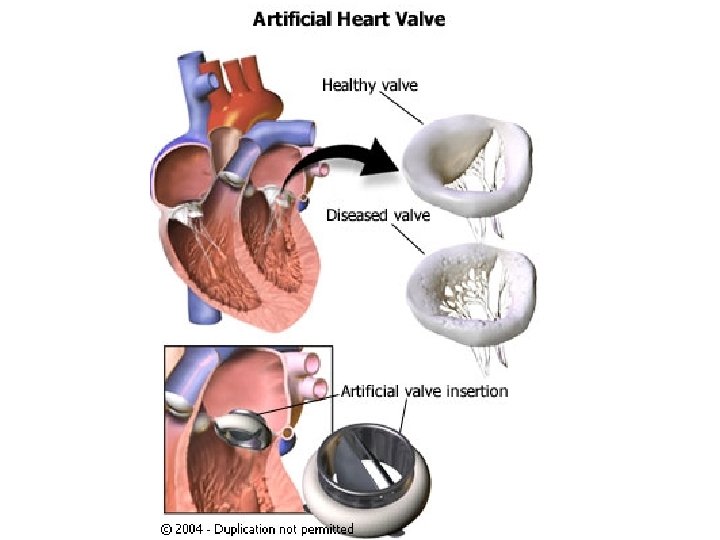
Problems with Heart and Vessels • Heart Valve Problem-Heart Murmur Can also have murmur from valves not opening well=Valvular Stenosis
Problems with Heart and Vessels • Angina=
Problems with Heart and Vessels • Heart Attack (Myocardial Infarction, Coronary Thrombosis)
Angioplasty Bypass Stents
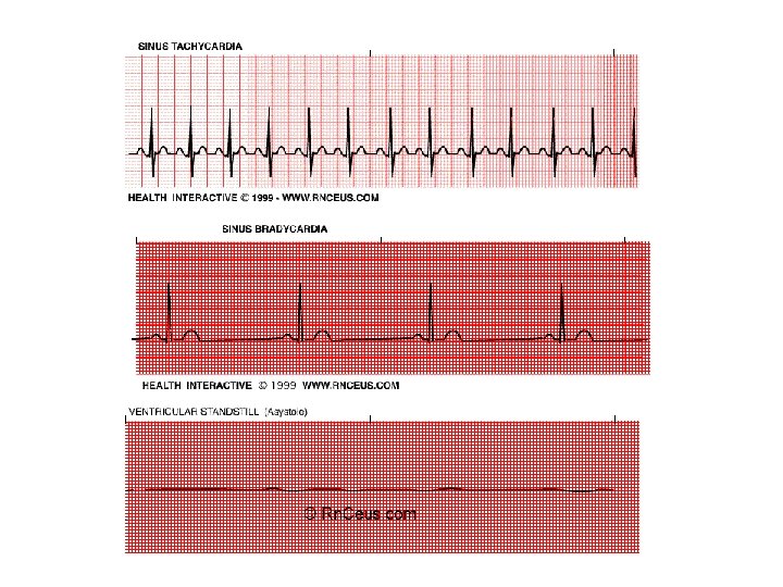
Problems with Heart and Vessels • Fibrillation (and Circus Movement) from heart damage or bad Nodes
Defibrillation Stop heart and let it restart in hopefully normal rhythm
Node Replacement
High Blood Pressure (Hypertension) • Causes: – Atherosclerosis (Hardening of Arteries) (Hardened cholesterol deposits called plaques with blood clot formation) • Problem: Can lead to heart attack and or stroke • Drugs: – Diuretics=increase urine output – β- Blockers=keep vessels dilated
Ways to avoid cardiovascular diseases • Don’t Smoke • Eat healthy, low fat foods • Monitor cholesterol levels (LDLs bad, HDLs good) • Don’t eat excessively salty foods (Sodium) • Exercise • Avoid excess alcohol • Monitor blood pressure if in “Risk Groups”
Blood and Immunity What is blood? Is blood a tissue?
Blood Functions • Transport – Nutrients – Wastes – Hormones • Regulation – Temperature regulation – p. H stabilization – Water balance • Protection – Specialized cells for defense – Clotting ability
Blood Components 1. Plasma – Liquid part – 55% of blood – Mostly water – Salts, glucose, amino acids, fatty acids, hormones, antibodies, vitamins, enzymes, metabolic wastes
Blood Components 2. Red Blood Cell (Erythrocytes) – We have about 30 Trillion – Transport Oxygen and CO 2 – Mature cells have no nucleus – Contains Iron-containing Hemoglobin protein – Made in bone marrow – Live about 120 days (2 million destroyed/sec) – Liver and spleen remove defective cells
Bone Marrow
Red Blood Cell
Problems related to Red Blood Cells • Anemia – Low RBC count and or low hemoglobin count – Low Iron amount or vitamin/mineral amount – Results in low blood oxygen amount – Pale, cold skin, low blood pressure, fatigue, dizziness
Problems related to Red Blood Cells • Sickle Cell Anemia – Genetic Disease of African ancestry – defective hemoglobin gene – Make misshaped RBC – Anemic symptoms – If two bad genes, death at early age – If one bad gene, pain at times – Protection against Malaria parasite
Sickled Cell Normal Cell Blockages
Blood Components 3. White Blood Cells (Leukocytes): • • • Protection Larger than RBCs Have nuclei Less in number compared to RBC Made in bone marrow 60 billion
White Blood Cell Types • Neutrophils and Monocytes – Phagocytic – Engulf bacteria, debris, virus-infected cells, cancer cells – Macrophages develop from monocytes and crawl around tissues searching for invaders
White Blood Cell Types • Eosinophils – Nonphagocytic – Involved in allergic reactions – Involved in parasitic infections
White Blood Cell Types • Basophil – Involved in inflammatory reactions – Involved in allergic reactions and parasitic infections – Related to Mast Cells
White Blood Cell Types • Lymphocytes – Nonphagocytic cells – Involved with Immunity – Subtypes: • B Cells: – Make Antibodies • T Cells: – Destroy virus-infected cells and cancer cells
Problems with White Blood Cells • Leukemia Normal Blood Smear Leukemia Blood Smear
Problems with White Blood Cells • Allergic reactions with possible Anaphylactic Shock (Overreaction to an invader)-Over-reaction
Problems with White Blood Cells • Autoimmune Diseases (Target Own Body) – Multiple Sclerosis (Target Brain) – Chrohn’s Disease (Target Gut) – Lupus (Target assorted body organs) – Psoriasis (Target Skin) – Rheumatoid arthritis (Target Joints) – Type 1 diabetes mellitus (Target Pancreas)
Blood Components 4. Platelets – Involved in blood clotting – Cell fragments – Formed in bone marrow – 1. 5 Trillion
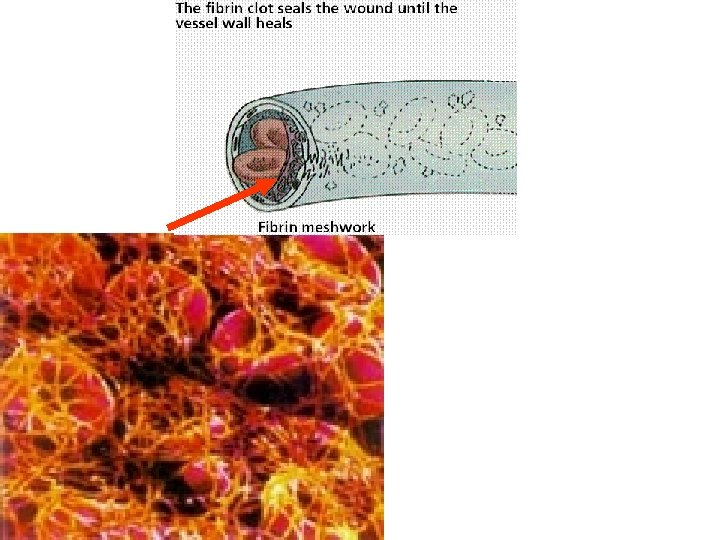
Clotting 1. Break in wall 2. Platelets stick 3. They release Thromboplastin
Clotting 4. Thromboplastin converts inactive Prothrombin (plasma protein, need Vit. K to make it) into active Thrombin 5. Active Thrombin activates inactive Fibrinogen into Fibrin Strands
Clot Busters • • TPA (Tissue plasminogen activator) Heparin Aspirin Plasmin
Problems with Clotting • Hemophilia – Can’t clot blood – Low platelets and/or – Low Vit. K and/or – Lack any one of the factors along the cascade – Genetic Disease –Sex Chromosome-Linked
Defense against Invaders • 1 st line Defense (Non Specific) – – – – Skin Sweat Tears Saliva Mucus membranes Stomach acid urine
What if the invader Breaks through 1 st line?
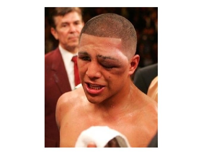
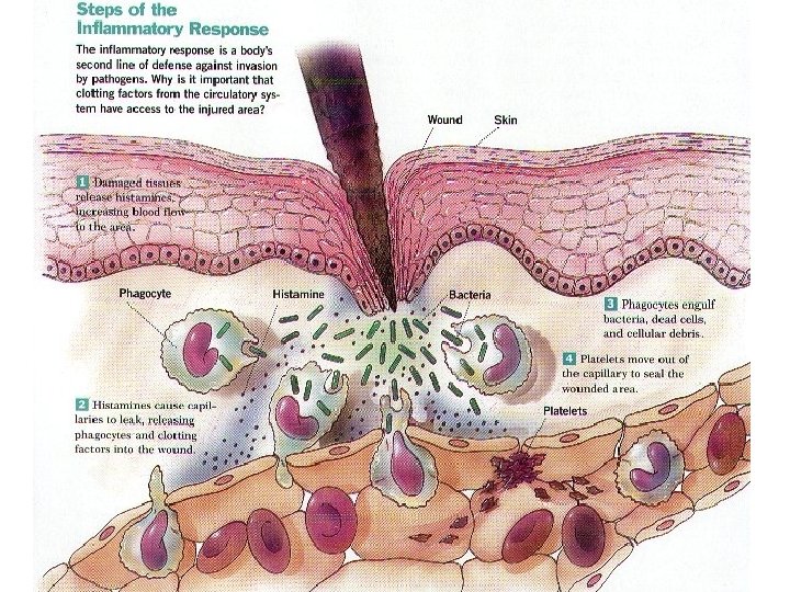
Defense against Invaders • 2 nd Line Defenses – Inflammatory Reaction (“setting on fire”) – Nonspecific Reaction • Infection area becomes – Swollen (more blood delivered to site because of damage cell Histamine release) – Red (More blood delivered to site) – Warm (Blood is warm) – Macrophages eat invaders and damaged cells – Macrophages die and become pus
Other 2 nd line Defenses • Fever • Interferon release by virally-infected cells – Causes neighbor cells to produce antiviral proteins
What if the invader Breaks through 2 nd line?
Immune Response • Specific recognition and destruction of foreign invaders – Players: • • • Bone Marrow WBC Lymph Nodes Tonsils Thymus Spleen
Immune Response • How does our body determine “self” from “nonself” ?
Suppose our body cells looked like this…
…and our bodies were invaded by this… Pathogenic Rubber Duckyus
How does our body recognize invaders?
Antigens! • Antigens – Components on an invader that are foreign to our body and lead to an immune attack
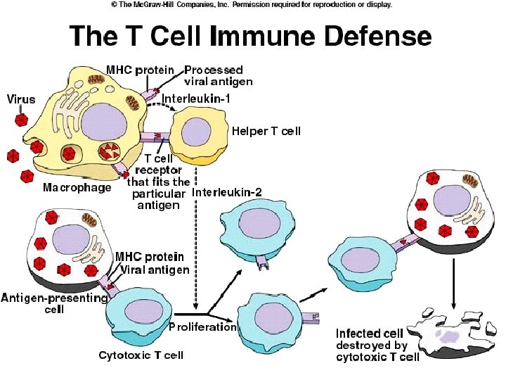
Stages of Immune Response • Primary Immune Response: – Antigen in body for 1 st time – Macrophage eats invader and displays invader's antigen on its surface
Stages of Immune Attack • Primary Immune Response – Helper T Cell binds to invader antigen with receptor (becomes semi-activated) – Helper T Cell binds to antigen on macrophage – Helper T Cell then becomes fully activated
Stages of Immune Attack • Primary Immune Response: – Activated Helper T Cell makes and secretes IL 2 (Interleukin 2) – Cytotoxic T Cells bind to antigen on antigenpresenting cells and become semi-activated – IL 2 fully activates Cytotoxic T Cells and these start to replicate – Cytotoxic T Cells bind infected cells and cancer cell and blow them up with toxins
Stages of Immune Attack • Primary Immune Response – Cytotoxic T Cells divide into 2 types of T Cells • More Cytotoxic T Cells • Memory Cytotoxic T Cells – Remain in body for many years – Don’t need priming to become activated • Cell-mediated attack • Secondary Immune Response – Second exposure, no need for T-Helper priming – Memory Cells ready to attack
B Cells • B cells secrete substances known as antibodies (Produce Humoral Response). • Each B cell is programmed to make one specific antibody. • Transported by the blood to lymph nodes and lymphatic organs
Antibodies • Alias: Immunoglobulins. • Proteins consisting of two heavy chains and two light chains • "Y" shaped molecule.
Antibodies • Antibodies have different binding abilities dependent on shapes to match the antigens • Neutralize invader • Agglutination • Tag for phagocytosis by macrophage Agglutination
How do our B cells know what an invader is? Antigen receptors: – protein molecules (antibodies) that stick out on B cells’ membranes that recognize invaders. Antigen-binding site: Specific region on the antibody molecule that “fits” the antigen determinants like a lock and key You have between 100 million and 200 billion different kinds of B cells and are Situated in the spleen, lymph Nodes and other parts of The lymphatic system
B cells Primary response: Humoral Response 1. 2. 3. 4. 5. 6. B cells come in contact with an antigen for the first time. Macrophage ingests the antigen and displays it on its membrane. B cell connects to antigen and becomes semiactivated B cell is stimulated by a helper T cell that recognizes the same antigen (Full activation) (IL 2). B cell produces plasma cells and memory B cells – Plasma cells make antibodies and release into blood & bind to antigen forming an antigen-antibody complex phagocytes engulf and destroy the antigenantibody complex
Secondary Response • Secondary exposure causes faster and stronger response • Memory cells are already in circulation and are ready to be activated by secondary response (Life time immunity)
Secondary response • Same antigen enters the body • Memory cells recognize it and divide rapidly • New plasma cells form producing large quantities of antibodies
Problem with B and T Cells • SCID – – Severe Combined Immunodeficiency Disorder Born with no B or T cells Open to attack Boy in Bubble Disease David-Dead at 12
Complement System • Primitive • Series of enzymes in blood • Anti-bacterial • Bacterium triggers a “Lego” effect of these enzymes • Enzymes drill hole into bacterium
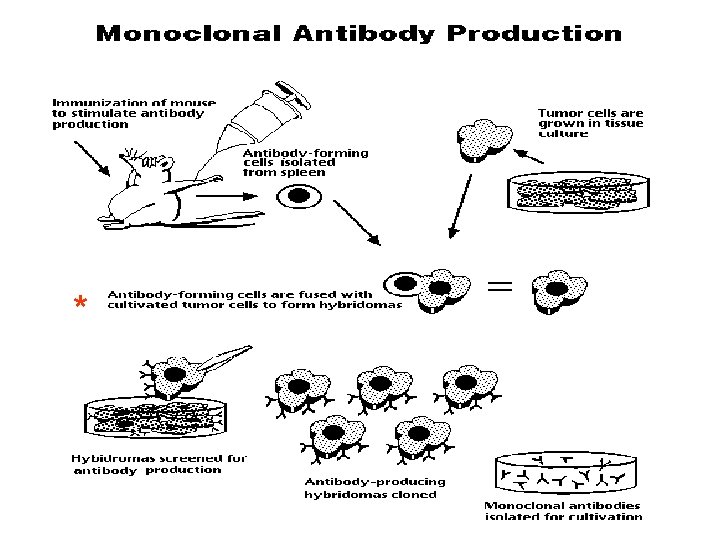
Monoclonal Antibodies and Cancer Treatment
Immunity and Vaccines 1. Active Immunity • • • Exposed to antigen Mount Immune Response (B Cell and T Cell) Produce B and T Memory Cells Immunity lasts long time Edward Jenner (Father of Vaccination) – Small pox vaccine • Can inject pure antigen or whole weakened or dead pathogen
Immunity and Vaccines • Passive Immunity – Inject pre-made antibodies against antigen – Temporary immunity – “Borrowed Immunity” – Ex. Maternal Immunity given to baby through breast milk
Immunity and Blood Types • ABO Blood Groups
Blood Types-Transfusions A B AB Person has Anti-B Antibodies Anti-A Antibodies Person has no antibodies against antigens Universal Receiver (42%) (12%) (3%) Person has Anti-A and Anti-B Antibodies Universal Donor (43%)
Blood Type Detection Agglutination Tests
Rh Factors Rh 85% Positive
What if…. . • Pregnant mom is Rh • Unborn baby is Rh+
Solution • Give Mom Rho. GAM drug – Destroys Anti-Rh antibodies of Mom
Transplants • Whole organs or limbs transfers from person to person • We all have unique proteins on organ surface • If organ antigens are foreign, organ rejected • Control rejection with immunosuppressive drugs • Stem Cell Research
AIDS • AIDS (Acquired Immune Deficiency Syndrome) • Due to the HIV (Human Immunodeficiency Virus) • Infects and destroys T-Helper Cells • Can remain dormant in body organs • Retrovirus (RNA virus that converts its RNA into DNA)-Reverse Transcriptase
HIV • • Mutates very quickly Person dies of Opportunistic Infections Spread via body fluid to body fluid Treatment: – AZT – Serine Protease Inhibitors
GLOBAL SUMMARY OF THE HIV/AIDS EPIDEMIC 2008 Globally, there were an estimated 33 million [30 million– 36 million] people living with HIV in 2007 The annual number of new HIV infections declined from 3. 0 million [2. 6 million– 3. 5 million] in 2001 to 2. 7 million [2. 2 million– 3. 2 million] in 2007. Overall, 2. 0 million [1. 8 million– 2. 3 million] people died due to AIDS in 2007, compared with an estimated 1. 7 million [1. 5 million– 2. 3 million] in 2001.