Transplantation Transplantation transfer of tissue or organ autologous


 phenotyping: Evaluation of HLA molecules using typing serums Typing antiserums =")

 ● determination of T lymphocytes alloreactivity ● mixed donor and")
")


 disease ● after bone marrow transplantation ● Gv. H also after")


 ● donor T lymphocytes react against residual leukemick")


 injection, this antibodies bind to Rh. D Ag on baby´s red")


 Cytotoxic")


 ● caused by Ig.")
 ● creation")

- Slides: 34
Transplantation
Transplantation = transfer of tissue or organ ● autologous - donor = recipient ● syngeneic - genetically identical donor and recipient (identical twins) ● allogeneic - genetically nonidentical donor of the same species ● xenogenic - the donor of another species ● implant - artificial tissue compensation
Allotransplantation ● differences in donor-recipient MHC gp and secondary histocompatibility Ag ● alloreactivity of T lymphocytes - the risk of rejection and graft-versus-host disease ● direct detection of alloantigens – recipient T lymphocytes recognize the different MHC gp and non-MHC molecules on donor cells ● indirect recognition of alloantigens - APC absorb different MHC gp from donor cells and present the fragments to T lymphocytes ● CD 8+ T cells recognize MHC gp I. ● CD 4+ T cells recognize MHC gp II.
Recognition of alloantigens
Testing before transplantation ● ABO compatibility -risk of hyperacute or accelerated rejection (= formation of Ab against A or B Ag on graft vascular endothelium) ● HLA typing (determining of MHC gp alelic forms) phenotyping and genotyping by PCR ● Cross-match - lymphocytotoxic test – detection of preformed Ab (after blood transfusions, transplantation, repeated childbirth) ● Mixed lymphocyte reaction - testing of T lymphocytes alloreactivity, monitor for reactivity of lymphocytes to allogeneic HLA
HLA typing a) phenotyping: Evaluation of HLA molecules using typing serums Typing antiserums = alloantiserums of multipar (created cytotoxic Ab against paternal HLA Ag of their children), serum of patients after repeated blood transfusions, monoclonal Ab - molecules HLA class I: separated T lymphocytes - molecules HLA class II: separated B lymphocytes b) genotyping: evaluation of specific alleles DNA typing of HLA class II: DR, DP, DQ by PCR
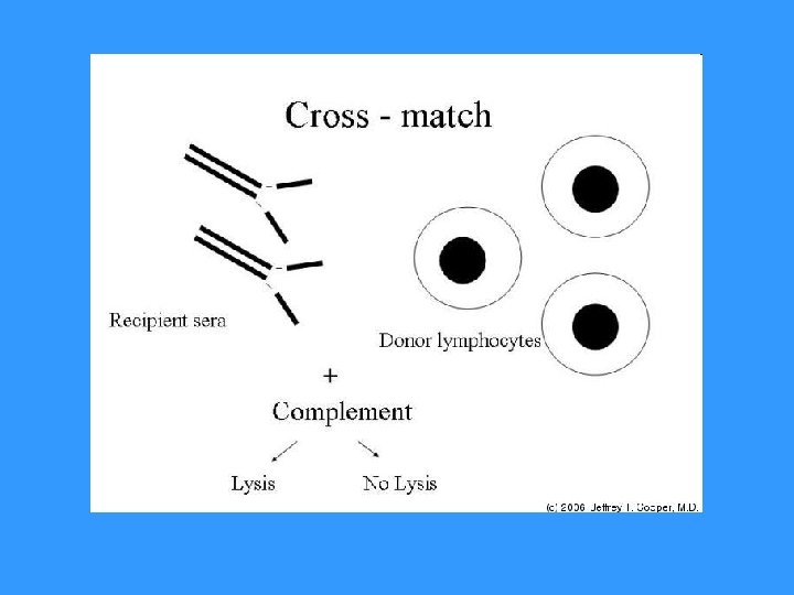
Cross-match test ● determination of preformed antibodies ● recipient serum + donor lymphocytes + rabbit complement → if cytotoxic Ab against donor HLA Ag are present in recipient serum (called alloantibodies = Ab activating complement) → lysis of donor lymphocytes. Visualization of dye penetration into lysis cells. ● positive test = the presence of preformed Ab → risk of hyperacute rejection! → contraindication to transplantation
Mixed lymphocyte reaction (MRL) ● determination of T lymphocytes alloreactivity ● mixed donor and recipient lymphocytes → T lymphocytes after recognition of allogeneic MHC gp activate and proliferate One-way MRL ● determination of recipient T lymphocytes reactivity against donor cells ● donor cells treated with chemotherapy or irradiated lose the ability of proliferation
Mixed lymphocyte reaction (MRL)
Immunologically privileged sites and tissues • Transplantation of some tissues don´t lead to the induction of allogeneic reactivity • minimal content of lecocytes • mechanisms that prevent to the development of injurious inflammation • Evolutionarily significant, protection of vital organs (brain, eye, gonads) • Factors protecting immunologically privileged structures • isolation from the immune system • preference of TH 2 reactoin, supression of TH 1 reaction • Fas. L expression • production of TGFb
Rejection Factors: ● The genetic difference between donor and recipient, especially in the genes coding for MHC gp (HLA) ● Type of tissue / organ - the strongest reactions against vascularized tissues containing many APC (skin) ● The activity of the recipient immune system - the immunodeficiency recipient has a smaller rejection reaction; immunosuppressive therapy after transplantation – suppression of rejection ● State of transplanted organ - the length of ischemia, the method of preservation, traumatization of organ at collection
Hyperacute rejection ● minutes to hours after transplantation ● humoral mediated immune response mechanism: ● if in recipients blood are present preformed or natural Ab (Ig. M anti-carbohydrate Ag) before transplantation → Ab + Ag of graft (MHC gp or endothelial Ag) → graft damage by activated complement (lysis of cells) ● the graft endothelium: activation of coagulation factors and platelets, formation thrombi, accumulation of neutrophil granulocytes prevention: ● negative cross match before transplantation, ABO compatibility
Accelerated rejection ● 3 to 5 days after transplantation ● caused by antibodies that don´t activate complement ● cytotoxic and inflammatory responses triggered by binding of antibodies to Fc-receptors on phagocytes and NK cells prevention: ● negative cross match before transplantation, ABO compatibility
Acute rejection ● days to weeks after the transplantation or after a lack of immunosuppressive treatment ● cell-mediated immune response mechanism: ● reaction of recipient TH 1 and TC cells against Ag of graft tissue ● infiltration by lymphocytes, mononuclears, granulocytes around small vessels → destruction of tissue transplant
Chronic rejection ● from 2 months after transplantation ● the most common cause of graft failure mechanism is not fully understood: ● non-immunological factors (tissue ischemia) and TH 2 responses with production alloantibodies, pathogenetic role of cytokines and growth factors (TGF β) ● fibrosis of the internal blood vessels of the transplanted tissue, endothelial damage →impaired perfusion of graft → gradual loss of its function dominating findings: vascular damage
Bone Marrow Transplantation ● Collection of hematopoietic stem cells ● Myeloablation ● Transplantation ● Engraftment ● Rejection ● Graft-versus-host reaction
Graft-versus-host (Gv. H) disease ● after bone marrow transplantation ● Gv. H also after blood transfusion to immunodeficiency recipients ● T-lymphocytes in the graft bone marrow recognize recipient tissue Ag as foreign (alooreactivity)
Acute Gv. H disease ● days to weeks after the transplantation of stem cells ● damage of liver, skin and intestinal mucosa ● prevention: appropriate donor selection, the removal of T lymphocytes from the graft and effective immunosuppression
Chonic Gv. H disease ● months to years after transplantation ● infiltration of tissues and organs by TH 2 lymphocytes, production of alloantibodies and cytokines → fibrosis ● process like autoimmune disease: vasculitis, scleroderma, sicca-syndrome ● chronic inflammation of blood vessels, skin, internal organs and glands, which leads to fibrosis, blood circulation disorders and loss of function
Graft versus leukemia effect (Gv. L) ● donor T lymphocytes react against residual leukemick cells of recipient (setpoint response) ● mechanism is consistent with acute Gv. H ● associated with a certain degree of Gv. H (adverse reactions)
Immunologic relationship between mother and allogenic fetus
Immunologic relationship between mother and allogenic fetus ● fetal cells have on the surface alloantigens inherited from his father ● pregnancy = "semiallogenic transplantation“ Tolerance of fetus by mother allow the following mechanisms: ● the relative isolation of the fetus from maternal immune system (no mixing of blood circulation) ● trophoblast - immune barrier witch protects against mother alloreactive T lymphocytes - don´t express classical MHC gp, expresses non-classical HLA-E and HLA-G ● transfer of small doses of fetal antigens in maternal circulation causes tolerance. . . suppressin of TH 1 and preference of TH 2 immune mechanisms in pregnancy
Rh incompatibility • Complications in pregnancy: production of anti-Rh. D antibodies by Rh. D- mother carrying an Rh. D+ fetus (hemolytic disease of newborns) • Fetal erythrocytes penetrate into the maternal bloodstream during pregnancy - a small amount, don´t immunize • During childbirth or abortion (after 8 weeks of gestation) fetal erythrocytes can penetrate into the bloodstream of mother → immunization, formation of anti-Rh. D antibodies • After childbirth, investigate Rh factor of born child, if is child Rh+, mother gets up to 72 hours after birth injection of anti-Rh antibodies (administered after abortion too)
Rh incompatibility
• Anti-Rh(D) injection, this antibodies bind to Rh. D Ag on baby´s red blood cells, this Ag than can´t bind to BCR and can´t activate B lymphocytes, this immune comlexes also actively inhibit B lymphocytes • During next childbirths, if fetus is Rh+ and mother produce anti-Rh antibodies, this Abb destroy red blood cells of fetus, which can lead to fetal death, or in severe postpartum anemia (anemia neonatorum) and neonatal jaundice (icterus gravis neonatorum) • For each pregnant woman during the first trimester investigate blod Rh factor and the presence of antibodies, in Rh- women performed a test for antibodies also in II. and III. trimester
Immunopathological reactions
Classification by Coombs and Gell Immunopathological reactions: immune response, which caused damage to the body (secondary consequence of defense responses against pathogens, inappropriate responses to harmless antigens, autoimmunity) IV types of immunopathological reactions: Type I reaction - a response based on Ig. E antibodies Type II reaction - a response based on antibodies, Ig. G and Ig. M Type III reaction - a response based on the formation of immune complexes Type IV reaction - cell-mediated response
Immunopathological reactions based on antibodies Ig. G and Ig. M (reaction type II) Cytotoxic antibodies Ig. G and Ig. M: ● complement activation ● ADCC ● binding to Fc receptors on phagocytes and NK cells
Examples of immunopathological reaction Type II • Transfusion reactions in administration of incompatibile blood: binding of antibodies to antigens on erythrocytes → activation of the classical way of complement → cell lysis • Hemolytic disease of newborns: caused by antibodies against Rh. D antigen
Autoimmune diseases: ● organ-specific cytotoxic antibodies (antibodies against erythrocytes, neutrophils, thrombocytes, glomerular basement membrane. . . ) ● blocking or stimulating antibodies Graves - Basedow's disease - stimulating antibodies against the receptor for TSH Myasthenia gravis - blocking of acetylcholin receptor→ blocking of neuromuscular transmission Pernicious anemia - blocking the absorption of vitamin B 12 Antiphospholipid syndrome - antibodies against fosfolipids Fertility disorder - antibodies against sperms or oocytes
Immunopathological reactions based on immune complexes formation (reaction type III) ● caused by Ig. G antibodies → bind to antigen → creation of immune complexes ● immunocomplexes - bind to Fc receptors on phagocytes - activate complement ● immune complexes, depending on the quantity and structure, are eliminated by phagocytes or stored in tissues ● pathological immunocomplexes response arises when is a large dose of antigen, or antigen in the body remains; arise 10 -14 days after aplication of Ag and induced inflamation can get to chronic state ● immune complexes are deposited in the kidneys (glomerulonephritis), on the surface of endothelial cells (vasculitis) and in synovie joint (arthritis)
Serum sickness ● therapeutic application of xenogeneic serum (antiserum to snake venom) ● creation of immune complexes and their storage in the vessel walls of different organs ● clinical manifestations: urticaria, arthralgia, myalgia Systemic lupus erythematosus ● antibodies against nuclear antigens, ANA, anti-ds. DNA Farmer's lung ● Ig. G antibody against inhaled antigens (molds, hay) Post-streptococcal glomerulonephritis, cryoglobulinemia, revmatoid arthritis, post-infectious arthritis
THANK YOU FOR YOUR ATTENTION