Translation Translation q Translation is the process by

Translation

Translation q Translation is the process by which a protein is synthesized from the information contained in a molecule of m. RNA. q During translation, an m. RNA sequence is read using the genetic code. q The genetic code defines how an m. RNA sequence is to be translated into the 20 -letter code of amino acids, which are the building blocks of proteins.
. Amino acid structure")
Proteins are polymers constructed out of 20 different amino acids (AA). Amino acid structure

Proteins Individual amino acids are linked together in linear chains by covalent amide bonds, called peptide bonds. Peptide bond formation between the amino group of one AA and the carboxyl group of another AA results in the release of a water molecule (dehydration reaction).

Proteins q A short chain of AA linked by peptide bonds and having a defined sequence is called a peptide. • Peptides generally contain fewer than 20 -30 AA. q Longer AA chains are referred to as polypeptides. § Polypeptides are often 200 - 500 AA residues long. The longest protein described today is the muscle protein titin with more than 35. 000 amino acid residues. Titin provides elasticity and stabilizes myosin.

The longest word in English is the full chemical name of the protein titin 189, 819 letters

Genetic code The genetic code is a three- letter code. There are 4 possible bases to choose from at each of the 3 base positions in a codon. There are 43 = 64 possible codons. But there are only 20 major types of amino acid.

Genetic code The genetic code is a degenerate code: each amino acid is specified by multiple codons (on average about 3 different codons). Only two amino acids have a single codon: Met (methionine) Trp (tryptophan) Leucine (Leu), Serine (Ser) and arginine (Arg) are each specified by 6 different codons.
.")
Genetic code Synthesis of all polypeptide chains begins with the amino acid methionine (Met). Ø The codon specifying the aminoterminal Met AUG is a start codon. Ø The three codons UAA, UGA and UAG are stop (termination) codons. Ø Stop codons do not specify AA.

Reading frame The sequence of codons that runs from a specific start codon to a stop codon is called a reading frame. • • • Gene sequence: AGGTCATGTCTAGGTATGCCC Frame 1: AGG TCA TGT CTA GGT ATG CCC Arg Ser Cys Leu Gly Met Pro Frame 2: A GGT CAT GTC TAG GTA TGC CC Gly His Val ---- Val Cys Pro Frame 3: AG GTC ATG TCT AGG TAT GCC C Val Met Ser Arg Tyr Ala Because the genetic code is a non-overlapping triplet code without divisions between codons, a particular m. RNA could be translated in 3 different reading frames, yielding 3 different peptides.

The genetic code is described as a universal code, meaning that the same code is used throughout all life forms. This is not strictly true because of alternative interpretation of codons. The genetic code has been found to differ for a few codons in mitochondria, chloroplasts or a single-celled plant (Acetabularia). Differences between the universal and mitochondrial genetic codes. Codon Universal code Human mitochondrial code UGA Stop Trp AGA Arg Stop AUA Ile Met AGG Arg Stop
- carries the information in")
The major participants in protein synthesis Messenger RNA (m. RNA)- carries the information in DNA to the ribosome for translation. Aminoacylated t. RNA- transports a specific amino acid to the ribosome. Ribosomes- a factories of protein synthesis.

Ribosomes are protein factories of the cell. Each cell contains thousands of ribosomes. Ribosomes carry out protein synthesis in two locations: Ø In the cytosol (free ribosomes). Ø On the outside of the endoplasmic reticulum (ER) or the nuclear envelope (bound ribosomes).

Look like small circular organelles. Ribosomes in the cytoplasm make proteins for use inside the cell (enzymes, hormones). Transmission electron microscopy (TEM) picture Ribosomes attached to endoplasmatic reticulum (ER) make proteins for export out of the cell or for use in lysosomes.

Ribosomes Ribosoms are large RNA- protein complexes composed of two subunits. 60 S 40 S S* - Svedberg units, a measure of sedimentation rate of macromolecules centrifuged under standard conditions.
")
Ribosome subunits Larger 60 S subunit contains: • 28 S r. RNA (large RNA) • 5. 8 S r. RNA Smaller 40 S subunit contains: 18 S r. RNA (small RNA) • 5 S r. RNA • more than 50 ribosomal proteins over 30 ribosomal proteins q RNA components are predominantly responsible for the catalytic function of the ribosome. • r. RNA molecules constitutes about 60% of the mass of a ribosome. q The protein components enhance the function of the r. RNA molecules.

Ribosomes Eukaryotic Prokaryotic

Ribosome The ribosome has an m. RNA binding site and three sites for t. RNA binding. The t. RNA binding sites are A, P and E.

Ribosome A-site The aminoacyl or acceptor site Ø Receives the t. RNA with the next amino acid to be added to the chain. P- site The peptidyl or donor site Ø Holds on aa-t. RNA and the growing chain of amino acids. E-site The exit site Ø Releases the used t. RNA back into the cytoplasm.

q During translation, a ribosome moves along m. RNA chain, interacting with various protein factors and t. RNA and undergoing large conformational changes. q The ribosome directs elongation of a polypeptide at a rate of 3 to 5 amino acids added per second. q Small proteins of 100 - 200 amino acids are made in minute or less. q It takes 2 -3 hours to make the largest known protein, titin, which contains more than 35. 000 amino acid residues.
 Transfer RNA (t RNA) show particularly high degrees of secondary")
Transfer RNA (t. RNA) Transfer RNA (t RNA) show particularly high degrees of secondary structure (cloverleaf). q Transfer RNA are the smalest of three major species of RNA molecules, containing 74 -95 nucleotide residues. q They transfer the amino acids from cytoplasm to the protein synthesizing machinery, hence the name t. RNA. Involved in process of translation. q. Each specific t. RNA contains a three-nucleotide sequence, an anticodon, that can base-pair with its complementary codon in the m. RNA.

Wooble pairing q Although there are 64 codons, the corresponding number of t. RNA molecules with different anticodons is less, just over 30 types of cytoplasmic t. RNA and only 22 types of mitochondrial t. RNA. q Many t. RNA can recognize more than one codon. q The interpretation of all 64 codons is possible because the normal base pairing rules are relaxed in the case of codon- anticodon recognition. q The wobble (relaxed base-pairing) hypothesis states that pairing of codon and anticodon follows the normal A-U and G-C rules for the first two base positions in a codon.

Wooble pairing q The exceptional woobles occur at the third position in a codon and the first base of the anticodon.
 How does the amino acids become attached to its corresponding")
Aminoacyl-t. RNA synthetases (ARS) How does the amino acids become attached to its corresponding t. RNA? Amino acid + t. RNA = aminoacyl- t. RNA (aat. RNA). This process is catalyzed by the enzyme aminoacyl-t. RNA synthetases. t. RNA molecule is reactivated many times (recycled).

Aminoacylation of t. RNA by aminoacyl-t. RNA synthetases. ARS catalyze covalent joining of amino acids to t. RNA in two- step reaction. Step 1 is formation of an aminoacyl adenylate (remains bound to the enzyme active site). Enzyme + amino acid + ATP Enzyme (aminoacyl-AMP) + PPi

Aminoacylation of t. RNA by aminoacyl-t. RNA synthetases. In Step 2 the aminoacyl group is transferred to t. RNA. Enzyme (aminoacyl-AMP) + t. RNA aminoacyl-t. RNA+ Enzyme + AMP For class I ARS enzymes, the aminoacyl group is transferred initially to the 2′-OH group of the 3′-terminal adenosine residue, then to the 3′-hydroxyl group by a transesterification reaction. For class II ARS enzymes, the aminoacyl group is transferred directly to the 3′-OH of the terminal adenosine residue.

For class I ARS enzymes, the aminoacyl group is transferred initially to the 2′-OH group of the 3′-terminal adenosine residue, then to the 3′-hydroxyl group by a transesterification reaction. For class II ARS enzymes, the aminoacyl group is transferred directly to the 3′-OH of the terminal adenosine residue.

t. RNA synthetases remove their own coupling errors through editing of incorrectly attached amino acids. The incorrect amino acid will be removed by the editing site.

Translation process Eukaryotic translation is similar to that of prokaryotes but a bit more complex. Differences in the translation process is due to differences in transcript structure and location of translation.

Cooperation between transcription and translation. Prokaryotes ü Protein synthesis in prokaryotes begins when m. RNA synthesis is still in progress. ü In genomic DNA, synthesis of the next m. RNA molecule starts before the previous m. RNA molecule is completely synthesized. ü Protein synthesis is also initiated using the incompletely synthesized m. RNA molecule. ü Occasionally, m. RNA degradation proceeds from the 5′terminus, while m. RNA synthesis is still in progress at the 3′ terminus. ü Thus, the half-life of m. RNA in E. coli is generally short, lasting only a few minutes.

Cooperation between transcription and translation. Eukaryotes ü The synthesized pre-m. RNA undergoes post-transcriptional processing. m. RNA ü The completed m. RNA is transported into the cytoplasm from the nucleus. ü Once in the cytoplasm, it is not necessarily immediately used in protein synthesis. ü In eukaryotes, the half-life of m. RNA ranges from dozens of minutes to very long periods. This spatial and temporal separation of transcription and translation is a major difference between eukaryotes and prokaryotes.

Steps in translation process 1. Initiation 2. Elongation 3. Termination

Molecules involved in translation initiation step 1. Mature m. RNA 2. Ribosome 3. Eukaryotic initiation factors (e. IFs) 4. Initiator t. RNA (Met-t. RNAi) 4. Guanosine triphosphate (GTP)

Differences between eukaryotic and prokaryotic translation initiation? The prokaryotic m. RNA has a ribosome binding site at its beginning just prior (within 4 -18 nucleotides ) to the initiation codon AUG. This sequence is known as Shine- Delgarno sequence (SD region). Shine- Delgarno sequence bind well with the 3’ end of 16 S r. RNA of the prokaryotic ribosome.

Initiation The first step in eukaryotic translation is the assembly of the translation initiation complex which consists of: Ø an m. RNA, Ø the small and large ribosomal subunits, Ø an initiator t. RNA carrying methionine – Met-t. RNA (the amino acid specified by the first codon, or start codon, of an m. RNA coding sequence).
 • The eukaryotic initiation factors (e. IFs) play")
The eukaryotic initiation factors (e. IFs) • The eukaryotic initiation factors (e. IFs) play a role in bringing together the initiator t. RNA, ribosome and m. RNA. • These soluble proteins help stabilize the formation of the functional ribosome around the start codon and also provide regulatory mechanisms in translation initiation. • There exist many more eukaryotic than prokaryotic initiation factors reflecting the greater biological complexity of eukaryotic cells. • Eukaryotic translation requires at least 12 eukaryotic initiation factors. • Initiation factors help assemble the initiation complex using energy provided by hydrolysis of GTP (guanosine triphosphate, a energy storage molecule similar to ATP).

Initiation Translation initiation complex. Ribosomes occur in the cytoplasm in dissociated condition, their smaller and larger subunits separated. • Initiator t. RNA (Met-t. RNA) gets loaded onto the 40 S small ribosomal subunit. • Eukaryptic initiation factors (e. IFs) binds as well to the 40 S ribosomal subunit. • A 43 S pre-initiation complex is formed containing 40 S- Met-t. RNA- e. IFs. Only Met-t. RNA can bind tightly to the small ribosomal subunit without the assembly of the entire ribosome.

Initiation Translation initiation complex. • The pre-initiation complex binds to the 5’ cap end of the m. RNA. • More e. IFs binds to 5’ Cap end of the m. RNA. • A 48 S pre-initiation complex is formed containing 40 S- Met-t. RNA- e. IFs-m. RNA.

Initiation Translation initiation complex. • The 48 S pre-initiation complex moves along the transcript in 5’- 3’ direction in search for the first AUG. • When first AUG is found, e. IFs dissociate. • The large ribosomal subunit (60 S) binds e. IF 6/e. IF 5 and assembles to the complex at the start codon. • When the large ribosomal subunit binds to the small subunit and the remaining e. IF are released, the initiation complex is complete. • The initiator t. RNA is positioned in the P- site of the ribosome and the A-site is available to receive the next charged t. RNA.

Differences between eukaryotic and prokaryotic translation initiation? Prokaryotic cell has modifed the initiation amino acid –formylated metionine. So in the prokaryote, there are two types of t. RNA for metionine: § t. RNAformyl. Met for initiation carrying formylated metionine. § t. RNAMet for carrying normal metionine to growing polypeptide. The initiation of protein synthesis consists in the recruitment of a ribosome·initiator t. RNA complex to the initiation codon of a messenger RNA. In prokaryotes, this process involves the direct interaction of the ribosomal RNA with the m. RNA. In contrast, eukaryotes have evolved a sophisticated mechanism that relies mostly on protein. RNA and protein-protein interactions.

Elongation Molecules involved in elongation step of translation 1. Aminoacyl-t. RNAs. 2. A set of proteins known as elongation factors (EFs). 3. An m. RNA: 80 S ribosome: peptidyl-t. RNA complex (peptidyl-t. RNA in the P site). 4. Guanosine triphosphate (GTP).

This bond is catalyzed by the enzyme peptidyl transferase.

Termination • Elongation continues until 80 S ribosome encounters one of the stop codons, UAA, UAG, or UGA, which signals chain termination and positioned in the ribosomal A-site. • There are no aminoacyl t. RNA molecules that recognize these sequences. • Proteins known as eukaryotic release factors (e. RF) recognize the stop codon at the A site and promote polypeptide release from the ribosome. • These release factors closely mimic the shape and charge distribution of t. RNAs such that they can bind to the ribosome, but they lack an amino acid. Therefore, the ribosome can no longer extend the growing chain of amino acids, causing the peptide chain to be released.
.")
Termination Stop codons are recognized by a protein, e. RF-1 (eukaryotic release factor 1). The activity of e. RF-1 is stimulated by e. RF-3, which is a GTP-binding protein that forms a complex with e. RF-1. This complex binds to ribosomes having a stop codon in the A-site. The ribosome-bound complex stimulates the cleavage of the bond (ester bond) between the peptide and the t. RNA (peptidyl-t. RNA) thereby releasing the new protein from the ribosome. The factors are released from the ribosome following GTP hydrolysis. The ribosome can now start a new translational cycle. In most cases the ribosome initiates a new round of translation on the same m. RNA.

Polysome Translation of a single eukaryotic m. RNA molecule to yield a typical-sized protein takes 1 -2 min. The efficiency of translation is significantly increased by Ø the simultaneous translation of a single m. RNA by multiple ribosomes, Ø rapid recycling of ribosomal subunits after they disengage from the 3’ end of m. RNA.

Polysome Polyribosomes or polysomes- a cluster of ribosomes linked together by a m. RNA and simultaneously translating the same transcript. Polysomes could be seen as beads on a string in electron micrographs.

Polysomes in prokaryotes Transcription of gene and translation of m. RNA occur simultaneously. The three-dimensional structure of the polysome has been elucidated in bacteria.
-binding protein (PABP) interact with")
Circular polyribosome in eukaryotic cells Multiple copies of the poly(A)-binding protein (PABP) interact with both an m. RNA poly (A) tail and the eukaryotic initiation factor e. IF 4 subunit 4 (e. IF 4 G). Since the e. IF 4 subunit E (e. IF 4 E) binds to the cap structure on the 5’ end of an m. RNA, the two ends of m. RNA molecule are bridged by the interventing proteins, forming a circular m. RNA.

Circular polyribosome in eukaryotic cells Because two ends of a polysome are relatively close together, ribosomal subunits that disengage from the 3’ end are positioned near the 5’ end, facilitating re-initiation by the interaction of the 40 S subunit and its associated initiation factors with e. IF 4 boung to the 5’ cap. Circular structure of eukaryotic m. RNA increases translation efficiency by facilitating ribosome recapture on circularized m. RNAs.

Ribosome life cycle q The ribosome subunits cycle between 80 S complexes and a pool of free subunits. q The ribosome subunits cycle rapidly through protein synthesis. q 80% of ribosomes are engaged in protein synthesis at any instant. q When 80 S ribosome dissociates from m. RNA, it separates into free 60 S and 40 S subunits. q Intact 80 S ribosomes are inactive in translation, because only 40 S subunit can interact with initiation factors.

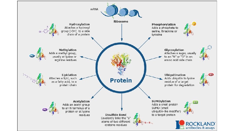
Post-tanslational modifications The translation of an m. RNA sequence into an amino acid sequence on the ribosome is not the end of the process of forming a protein. To function, the completed polypeptide chain must fold correctly into its three-dimensional conformation, bind any cofactors required. Many proteins also require covalent modifications of selected amino acids. Although the most frequent modifications are protein glycosylation and protein phosphorylation, more than 100 different types of covalent modifications are known.

Antibiotics inhibit m. RNA translation in bacteria

Could you translate the small eukaryotic m. RNA: 5’- GCUGAAGGAUGCAUAAGUCAGGCUAAGCUCCCAAAAAA -3’ 5’- UTR 3 ’- UTR

Could you translate the small eukaryotic m. RNA: 5’- GCUGAAGG AUG CAU AAG UCA GGC UAA GCUCCCAAAAAA -3’ 5’- UTR Met His Lys Ser Gly 3 ’- UTR

Regulation of translation q Gene expression is regulated not only on the transcription level but it is also regulated in translational level. q Translational control of gene expression can permit a more rapid response to altered environmental stimuli than the alternative of activating transcription. q As with other types of regulation, translation control typically functions at the level of initiation.

Global regulation of gene translation Global regulation of translation can involve regulation of translation initiation factors and occurs by two main mechanisms: ü disruption of the closed loop containing m. RNA, e. IF 4 complex and PABP ü decreased ternary complex formation

Global regulation of gene translation Disruption of the closed loop q is achieved by a group of inhibitor proteins that compete with e. IF 4 G for a binding site on e. IF 4 E, thereby preventing this critical interaction. q This group of e. IF 4 E-binding proteins, called 4 E-BP 1, 4 E-BP 2 and 4 E-BP 3, are regulated by phosphorylation, in a pathway downstream of the ‘mammalian target of rapamycin’ (m. TOR) kinase. q Signalling through m. TOR leads to the phosphorylation of 4 E-BPs, which are then unable to bind e. IF 4 E, allowing it to interact with e. IF 4 G.

Global regulation of gene translation Decreased ternary complex formation q The second major mechanism of global regulation is mediated by the phosphorylation of the e. IF 2 αsubunit on serine 51 by four e. IF 2α-kinases, GCN 2, PERK, HRI and PKR. q Phosphorylation on serine 51 leads to increased affinity of the protein for e. IF 2 B, preventing it from carrying out guanosine nucleotide exchange on other e. IF 2 molecules. q This results in a reduction in the levels of ternary complex and global inhibition of translation. q Global inhibition of translation occurs under conditions of cellular stress, such as amino acid starvation, viral infection and hypoxia and DNA damage.

m. RNA- specific regulation of translation In addition to the global mechanisms of translation, there are other m. RNA-specific mechanisms at work. q. Most examples of message-specific regulation are dependent upon sequence elements, which may or may not be structured, within the 5′ and 3′ UTRs. q. These are ü Internal Ribosome Entry Segments- IRESs, ü Upstream Open Reading Frames – u. ORFs and ü micro RNA target sites- mi. R-binding sites. q. They may function independently or in combination.

Elements in the 5′ or 3′ UTRs that regulate m. RNA translation Internal ribosome entry segments (IRES) q IRESs are typically highly structured RNA elements in the 5′ UTR that allow binding of ribosomes at or near the AUG start codon, independent of cap recognition. q Have been shown to activate or maintain translation following a range of cellular stresses that compromise the cap-binding complex. q They are assisted by proteins, IRES transacting factors (ITAFs), which bind to the IRES and modify its structure and/or interact with other elements of the translation machinery. IRES q It is estimated that 10% of m. RNAs contain IRES elements within their 5′ UTRs.

Elements in the 5′ or 3′ UTRs that regulate m. RNA translation Upstream Open Reading Frames – u. ORFs q Upstream open reading frames (u. ORFs) are sequences defined by an initiation codon in frame with a termination codon located upstream or downstream of the main AUG. They are located in the 5′ UTR. q u. ORFs correlate with significantly reduced protein expression levels because they reduce the efficiency of translation initiation of the main downstream ORF in unstressed conditions.

Upstream Open Reading Frames – u. ORFs u. ORF-mediated translational control can occur through different mechanisms. (A) The leaky scanning mechanism is dependent on the efficiency of u. AUG recognition; sometimes the ribosome can translate the u. ORF, but other times the scanning machinery bypasses the u. AUG, recognizing the downstream AUG and translating the main ORF. (B) When a scanning ribosome recognizes and translates a functional u. ORF, there is synthesis of a small peptide; if translation termination of the u. ORF is efficient, both 60 S and 40 S ribosomal subunits might dissociate from the transcript and the main ORF is not translated. (C) A u. ORF can repress translation of the main ORF in a peptide-dependent manner; in this case, the u. ORF-encoded peptide interacts with the translating machinery and promotes ribosome blockage. (E) After translation termination of the u. ORF, the 40 S ribosomal subunit can remain associated with the transcript, resume scanning, and recognize the downstream main AUG—a mechanism designated as translation reinitiation.

Elements in the 5′ or 3′ UTRs that regulate m. RNA translation q Both IRESs and u. ORFs are examples of m. RNA elements that drive translation under conditions in which global translation is inhibited. q However, other regulatory elements permit specific m. RNA regulation under physiological conditions, such as the micro-RNA (mi. R) regulation pathway.
 q Micro. RNAs are genomically- encoded, small single-stranded RNA molecules.")
Micro. RNAs (mi. RNA) q Micro. RNAs are genomically- encoded, small single-stranded RNA molecules. q These mi. RNA are on average 20 -25 nucleotides in length. q They can bind m. RNA and degrade m. RNA or block it’s translation.

mi. RNA qmi. RNA target sites are generally located in the 3′ UTRs of m. RNAs. q Binding of the mi. RNA to its target site with imperfect complementarity inhibits protein synthesis at either the initiation stage of translation or a post-initiation event. q More than 1. 400 mi. RNAs have been described in humans, each of which is predicted to regulate numerous different m. RNAs. q. This results in a highly complex regulatory network that is estimated to regulate around 30 - 60% of all protein-coding genes.

Mutations in global translation factors One of the best- characterized neurological diseases cause by dysfunctional translational control is leukoencephalopathy with vanishing white matter or vanishing white matter disease (VWM). • VWM is an autosomal recessive disease. • Children most often suffer from genetic VWM. The overall incidence of WMDs in children is ~1: 1000, similar to that of Multiple Sclerosis (MS) in adults. • WM damage leads to motor, cognitive and behavioral dysfunction. • The affected cells are glia (astrocytes and oligodendrocytes) and myelin is either not formed or lost. For most VWM no cure is available. Children with leukodystrophy die early.

Mutations in global translation factors • Mis-sense mutations in any one of the five subunits of e. IF 2 B, the guanosine nucleotide-exchange factor responsible for recharging e. IF 2, can cause VWM. • e. IF 2 B is one of the key regulators of translation initiation, being sequestered by phosphorylated e. IF 2α during conditions of cell stress, which leads to global translational inhibition. • It is notable that symptoms typically appear following mild infections or head trauma, indicating a role for cell stress in the pathology of this disease.

Translational errors Errors arise at all steps of protein synthesis, from transcription to protein folding, and have widespread phenotypic consequences. • Amino-acid misincorporations during translation are estimated to occur once in every 1, 000 to 10, 000 codons translated. At this error rate, 15% of average-length protein molecules will contain at least one misincorporated amino acid. • Polypeptide errors can induce protein misfolding, aggregation and cell death. • Misfolded proteins underlie a broad array of neurogenerative diseases and multiple sclerosis.
- Slides: 69