Translation RNA DIRECTED SYNTHESIS OF A POLYPEPTIDE Translation


 determines protein primary structure (amino acid")


- Slides: 43
Translation RNA DIRECTED SYNTHESIS OF A POLYPEPTIDE
Translation �In the process of translation, a cell interpets a genetic message by reading the codons on a strand of m. RNA, and builds a polypeptide accordingly �The interpreter of the of this message is a molecule called transfer RNA or t. RNA The job of t. RNA is to transfer amino acids from the cytoplasmic pool of amino acids to ribosomes �The ribosome than catalyzes the covalent linkage of 1 amino acid to another through a condensation reaction, forming a peptide bond
t. RNA �Not all t. RNA molecules are the same �There almost as many t. RNA molecules as there are codons (45) �Each t. RNA molecule has a nucleotide triplet called an anticodon which is complementary to, and so base pairs with the codon
�Made of RNA off of a DNA template t. RNA �Can be reused over and over �About 80 nucleotides long with stretches of complementary bases which allows for hydrogen bonding between complementary bases giving it its characteristic shape
Aminoacyl-t. RNA Synthetases �A family of related enzymes whose job it is to correctly match up amino acids with the correct t. RNA molecule �The active site of each aminoacyl-t. RNA synthetase molecule fits only one combintaion of amino acid and t. RNA �There are 20 different synthetases, one for each amino acid This means the synthetase is able to bind all t. RNA’s with anticodons for their amino acid
Aminoacyl-t. RNA Synthetases �The Aminoacyl-t. RNA Synthetases catalyzes the covalent linkage of the amino acid to the t. RNA through hydrolysis of ATP �Once an amino acid is attached to a t. RNA, it is called a charged t. RNA �Charged t. RNA’s are released from the Aminoacylt. RNA Synthetases and available to deliver its amino acid to a ribosome to be incorporated into a growing polypeptide
Wobble � 1 st recognition process involved matching appropriate a. a. with correct t. RNA � 2 nd recognition process involves matching correct t. RNA with codon �Wobble: the flexible base pairing at the 3 rd codon position Because there are only 45 t. RNA, there must be some versatility in base pairing allowed This occurs with the 3 rd base on the codon & anticodon Base pairing rules are relaxed here This allows one anticodon to base pair w/ several different codons, as long as the 1 st 2 bases pair correctly
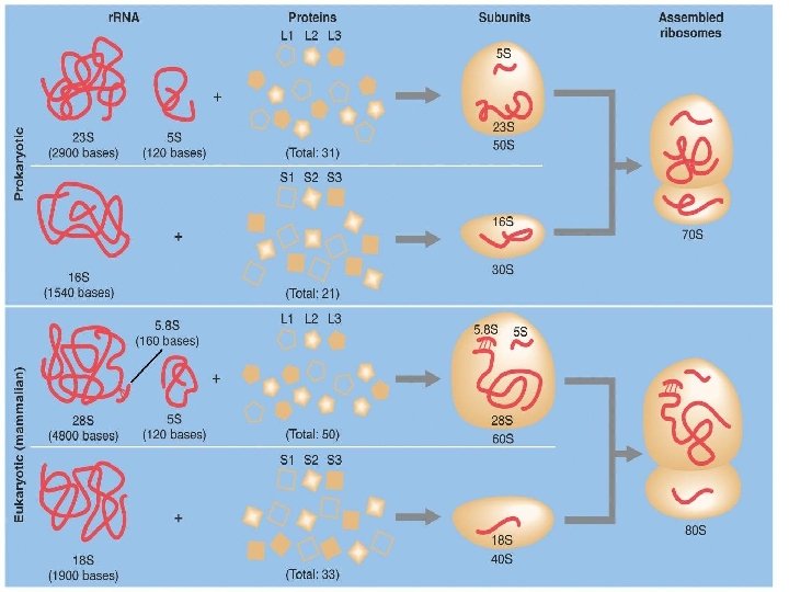
Ribosomes �Facilitate the specific coupling of t. RNA anticodons with m. RNA codons during protein synthesis �There are 2 ribosomal subunits, each composed of ribosomal RNA or r. RNA Genes on chromosomal DNA are transcribed, and the RNA is processed and assembled with proteins imported from the cytoplasm This occurs in the nucleolus The completed ribosomal subunits are then exported through a nuclear pore
Ribosomes �In both prokaryotes and eukaryotes both ribosomal subunits only join to form a complete functional ribosome when attached to an m. RNA
Ribosomes are always composed of 2 subunits, a large subunit and a small subunit Prokaryotic Eukaryotic �Large subunit is called the 50 s Composed of 2 strands of r. RNA �Small subunit is called the 30 s Composed of 1 strand of r. RNA �= a complete ribosome of ____? _____ � 70 s ribosome the 60 s Composed of 3 strands of r. RNA �Small subunit is called the 40 s Composed of 1 strand of r. RNA �= a complete ribosome of ____? _____ � 80 s ribosome
Significance �The differences between prokaryotic and eukaryotic ribosomes are small but significant �Many of our antibacterial drugs work by inactivating bacterial ribosomes without inhibiting eukaryotic ribosomes Tetracycline Streptomycin � Binds to the S 12 Protein of the 30 S subunit of the bacterial ribosome, interfering with the binding of formyl-methionyl-t. RNA to the 30 S subunit. � This prevents initiation of protein synthesis and leads to death of microbial cells
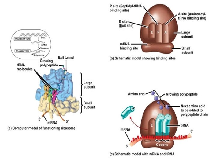
Structure of a Ribosome �In addition to a binding site for m. RNA, each ribosome has 3 binding sites for t. RNA �P site: peptidyl t. RNA site Holds the t. RNA carrying the growing polypeptide chain while… �A site Aminoacyl t. RNA site Holds the t. RNA carrying the next amino acid to be added �E site: exit site for discharged t. RNA’s
Ribozymes �Because it is the r. RNA that is responsible for the catalytic abilities of the ribosome, the entire thing can be regarded as one large ribozyme �The associated proteins in a ribosome are there mainly for structural support and have little or no enzymatic abilities
3 Stages to Building a Polypeptide �Initiation �Elongation �Termination
Translation: Initiation Prokaryotes Eukaryotes � 1 st the small ribosomal �Small subunit with subunit binds the m. RNA just upstream of the start codon, along with the t. RNA carrying methionine �Can bind in any order Ribosome may bind t. RNA 1 st or m. RNA 1 st initiator t. RNA already bound, binds the 5’ cap of m. RNA �It then scans downstream along the m. RNA until it reaches the AUG start codon This is important cause it establishes the codon reading frame
Translation Initiation Complex �So basically, initiation brings together m. RNA, the 2 subunits of the ribosome, and a t. RNA bearing the 1 st amino acid of a polypeptide �The union of the above is followed by attachment of the large ribosomal subunit �This completes the translation initiation complex �Proteins called initiation factors are required to bring all of these components together
Translation Initiation Complex
Energy �The cell expends energy forming the translation initiation complex �The energy comes from hydrolysis of the GTP molecule, or guanosine triphosphate
Translation: Elongation �In the elongation phase, amino acids are added 1 by 1 to the preceding amino acid �Each addition involves participation of proteins called elongation factors �Elongation occurs in a 3 step cycle Energy expenditure by hydrolysis of GTP occurs in the 1 st and 3 rd steps
3 Steps of Elongation � 1 st is Codon Recognition Requires hydrolysis of GTP � 2 nd Peptide Bond Formation between amino acids in the A and P sites, catalyzed by the ribosome Removes the amino acid from the t. RNA that carried it � 3 rd Translocation of t. RNA in the A site to the P site, while at the same time moving the t. RNA in the P site to the E site m. RNA is moved through the ribosome 5’end 1 st
Termination of Translation �Once a stop codon reaches the A site of the ribosome they signal the end of translation �Stop codons do not code for an amino acid and include UAG UAA UGA �A protein called a release factor binds directly to the stop codon in the A site
Central Dogma: A Closer Look
Release Factors � Causes addition of a water molecule instead of an amino acid �This reaction hydrolyzes (breaks) the bond between the completed polypeptide and the t. RNA in the P site, releasing the polypeptide through the exit tunnel of the ribosome Exit tunnel is NOT the E site Exit tunnel is a special tunnel in the ribosome through which the polypeptide is released
Polyribosomes �A single ribosome can make an average polypeptide in less then a minute �Typically, many ribosomes translate a single m. RNA at the same time A single m. RNA is used to make many copies of a polypeptide simultaneously �Once a ribosome moves past the start codon, another can bind �Strings of ribosomes on a single m. RNA are called polyribosomes
Polyribosomes Ribosomes Polypeptide chains �As seen with an electron microscope m. RNA
What kind of cell is this & how do you know?
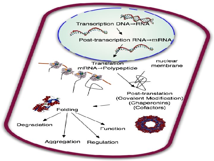
Completing & Targeting the Functional Protein �Translation alone is not sufficient to make a functional protein �Proteins must undergo posttranslational modifications and correct folding be functional
Protein Folding �During translation a polypeptide begins to spontaneously fold and coil as a direct consequence of the properties of its amino acids and how they are strung together (primary structure) �This creates a protein with a specific 3 -D shape (secondary & tertiary structure) �Chaperone proteins are necessary to help the protein fold correctly
Important �The sequence of DNA (or the gene) determines protein primary structure (amino acid sequence) �Protein primary structure then determines secondary and tertiary structure �So… DNA is ultimately responsible for protein structure and so protein function
Post-Translational Modifications �Sometimes even correctly folded proteins must be modified before the protein can do its job Amino acids may be chemically modified through � Attachment of sugars, phosphate groups, lipids… Enzymes may remove one or more amino acids from the leading end A polypeptide chain be enzymaticaly cleaved into 2 or more pieces Sometimes 2 separate polypeptides must work together (quarternary structure) hemoglobin
Example: Insulin �Insulin is 1 st synthesized as a single polypeptide chain, but only becomes active after an enzyme cuts out a central part of the chain �This leaves a protein composed of 2 polypeptide chains connected through disulfide bridges
Ribosomes Bound ribosomes Free ribosome �Suspended in the cytosol �Make proteins that will remain in the cytosol for internal cellular use �Bound to the cytosolic face of the RER or nuclear envelope �Make proteins of the: Endomembrane System � Nuclear envelope � Golgi � Lysosome � Vacuoles � Plasma membrane As well as proteins that are to be secreted (like insulin)
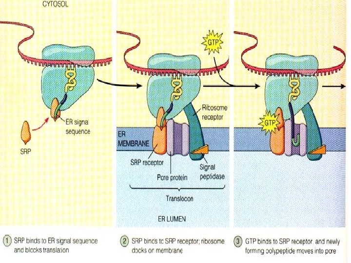
Targeting Polypeptides to Specific Locations �Protein synthesis always begins in the cytosol where a free ribosome begins translation of an m. RNA molecule �There the process continues until completion unless the growing polypeptide itself signals the ribosome to attach to the ER �Polypeptides destined for the endomembrane system or secretion are marked by signal peptides
Signal Peptides �Target the protein to the ER �About 20 a. a. ’s long at or near the leading end of the polypeptide �Signal peptides are recognized as they emerge from the ribosome by a complex called a signalrecognition particle (SRP) � SRP’s bring the ribosome to a receptor protein built into the ER which is part of a translocation complex
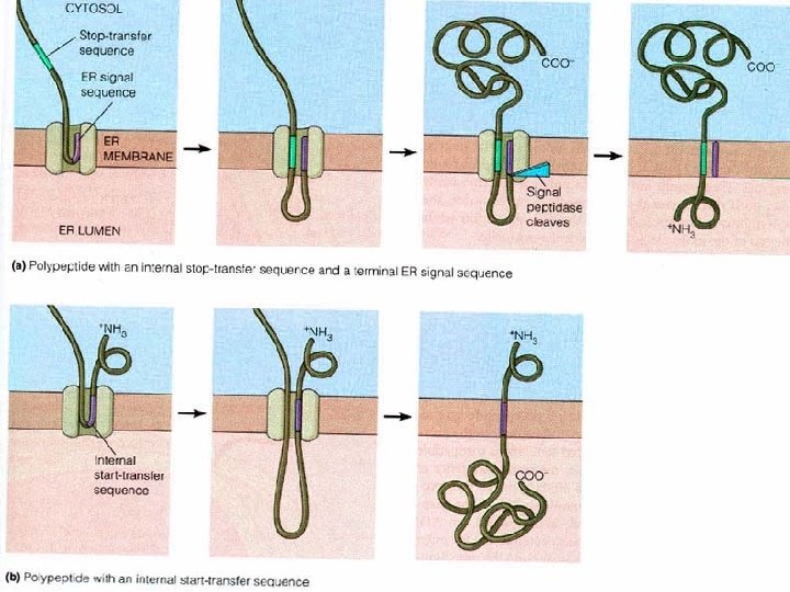
Translation �Translation then continues on the ribosome bound to the translocation complex until the growing polypeptide snakes across the ER membrane into the lumen via a protein pore If it to be a membrane bound protein, only part of the peptide will enter the lumen, while another part remains embedded in the membrane If it will be secreted, the entire polypeptide is released to the lumen �Signal peptides are usually removed by an enzyme
Targeting to Other Organelles �Different signal sequences are used to target proteins to different organelles �Translation is always completed before a protein is targeted to an organelle other than the ER Mitochondria Chloroplast Interior of the nucleus Lysosome