TRANSFUSION REACTIONS HEMOLYTIC TRANSFUSION REACTION Dr N NASSEH

 Delayed")
 Etiology Therapy Prevention Febrile")

 was")


 • In many cases of hemolytic transfusion")
 • TRALI is now the leading cause of transfusion-related")
")


- Slides: 22
-TRANSFUSION REACTIONS- HEMOLYTIC TRANSFUSION REACTION Dr N. NASSEH Qazvin university medical science- anesthesiology department
TRANSFUSION REACTIONS • HEMOLYTIC TRANSFUSION REACTION • • Acute hemolytic transfusion reaction (AHTR) Delayed hemolytic transfusion reaction (IMMUNE EXTRAVASCULAR REACTION) • TRANSFUSION-RELATED ACUTE LUNG INJURY • NONHEMOLYTIC TRANSFUSION REACTIONS • Febrile nonhemolytic transfusion reaction • Allergic reactions can be minor, anaphylactoid, or anaphylactic. • Noninfectious Serious Hazards of Transfusion (NISHOT) 1. Transfusion-associated circulatory overload (TACO): 2. Transfusion-related immunomodulation (TRIM): 3. Microchimerism: Chimerism refers to more than one cell line in an individual organism. Specifically, donor lymphocytes may persist in a patient 4. Posttransfusion purpura: . 5. Hypotensive transfusion reactions: 6. Transfusion-associated graft-versus-host disease (GVHD): 7. Transfusion-related AKI. 8. Alloimmunization: 9. HLA alloimmunization and human platelet antigen (HPA) alloimmunization: 10. Undertransfusion and overtransfusion: 11. Iron overload: • INFECTIOUS COMPLICATION
NONINFECTIOUS HAZARDS OF TRANSFUSION Transfusion Reaction Incidence (per 105 Transfusions) Etiology Therapy Prevention Febrile All components: 70 -6800 Storage-generated Proinflammatory Cytokines Patient antileukocyte antibodies bind to donor leukocytes Stop transfusing Give antipyretics Prestorage leukoreduction All components: 16. 8 -8000 Circulatory overload Patients with cardiac or renal disease, infants, and the critically ill are at increased risk Stop transfusing Give diuretics Identify patients at high Risk Transfuse slowly transfusion associated circulatory overload TACO Practice-dependent Supportive care Oxygen transfusion-related acute lung injury Erythrocytes: 10 -20 Platelets/plasma: 50 -100 Passive transfusion of donor antibodies Storage-generated toxic lipids Supportive care Remove high-risk donors from the donor pool Allergic All components: 3000 mild, 2 anaphylactic Mild reactions: Transfusion of soluble antigens in donor Plasma Anaphylaxis: Ig. A deficiency or other recipient protein deficiency Stop transfusing ASA monitors Pretransfusion antihistamine use remains common practice despite limited evidence Erythrocytes: 1. 1 -9. 0 Donor antibodies bind to patient erythrocytes Patient antibodies bind to donor erythrocytes Stop transfusing Repeat matching Standard operating procedures transfusion-related mmunomodulation. Unknown The mechanism is unknown but may depend on the presence of donor eukocytes Treat complications (e. g. , infection, malignancy) Prestorage leukocyte reduction may be beneficial, but this approach is controversial Microchimerism All components: 500010, 000 massive transfusion Permanent residence of donor cells in recipient Unknown Post transfusion purpura All components: 2 Recipient alloantibodies attack donor platelet antigens IVIG Avoid units positive for implicated HPA antigens in patients with a history of PTP Hypotensive Unknown Production of kinins by the activation of the contact system Patients on ACE inhibitors are at increased risk Stop transfusing ASA monitors Large- Avoid the use of negatively charged leukocyte reduction filters Transfusion into Immunocompromised host Transfusion of donor cells closely matching HLA type No consensus exists Consider bone marrow transplant Gamma irradiation of cellular products TRALI Hemolytic TRIM PTP Graft-versus-host Varies by patient population Large-bore IV access Epinephrine. Antihistamines- Supportive care Treat DIC bore IV access -Supportive care
• The most common complications of transfusion are febrile nonhemolytic and chill-rigor reactions. • The most serious complications are acute hemolytic reaction due to ABO incompatible transfusion and transfusion-related acute lung injury, which have very high mortality rates • Early recognition of symptoms suggestive of a transfusion reaction and prompt reporting to the blood bank are essential. • The most common symptoms are chills, rigors, fever, dyspnea, light-headedness, urticaria, itching, and flank pain. • If any of these symptoms (other than localized urticaria and itching) occur, the transfusion should be stopped immediately and the IV line kept open with normal saline. • The remainder of the blood product and clotted anticoagulated samples of the patient's blood should be sent to the blood bank for investigation.
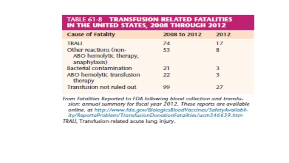
HEMOLYTIC TRANSFUSION REACTION • From 2008 to 2012, transfusion-related acute lung injury (TRALI) was the common cause of transfusion-related fatalities. • Since 1975, the FDA has required that all fatal reactions occurring in blood recipients or donors be reported within 24 hours by telephone or within 7 days in writing by all FDA-registered transfusion services. • From 1976 to 1985, 328 deaths were reported analyzed. Of these deaths, 159 were acute from hemolytic reactions and 23 from delayed reactions. • In 2011, the incidence of an acute hemolytic transfusion reaction resulting from ABO incompatibility was 1: 1200 to 1: 190, 000. (Anesthesiology News reported an incidence of 1: 606, 978). • Of the 159 deaths from acute hemolytic reaction, 137 were caused by errors involving ABO incompatibility. More than half of these mistakes occurred after the blood had been issued by the blood bank and were committed by nurses and physicians in the operating room, emergency department, or ward.
• One of the most catastrophic transfusion reactions is that arising from intravascular hemolysis. Intravascular hemolysis occurs when there is a direct attack on transfused donor cells by recipient antibody and complement. Such a reaction can occur from infusion of as little as 10 m. L of blood. • If properly treated, death is rare. However, prevention of kidney failure and a coagulopathy (DIC) is crucial. • Hemolytic transfusion reactions involving extravascular RBC destruction are generally less serious than those of the intravascular variety. • In these cases, recipient antibody coats but does not immediately hemolyze the transfused RBCs. Destruction occurs primarily in the reticuloendothelial system.
Signs and Symptoms • The clinical consequences of incompatible blood transfusions are very serious but quite variable. Factors include • • volume of transfused blood, number of antigenic sites on the red cell membrane, activity of the reticuloendothelial system properties of the antibody, (concentration and ability to activate complement) • The classic signs and symptoms of a hemolytic transfusion reaction—chills, fever, chest and flank pain, and nausea—are masked by anesthesia. • Under general anesthesia, the only signs may be hemoglobinuria, bleeding diathesis, or hypotension. • The presenting sign is usually hemoglobinuria.
• As little as 50 m. L of incompatible blood may exceed the binding capacity of Haptoglobin, which is a protein that can bind approximately 100 mg of Hb/100 m. L of plasma. • When Hb not exceeding this amount is injected or liberated into the bloodstream, the Hb circulates as a complex with haptoglobin, which is cleared by the reticuloendothelial system. • A sample of plasma that contains 2 mg/d. L of Hb is faintly pink or light brown. • When the level of Hb reaches 100 mg/d. L, the plasma is red. • When the level of plasma Hb reaches 150 mg/d. L, hemoglobinuria occurs. • In general, the quantity of the free Hb in the plasma correlates with the volume of incompatible blood transfused.
• The symptoms can be so alarming that cessation of blood is indicated, even if Hb is not seen in plasma. • Laboratory tests that should be performed if hemolytic transfusion reaction is suspected include Ø serum haptoglobin (levels are very low ) Ø plasma and urine Hb (hemoglobinemia , hemoglobinuria ) Ø Bilirubin (Hyperbilirubinemia ) Ø Direct antiglobulin (direct Coombs test) determinations. ( positive ) === can confirm the presence of hemolytic transfusion reaction because it shows that antibody is attached to transfused donor RBCs (The direct Coombs test is used to detect antibodies that are stuck to the surface of red blood cells. The indirect Coombs test looks for antibodies that are floating in the blood. These antibodies could act against certain red blood cells. ) Ø repeat compatibility testing, Ø repeat other serologic tests (i. e. , ABO and Rh) • The blood bank should check all paperwork to ensure that the correct blood component was transfused to the patient. • Although several consequences of intravascular hemolysis are possible, mainly the renal and coagulation systems are affected.
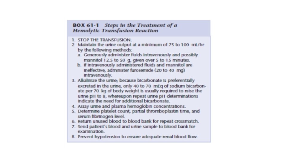
Treatment • The cause of acute renal failure from intravascular hemolysis is likely that Hb in the form of acid hematin precipitates in the distal tubule and causes mechanical tubular blockage. • The magnitude of the precipitation probably is inversely related to the volume of urine flow and its p. H. • The primary emphasis of therapy should be directed toward maintaining urinary output in excess of 75 -100 m. L/hr by generous administration of intravenous fluids and diuretics. • the administration of crystalloids to maintain the CVP between 10 and 15 cm H 2 O while initially administering 12. 5 to 50 g of Mannitol. • If this is ineffective, the dose of mannitol may be increased or the use of more potent diuretics, such as furosemide (20 - 40 mg) • . Alkalinization of the urine to prevent precipitation of acid hematin in the distal tubules is of questionable value but is easy and therefore recommended. (Na bicarbonate 0. 5 - 1 meq/kg) • Another approach to treatment of a severe hemolytic transfusion reaction who postulated that the kidneys might be spared from exposure to massive amounts of hemolyzed red cells(3000 ml) by removing all blood from a patient and replacing it with compatible blood. use of an extracorporeal circuit.
• DIC commonly occurs with hemolytic transfusion reactions, probably because RBC stroma is severed, releasing erythrocytin, which activates the intrinsic system of coagulation. • This activated coagulation leads to fibrin formation. • Subsequently, platelets and factors I, II, V, and VII are consumed. • As soon as a hemolytic transfusion reaction is recognized, platelet count, PT, and PTT should be determined to provide baseline values with which subsequent laboratory values can be compared. • Hypotension during a hemolytic transfusion reaction may result from activation of the kallikrein system.
DELAYED HEMOLYTIC TRANSFUSION REACTION (IMMUNE EXTRAVASCULAR REACTION) • In many cases of hemolytic transfusion reaction, the transfused donor cells may survive well initially, but after a variable delay (2 to 21 days) they are hemolyzed. • This type of reaction occurs mainly in recipients sensitized to RBC antigens by previous blood transfusions or pregnancy ===> more common in females who have a known disposition of alloimmunization. • often manifested only by a decrease in the post transfusion Hct value. • jaundice and hemoglobinuria can occur in these patients and can cause some impairment in renal function, but only rarely do they lead to death. • Unlike immediate reactions, antibodies most commonly involved in delayed hemolytic reactions are those in the Rh and Kidd systems rather than the ABO system. • The delayed hemolytic reaction may not be preventable, because pre transfusion testing is unable to detect very low levels of antibody present in potential blood recipients. • A delayed hemolytic transfusion reaction should include in differential diagnosis in any patient who has an unexplained decrease in Hct 2 to 21 days after a transfusion, even without obvious manifestation of hemolysis. • This is especially important in a postoperative patient when the decrease in Hct value is thought to be from blood loss and may be an important criterion as to whether additional surgery is necessary.
TRANSFUSION-RELATED ACUTE LUNG INJURY (TRALI) • TRALI is now the leading cause of transfusion-related mortality , although it probably is underdiagnosed and underreported. • This injury manifests as noncardiogenic pulmonary edema. • TRALI occurs in the absence of excessive intravascular volume and cardiac failure. • Clinically, symptoms and signs appear 1 to 2 hours after transfusion and are in force within 6 hours. Fever, dyspnea, fluid in the endotracheal tube, and severe hypoxia are typical. • CXR is characteristic of pulmonary edema, excessive circulatory volume (i. e. , LA hypertension) is not present. • • During anesthesia, a persistent ↓ in blood O 2 saturation can be the manifesting sign. • All blood components, especially FFP, are implicated as caustic factors. • The only specific therapy is stopping the transfusion and instituting critical care supportive measures. • blood bank notified for a different donor and quarantining all units from that donor. • All records should be reexamined, and the results of the patient’s human leukocyte antigen (HLA) testing should be evaluated if possible.
Count … TRALI • Risk factors TRALI include : • • higher interleukin-8 (IL-8) levels, liver surgery, chronic alcohol abuse, shock, higher peak airway pressures while being mechanically ventilated, smoking, positive fluid-balance. receipt of plasma or whole blood from female donors • The decreased use of plasma from female donors reduced the incidence of TRALI. • A related finding is that transfusion of PRBCs in the ICU to patients who already have ALI was not associated with 90 -day mortality.
NONHEMOLYTIC TRANSFUSION REACTIONS • • • Nonhemolytic reactions to blood transfusions usually are not serious and are febrile or allergic. Occasionally, fever may be the first sign of a hemolytic reaction or of bacterial contamination. The most common adverse reactions to blood transfusions are the less serious febrile reactions. The symptoms consist of chills, fever, headache, myalgia, nausea, and nonproductive cough occurring shortly after blood transfusion, caused by pyrogenic cytokines and intracellular contents released by donor leukocytes. Use of leukoreduced blood lowered the incidence of febrile reactions. Less frequently ===> hypotension, chest pain, vomiting, and dyspnea. Even pulmonary infiltrations with radiographic evidence of prehilar nodule formation and lower lung infiltrates along with overt pulmonary edema have been reported. Because febrile reactions obviously involve fever, they can be easily confused with a hemolytic transfusion reaction. A direct antiglobulin test readily differentiates a hemolytic reaction from a febrile reaction because this test rules out the attachment of an RBC antibody to transfused donor RBCs. No clear consensus exists on whether the transfusion should be terminated when a febrile reaction occurs.
• Allergic reactions can be minor, anaphylactoid, or anaphylactic. • An anaphylactoid reaction is clinically similar to anaphylaxis, but it is not mediated by Ig. E. • Most allergic transfusion reactions are minor and caused by the presence of foreign protein in the transfused blood. • The most common symptom is urticaria associated with itching. • Occasionally, the patient has facial swelling. When these reactions are clearly not a serious hemolytic reaction, the transfusion does not need to be discontinued. • Antihistamines are used to relieve the symptoms of the allergic reaction.
• Infrequently, a more severe form of allergic reaction involving Anaphylaxis occurs in which the patient has dyspnea, hypotension, laryngeal edema, chest pain, and shock. • These are anaphylactic reactions caused by the transfusion of Ig. A to patients who are Ig. A deficient and have formed anti-Ig. A. • This type of reaction does not involve red cell destruction and occurs very rapidly, usually after the transfusion of only a few milliliters of blood or plasma. • Patients who experience these anaphylactic reactions can be given transfusions with washed RBCs from which all traces of donor Ig. A have been removed or with blood lacking the Ig. A protein.
THANK YOU FOR YOUR ATTENTION