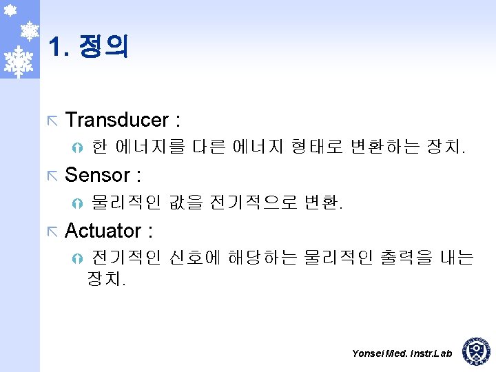
Transducer What is transducer A transducer is an
 What is transducer ? A transducer is an electronic device that converts")
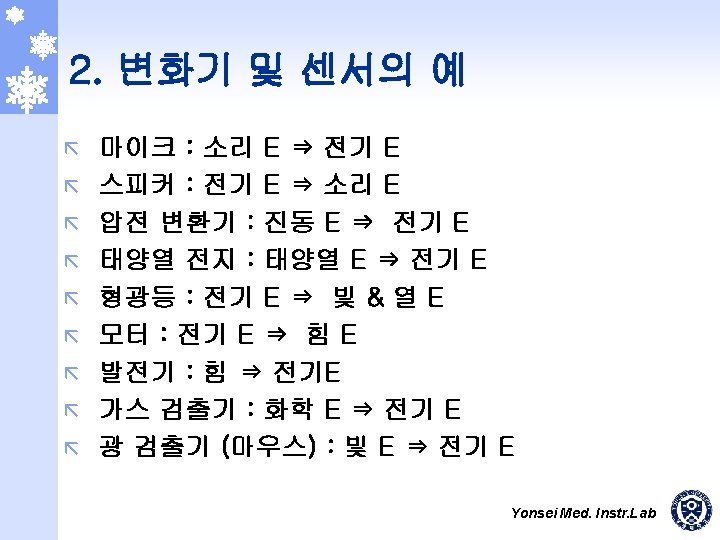
 측정 Ý Resistance(potentiometer) Ý Inductance(LVDT: Linear Variable Differential Transformer) Ý Capacitance Ý 압전효과(piezoelectric)")

 Translational. (b) Single-turn). (c) Multi-turn.")
 Resistance-wire type. (b) Foil type. (c) Helical-wire type. Arrows")
 d (b) c c d d")


 ã Urodynamic Testing Ý Uroflowmetry Ý Urethral Pressure Measurement Ý Water Cystometry")


 Metal-plate electrode used for application to limbs. (b) Metal-disk electrode")

 (b) Snap")
 Carbonfilled silicone rubber electrode. (b) Flexible thin-film neonatal electrode (after")
 Insulated needle electrode. (b)")
 Suction")

 Section of fine-bore glass")

 Ý Strainguage Ý Piezosensor Ý")
- Slides: 42
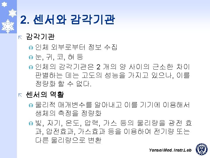
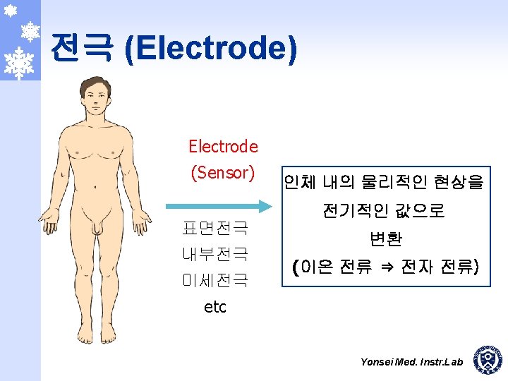
변환기 (Transducer) What is transducer ? A transducer is an electronic device that converts energy from one form to another. Yonsei Med. Instr. Lab
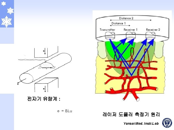
길이(변위) 측정 Ý Resistance(potentiometer) Ý Inductance(LVDT: Linear Variable Differential Transformer) Ý Capacitance Ý 압전효과(piezoelectric) ex) 심음기록 : phonocardiography, Ý Strain gauge 힘 측정 Ý LVDT, Strain gauge, Load cell, Torque meter ex) 점성도, 무게, 동작 분석의 force plate Yonsei Med. Instr. Lab
압력 측정 Ý Capacitive Ý Piezoresistive Ý LVDT Ý Silicon micro-machined sensor Ý Strain gauge Ý Vibrating wire(Diaphragm) 연속적 혈압측정 Yonsei Med. Instr. Lab
Three types of potentiometric devices for measuring displacements (a) Translational. (b) Single-turn). (c) Multi-turn. (From Measurement Systems: Application and Design, by E. O. Doebelin. Copyright 1990 by Mc. Graw-Hill, Inc. Used with permission of Mc. Graw-Hill Book Co. ) Yonsei Med. Instr. Lab
Typical bonded strain-gage units (a) Resistance-wire type. (b) Foil type. (c) Helical-wire type. Arrows above units show direction of maximal sensitivity to strain. [Parts (a) and (b) are modified from Instrumentation in Scientific Research, by K. S. Lion. Copyright 1959 by Mc. Graw-Hill, Inc. Used with permission of Mc. Graw-Hill Book Co. ] Yonsei Med. Instr. Lab
a c c a b b d (a) d (b) c c d d d b e (c) Inductive displacement sensors (a) Self-inductance. (b) Mutual inductance. (c) Differential transformer. Yonsei Med. Instr. Lab
Capacitance sensor for measuring dynamic displacement changes C = k A / x Yonsei Med. Instr. Lab
Pneumatic and impedance plethysmography of the digit Yonsei Med. Instr. Lab
Infrared LED Red LED Blood vessel Catheter Photosensor 1 2 CRT Photoplethysmography system measures oxygen saturation in vivo, using red and infrared light emitting diodes (LEDs) and a photosensor. The red and infrared LEDs are alternately pulsed in order to use a single photosensor. Yonsei Med. Instr. Lab
UDS(Urodynamic System) ã Urodynamic Testing Ý Uroflowmetry Ý Urethral Pressure Measurement Ý Water Cystometry Ý Gas Cystometry Ý Voiding Cystometry Ý Video Urodynamics Ý Impotence Studies : Cavernosometry Yonsei Med. Instr. Lab
UDS - DANTEC Yonsei Med. Instr. Lab
Electrode-electrolyte interface The current crosses it from left to right. The electrode consists of metallic atoms C. The electrolyte is an aqueous solution containing cations of the electrode metal C+ and anions A-. Yonsei Med. Instr. Lab
3. 전극의 전기적 등가모델 Equivalent circuit for a biopotential electrode in contact with an electrolyte Ehc is the half-cell potential, Rd and Cd make up the impedance associated with the electrode-electrolyte interface and polarization effects, and Rs is the series resistance associated with interface effects and due to resistance in the electrolyte. Yonsei Med. Instr. Lab
Magnified section of skin, showing the various layers (Copyright © 1977 by The Institute of Electrical and Electronics Engineers. Reprinted with permission, from IEEE Trans. Biomed. Eng. , March 1977, vol. BME-24, no. 2, pp. 134 -139. ) Yonsei Med. Instr. Lab
Ehe Electrode Cd Rd Gel Sweat glands and ducts Rs Ese EP Epidermis Ce Dermis and subcutaneous layer Re CP RP Ru A body-surface electrode is placed against skin, showing the total electrical equivalent circuit obtained in this situation. Each circuit element on the right is at approximately the same level at which the physical process that it represents would be in the lefthand diagram. 극펫 Yonsei Med. Instr. Lab
Body-surface biopotential electrodes (a) Metal-plate electrode used for application to limbs. (b) Metal-disk electrode applied with surgical tape. (c) Disposable foam-pad electrodes, often used with electrocardiograph monitoring apparatus. Yonsei Med. Instr. Lab
A metallic suction electrode is often used as a precordial electrode on clinical electrocardiographs. Yonsei Med. Instr. Lab
Insulating package Double-sided Adhesive-tape ring Metal disk Electrolyte gel in recess (a) (b) Snap coated with Ag-Ag. Cl Plastic cup External snap Gel-coated sponge Plastic disk Dead cellular material Foam pad Tack Capillary loops Germinating layer (c) Examples of floating metal body-surface electrodes (a) Recessed electrode with top-hat structure. (b) Cross-sectional view of the electrode in (a). (c) Cross-sectional view of a disposable recessed electrode of the same general structure shown in Figure 5. 9(c). The recess in this electrode is formed from an open foam disk, saturated with electrolyte gel and placed over the metal electrode. Yonsei Med. Instr. Lab
Flexible body-surface electrodes (a) Carbonfilled silicone rubber electrode. (b) Flexible thin-film neonatal electrode (after Neuman, 1973). (c) Cross-sectional view of the thin-film electrode in (b). [Parts (b) and (c) are from International Federation for Medical and Biological Engineering. Digest of the 10 th ICMBE, 1973. ] Yonsei Med. Instr. Lab
Needle and wire electrodes for percutaneous measurement of biopotentials (a) Insulated needle electrode. (b) Coaxial needle electrode. (c) Bipolar coaxial electrode. (d) Finewire electrode connected to hypodermic needle, before being inserted. (e) Crosssectional view of skin and muscle, showing coiled finewire electrode in place. Yonsei Med. Instr. Lab
Electrodes for detecting fetal electrocardiogram during labor, by means of intracutaneous needles (a) Suction electrode. (b) Cross-sectional view of suction electrode in place, showing penetration of probe through epidermis. (c) Helical electrode, which is attached to fetal skin by corkscrew type action. Yonsei Med. Instr. Lab
The structure of a metal microelectrode for intracellular recordings. Structures of two supported metal microelectrodes (a) Metalfilled glass micropipet. (b) Glass micropipet or probe, coated with metal film. Yonsei Med. Instr. Lab
A glass micropipet electrode filled with an electrolytic solution (a) Section of fine-bore glass capillary. (b) Capillary narrowed through heating and stretching. (c) Final structure of glass-pipet microelectrode. Yonsei Med. Instr. Lab
To oximetry instrument CVP injection port Thermistor Balloon Cardiac output computer connector Transmitting fiber-optic Receiving fiber-optic Optical module Proximal (CVP) lumen Distal (PA) lumen Balloon inflation lumen Sampling and pressure monitoring lumen The catheter used with the Abbott Opticath Oximetry System transmits light to the blood through a transmitting optical fiber and returns the reflected light through a receiving optical fiber. The catheter is optically connected to the oximetry processor through the optical module. (From Abbott Critical Care Systems. Used by permission. ) Yonsei Med. Instr. Lab
실습 ã 센서 가변저항 Ý Bendable potentiometer sensor (Flexpoint) Ý Strainguage Ý Piezosensor Ý ã 전극 Ý Ý Ý ECG surface electrode ECG suction electrode EMG surface electrode EEG ball electrode GSR mesh electrode Yonsei Med. Instr. Lab