Transcytosis Endocytosis followed by exocytosis Transports a substance

Transcytosis • Endocytosis followed by exocytosis • Transports a substance rapidly through a cell • HIV crossing a cell layer Copyright © The Mc. Graw-Hill Companies, Inc. Permission required for reproduction or display. HIV-infected white blood cells Anal or vaginal canal V iruses bud HIV Receptor-mediated endocytosis Lining of anus or vagina (epithelial cells) Cell membrane Exocytosis Receptor-mediated endocytosis Virus infects white blood cells on other side of lining

The cell cycle

Cell cycle • Cells normally perform their functions, they spend the bulk of their time doing that • Sometimes they need to reproduce and form new cells • Cell cycle is composed of two main phases, interphase and mitosis • Mitosis is composed of four stages

Copyright © The Mc. Graw-Hill Companies, Inc. Permission required for reproduction or display. G 2 phase o Pr e as h p se ha Metap S phase: genetic material replicates Anapha se Te lop G 1 phase cell growth ha se

Interphase • Performs it’s normal function (e. g. , muscle cells contract, stomach cells secrete, etc. ) • DNA (chromosomes) replicates during this phase, in preparation for dividing the cell into two cells • Now we have 92 chromosomes instead of the usual 46

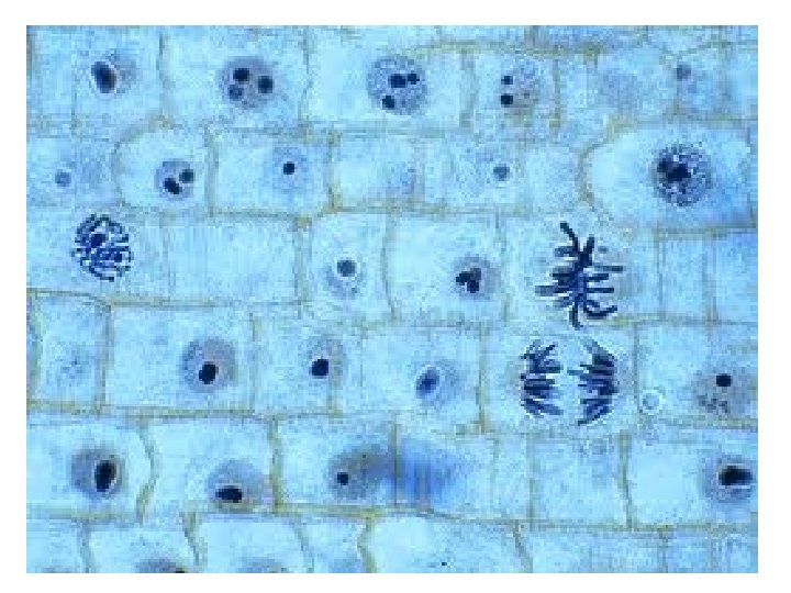
Mitosis • Occurs in every cell in your body, besides egg and sperm • A cell that divides and forms two new daughter cells • The two new cells need exact copies of everything in the old cell (including chromosomes)

Prophase • • “Pro”- before Replicated chromosomes condense Microtubules attach to chromosomes Nuclear membrane breaks down

Metaphase • “Meta”- after • Replicated chromosomes align across the equator of the cell, in preparation for separation

Anaphase • “Ana”- up, towards • Proteins pull sister chromosomes to opposite poles of the cell

Telophase • “Telo”- end • Nuclear membrane reforms • Chromosomes uncondense

• http: //www. youtube. com/watch? v=Vl. N 7 K 19 QB 0

Apoptosis • Programmed cell death • If cell cannot move into next phase, cell undergoes programmed cell death • Damaged cells that can’t be repaired

Now that we know about the cell cycle and mitosis, let’s look at what happens when mitosis goes wrong…

What is cancer? • Begins when cells divide when they shouldn’t • Unchecked cell division can lead to tumors • These tumors can be either malignant or benign • Malignant tumors are considered cancerous

What is cancer? Normal Ovarian Tissue Benign Ovarian Tumor Malignant Ovarian Tumor

Properties of Cancer Cells Breast cancer cell Brain cancer cell 1. Divide when they should not 2. Invade surrounding tissues 3. Travel to other locations in the body (metastasize)

Metastasis of Cancer

Why Does Cancer Start? The Cell Cycle Every cell in the body has a cell cycle and undergoes cell division at some stage in its life. The events of cell division is called the The Cell Cycle: cell cycle: 1. Interphase a) G 1 (gap) phase b) S (synthesis) phase c) G 2 (gap) phase 2. Mitosis—division of the nucleus 3. Cytokinesis—division of the cytoplasm

Why Does Cancer Start? Checkpoints of The Cell Cycle Cell division is tightly controlled and proceeds only if certain conditions have been met. Thus the cell has checkpoints. 1 2 Gap 2 3 MITOSIS CYTOKINESIS • Is the cell large enough? • Are sufficient nutrients available? Cell growth • Are growth factors present? and division preparation 2 • Was DNA copied correctly? • Is the cell large enough? Gap 1 Synthesis INTERPHASE 3 • Are chromosomes attached to microtubules? Cell growth 1

Cancer overrides these checkpoints • Normal proteins, produced by normal DNA, regulate these checkpoints, thus regulate cell division • Mutations of genes on the DNA produce mutant proteins that don’t play by the rules, leading to uncontrolled cell division and cancer • Some genes are more susceptible to mutation than others

Mutant Proteins

Mutant Proteins

• http: //www. youtube. com/watch? v=qjj. HKDn 1 2 q. I

Lifestyle and Genetic Factors Contribute to Cancer Inheritance of Breast Cancer genes, BRCA 1 and BRCA 2 contribute to these cancers.

Conventional Cancer Treatments Conventional Therapies—designed to stop cell division • Chemotherapy—chemical drugs target quickly dividing cells Many chemotherapeutic agents interfere with microtubule formation during prophase. • Radiation—high energy particles that damage DNA Problem: Chemotherapy and radiation do not distinguish between cancerous and non-cancerous cells. Quickly-dividing non-cancerous cells are destroyed as well. Cancerous cells also develop resistance.

Experimental Cancer Treatments 1. Radio-immunotherapy—cancer cell specific, radioactive antibodies deliver lethal doses of radiation to tumor cells Normal cell (no markers) Cancer cell (tumor markers) Radioactive antibody 2. Cancer vaccines—vaccines made of tumor substances stimulate immune system to better recognize and eliminate cancer cells. E. g. , Gardasil 3. Angiogenesis inhibitors—prevent a tumor from obtaining a blood supply. • Angiogenesis—the formation of new blood vessels from existing vessels • Once a tumor grows to a certain size it requires its own blood supply to deliver oxygen and nutrients for continued growth.
- Slides: 27