
Traffico vescicolare COME VENGONO TRATTENUTE LE PROTEINE RESIDENTI


- Slides: 56
• Traffico vescicolare
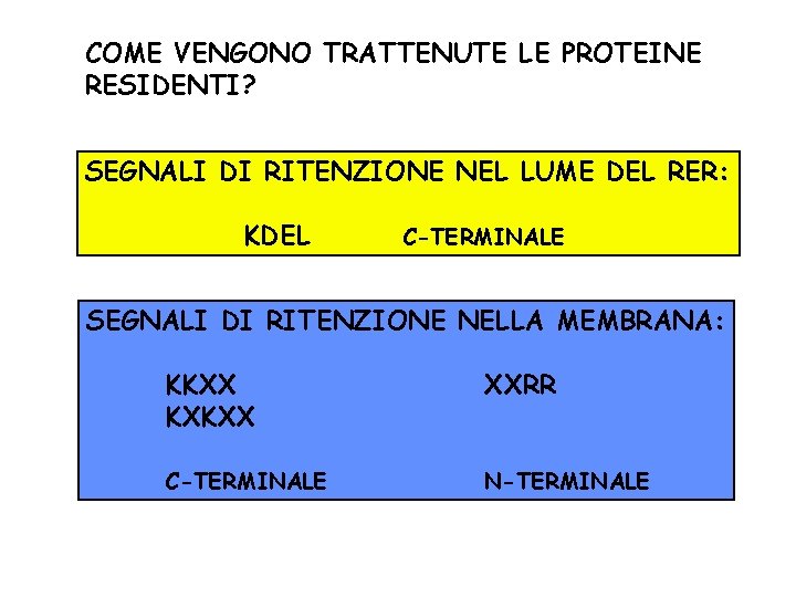
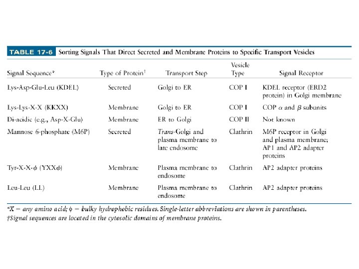
COME VENGONO TRATTENUTE LE PROTEINE RESIDENTI? SEGNALI DI RITENZIONE NEL LUME DEL RER: KDEL C-TERMINALE SEGNALI DI RITENZIONE NELLA MEMBRANA: KKXX KXKXX XXRR C-TERMINALE N-TERMINALE
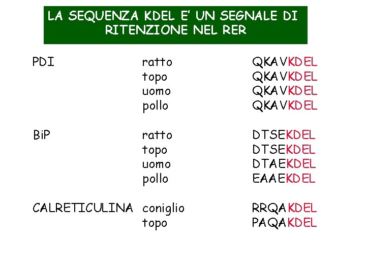
LA SEQUENZA KDEL E’ UN SEGNALE DI RITENZIONE NEL RER PDI ratto topo uomo pollo QKAVKDEL Bi. P ratto topo uomo pollo DTSEKDEL DTAEKDEL EAAEKDEL CALRETICULINA coniglio topo RRQAKDEL PAQAKDEL
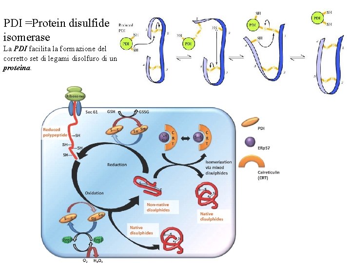
PDI =Protein disulfide isomerase La PDI facilita la formazione del corretto set di legami disolfuro di una proteina.
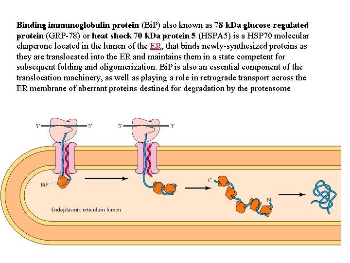
Binding immunoglobulin protein (Bi. P) also known as 78 k. Da glucose-regulated protein (GRP-78) or heat shock 70 k. Da protein 5 (HSPA 5) is a HSP 70 molecular chaperone located in the lumen of the ER, that binds newly-synthesized proteins as they are translocated into the ER and maintains them in a state competent for subsequent folding and oligomerization. Bi. P is also an essential component of the translocation machinery, as well as playing a role in retrograde transport across the ER membrane of aberrant proteins destined for degradation by the proteasome
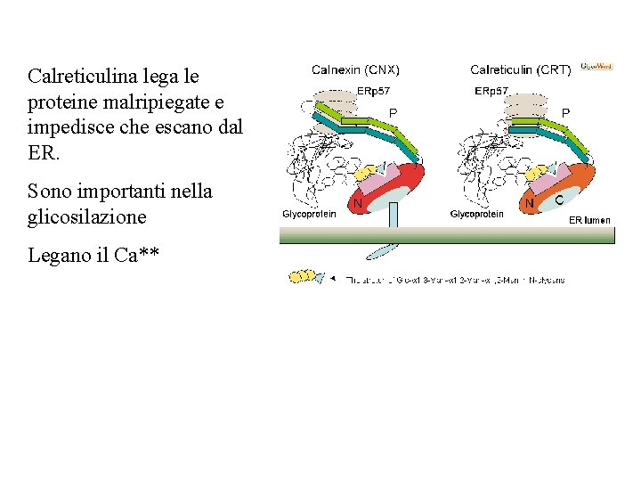
Calreticulina lega le proteine malripiegate e impedisce che escano dal ER. Sono importanti nella glicosilazione Legano il Ca**
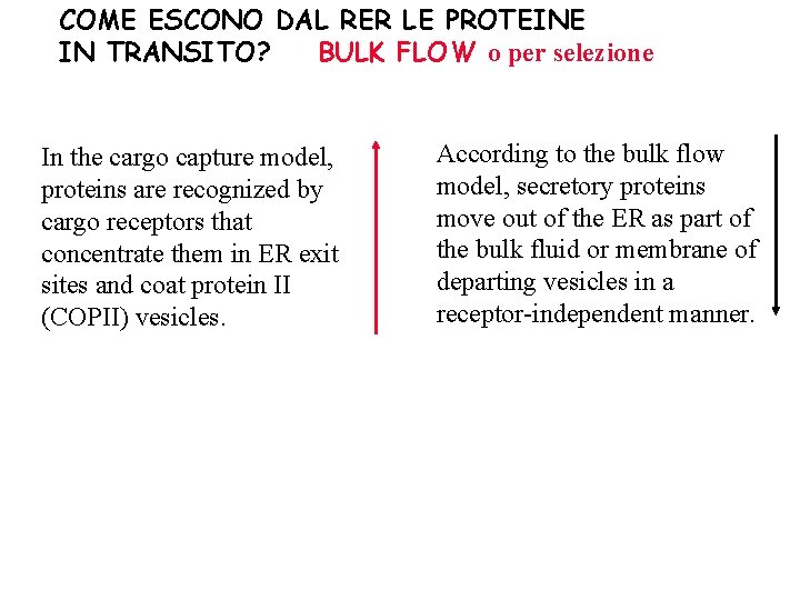
COME ESCONO DAL RER LE PROTEINE IN TRANSITO? BULK FLOW o per selezione In the cargo capture model, proteins are recognized by cargo receptors that concentrate them in ER exit sites and coat protein II (COPII) vesicles. According to the bulk flow model, secretory proteins move out of the ER as part of the bulk fluid or membrane of departing vesicles in a receptor-independent manner.
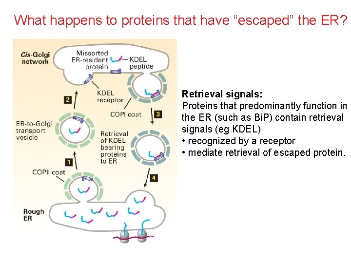
What happens to proteins that have “escaped” the ER? Retrieval signals: Proteins that predominantly function in the ER (such as Bi. P) contain retrieval signals (eg KDEL) • recognized by a receptor • mediate retrieval of escaped protein.
COME ESCONO LE PROTEINE DAL RER • VESCICOLE, VESCICOLE, VESCICOLE, VESCICOLE, VESCICOLE, VESCICOLE,
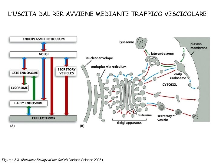
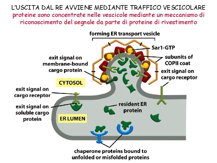
L’USCITA DAL RER AVVIENE MEDIANTE TRAFFICO VESCICOLARE Figure 13 -3 Molecular Biology of the Cell (© Garland Science 2008)
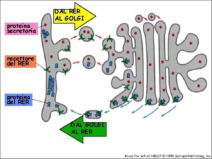
proteina secretoria DAL RER AL GOLGI recettore del RER proteina del RER DAL GOLGI AL RER
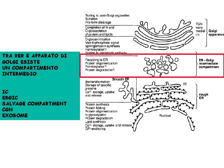
TRA RER E APPARATO DI GOLGI ESISTE UN COMPARTIMENTO INTERMEDIO IC ERGIC SALVAGE COMPARTMENT CGN EXOSOME
COME SI FORMA UNA VESCICOLA? QUANTI TIPI DI VESCICOLE VI SONO? COME SI DETERMINA SELETTIVITA’ DEL CONTENUTO? COME UNA VESCICOLA RICONOSCE IL BERSAGLIO? COME AVVIENE LA FUSIONE? COME VIENE REGOLATO L’INTERO PROCESSO?
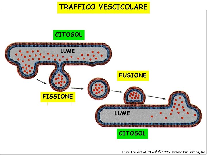
TRAFFICO VESCICOLARE CITOSOL LUME FUSIONE FISSIONE LUME CITOSOL
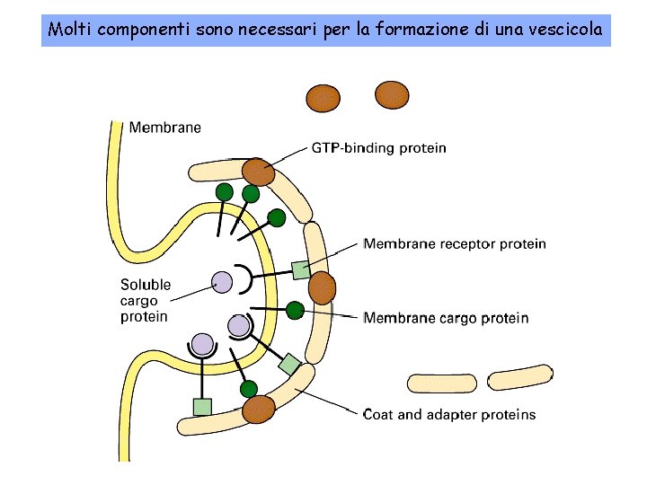
Molti componenti sono necessari per la formazione di una vescicola
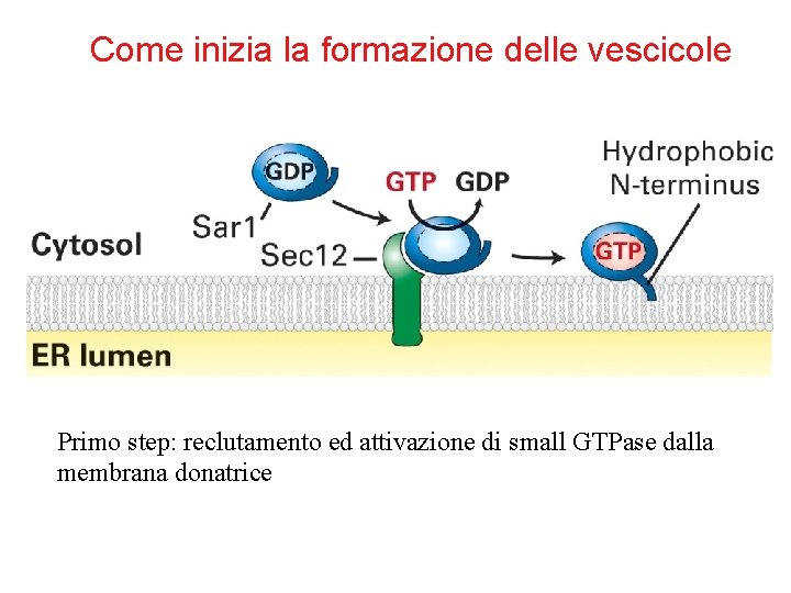
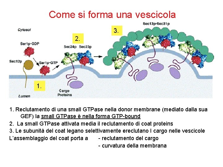
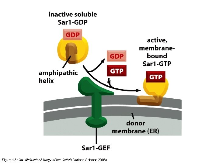
Come inizia la formazione delle vescicole Primo step: reclutamento ed attivazione di small GTPase dalla membrana donatrice
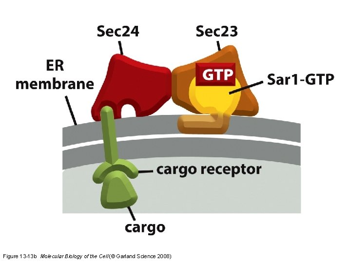
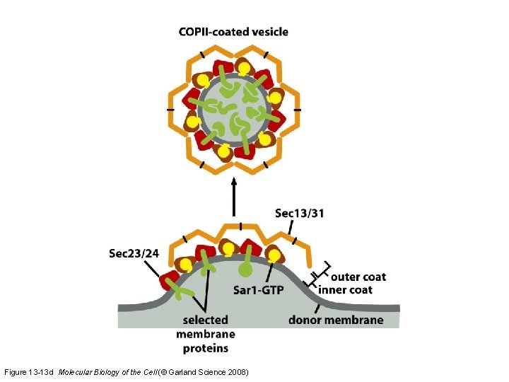
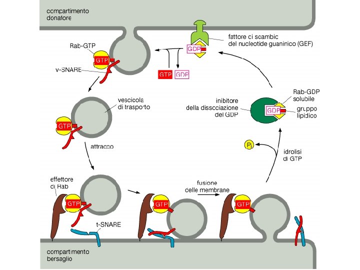
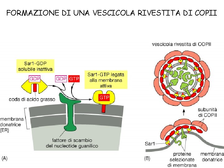
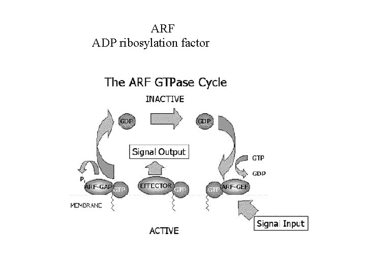
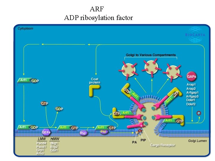
Come si forma una vescicola 3. 2. 1. Reclutamento di una small GTPase nella donor membrane (mediato dalla sua GEF) la small GTPase è nella forma GTP-bound 2. La small GTPase attivata media il reclutamento di coat proteins 3. Le subunità del coat legano selettivamente ereclutano I cargo nelle vescicole L’assemblaggio del coat porta a - reclutamento del cargo - curvatura della membrana
L’USCITA DAL RE AVVIENE MEDIANTE TRAFFICO VESCICOLARE proteine sono concentrate nelle vescicole mediante un meccanismo di riconoscimento del segnale da parte di proteine di rivestimento
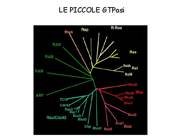
LE PICCOLE GTPasi
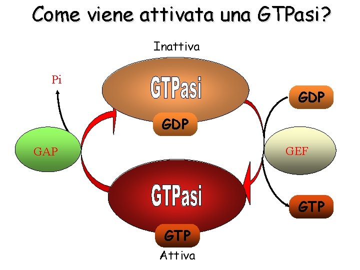
Come viene attivata una GTPasi? Inattiva Pi GDP GEF GAP GTP Attiva
Figure 13 -13 a Molecular Biology of the Cell (© Garland Science 2008)
Figure 13 -13 b Molecular Biology of the Cell (© Garland Science 2008)
Figure 13 -13 d Molecular Biology of the Cell (© Garland Science 2008)
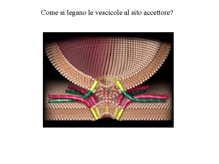
Come si legano le vescicole al sito accettore?
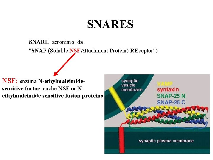
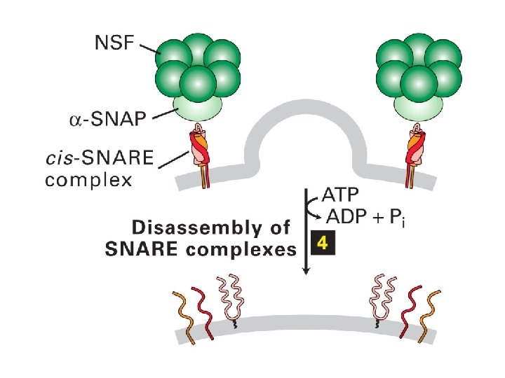
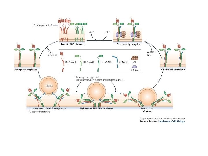
SNARES SNARE acronimo da "SNAP (Soluble NSFAttachment Protein) REceptor") NSF: enzima N-ethylmaleimidesensitive factor, anche NSF or Nethylmaleimide sensitive fusion proteins
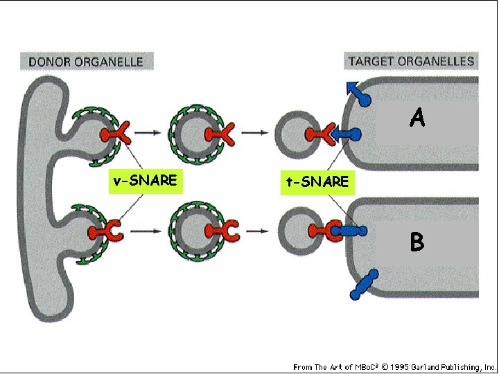
DONATORE ACCETTORI A v-SNARE t-SNARE B
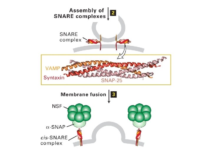
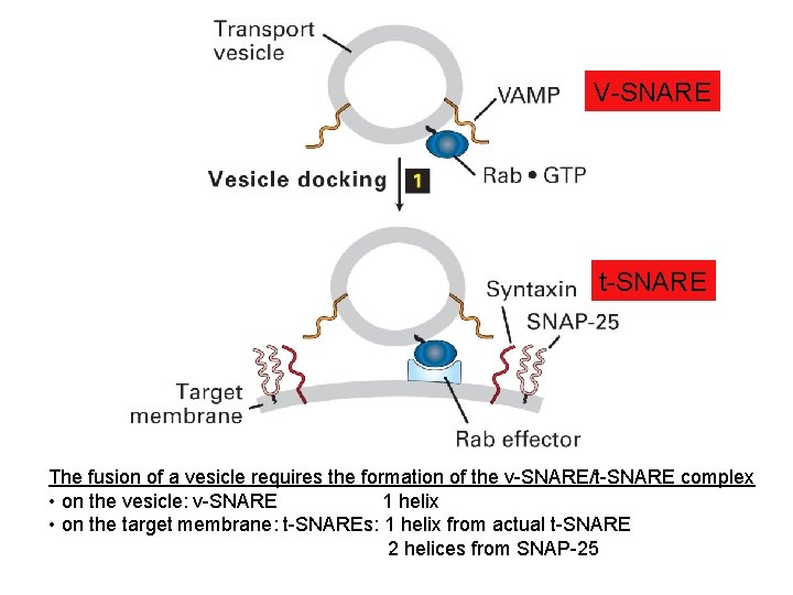
V-SNARE t-SNARE The fusion of a vesicle requires the formation of the v-SNARE/t-SNARE complex • on the vesicle: v-SNARE 1 helix • on the target membrane: t-SNAREs: 1 helix from actual t-SNARE 2 helices from SNAP-25
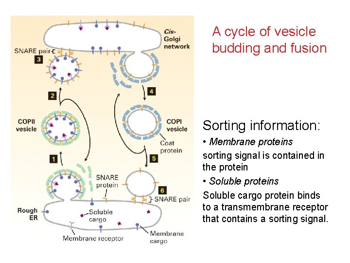
A cycle of vesicle budding and fusion Sorting information: • Membrane proteins sorting signal is contained in the protein • Soluble proteins Soluble cargo protein binds to a transmembrane receptor that contains a sorting signal.
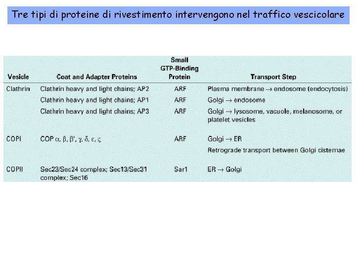
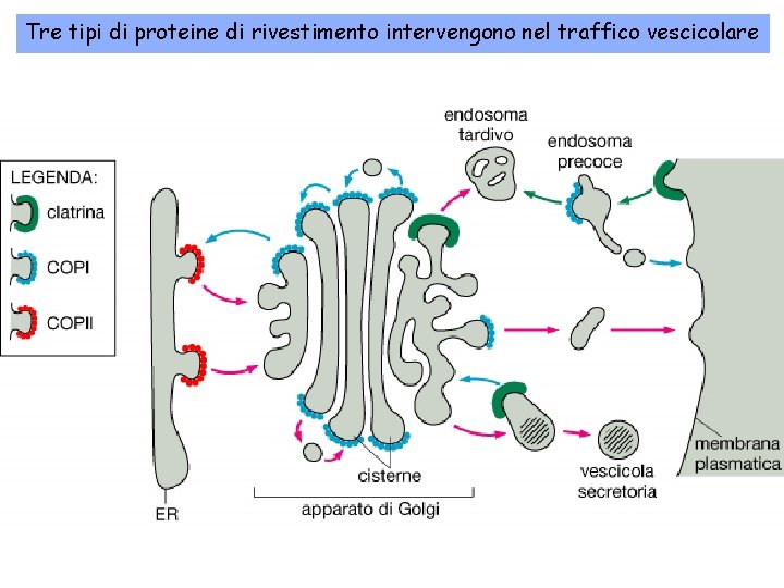
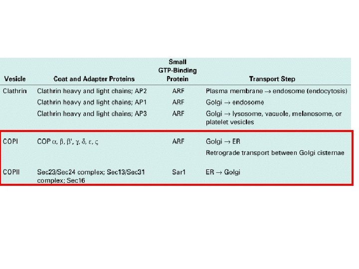
Tre tipi di proteine di rivestimento intervengono nel traffico vescicolare
Tre tipi di proteine di rivestimento intervengono nel traffico vescicolare
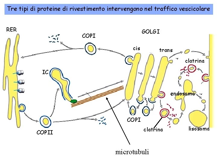
Tre tipi di proteine di rivestimento intervengono nel traffico vescicolare RER GOLGI COPI cis trans clatrina IC endosoma COPII clatrina microtubuli lisosoma
FORMAZIONE DI UNA VESCICOLA RIVESTITA DI COPII
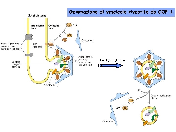
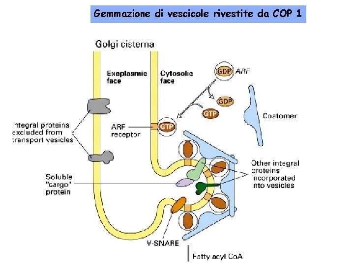
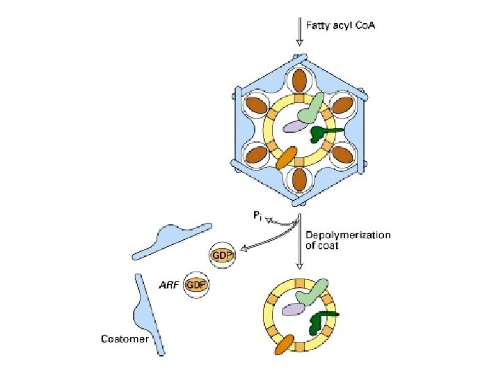
Gemmazione di vescicole rivestite da COP 1 Fatty acyl Co. A
Gemmazione di vescicole rivestite da COP 1
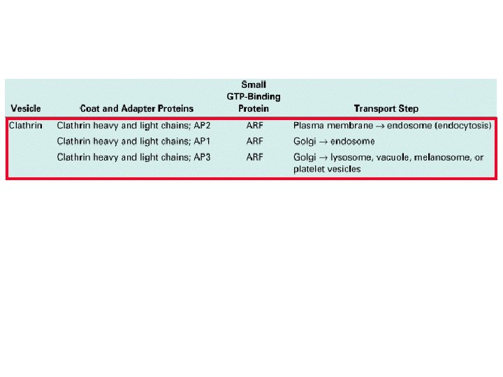
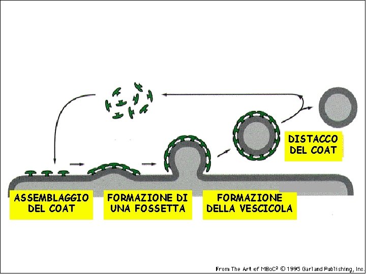
LE VESCICOLE RICOPERTE DI CLATRINA
DISTACCO DEL COAT ASSEMBLAGGIO DEL COAT FORMAZIONE DI UNA FOSSETTA FORMAZIONE DELLA VESCICOLA
ARF ADP ribosylation factor
ARF ADP ribosylation factor
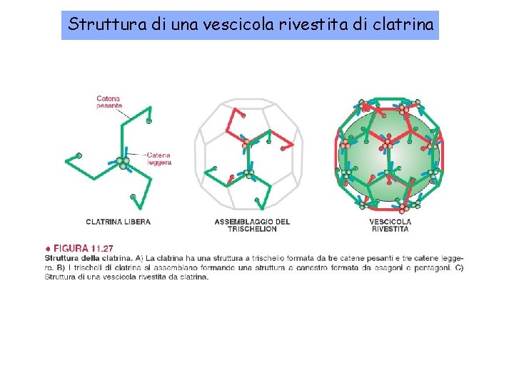
Struttura di una vescicola rivestita di clatrina
Movie
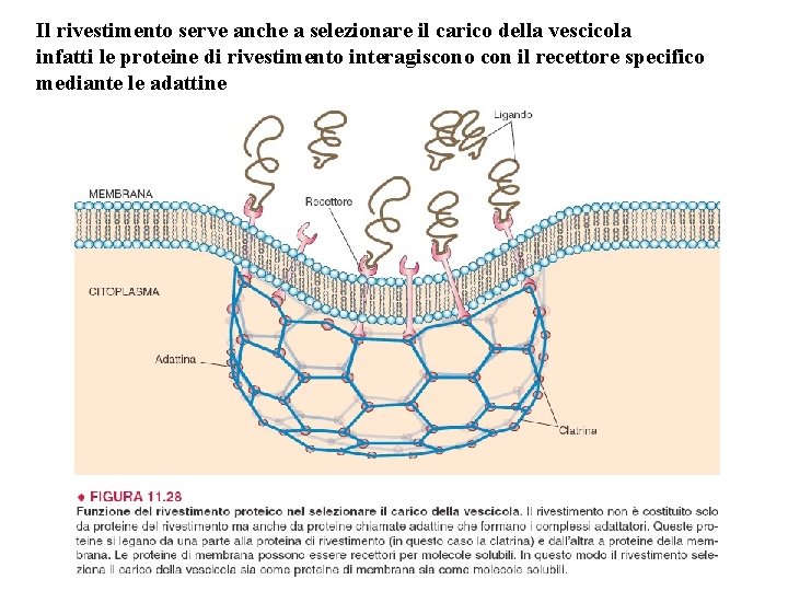
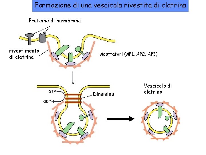
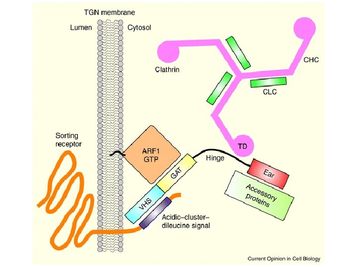
Il rivestimento serve anche a selezionare il carico della vescicola infatti le proteine di rivestimento interagiscono con il recettore specifico mediante le adattine
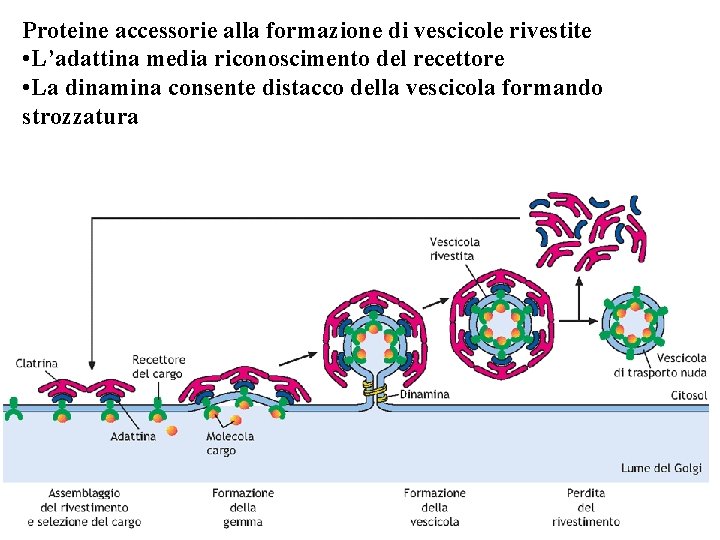
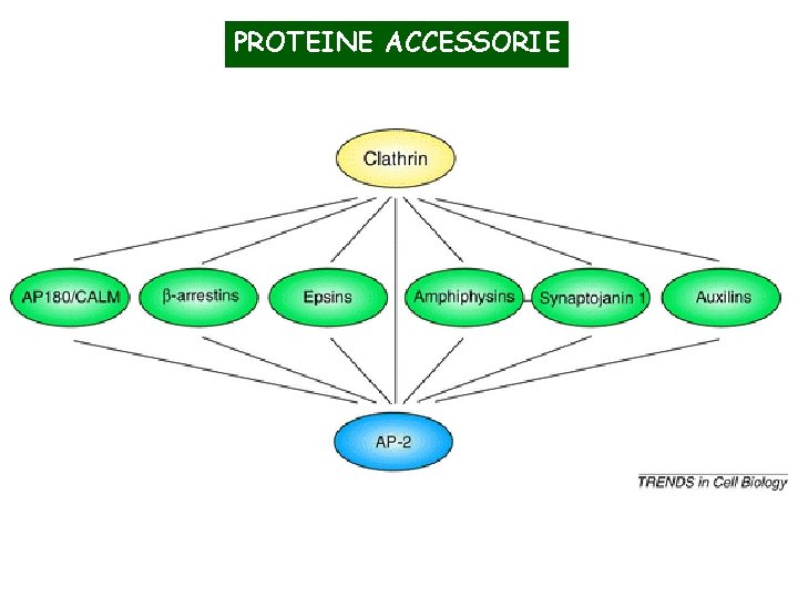
Proteine accessorie alla formazione di vescicole rivestite • L’adattina media riconoscimento del recettore • La dinamina consente distacco della vescicola formando strozzatura
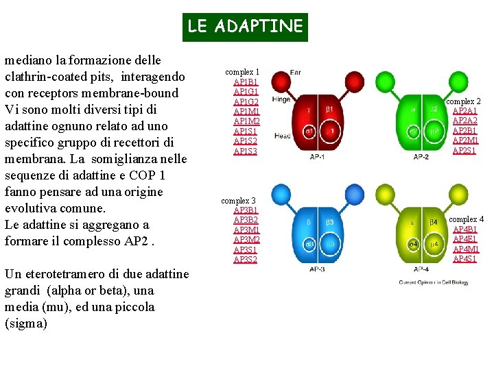
LE ADAPTINE mediano la formazione delle clathrin-coated pits, interagendo con receptors membrane-bound Vi sono molti diversi tipi di adattine ognuno relato ad uno specifico gruppo di recettori di membrana. La somiglianza nelle sequenze di adattine e COP 1 fanno pensare ad una origine evolutiva comune. Le adattine si aggregano a formare il complesso AP 2. Un eterotetramero di due adattine grandi (alpha or beta), una media (mu), ed una piccola (sigma) complex 1 AP 1 B 1 AP 1 G 2 AP 1 M 1 AP 1 M 2 AP 1 S 1 AP 1 S 2 AP 1 S 3 complex 2 AP 2 A 1 AP 2 A 2 AP 2 B 1 AP 2 M 1 AP 2 S 1 complex 3 AP 3 B 1 AP 3 B 2 AP 3 M 1 AP 3 M 2 AP 3 S 1 AP 3 S 2 complex 4 AP 4 B 1 AP 4 E 1 AP 4 M 1 AP 4 S 1
PROTEINE ACCESSORIE
Formazione di una vescicola rivestita di clatrina Proteine di membrana rivestimento di clatrina Adattatori (AP 1, AP 2, AP 3) Dinamina Vescicola di clatrina
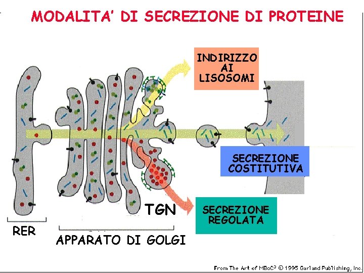
COME ESCONO LE PROTEINE DAL GOLGI? TGN
MODALITA’ DI SECREZIONE DI PROTEINE INDIRIZZO AI LISOSOMI SECREZIONE COSTITUTIVA TGN RER APPARATO DI GOLGI SECREZIONE REGOLATA