Topics of today lectures Hemostasis Meaning of hemostasis


 • Nervous reflexes - initiated by pain or other sensory impulses")
")


 Platelets produce : • Clotting factors like Fibrin Stabilizing Factor")


 • Plasma protein of globulin type. • MW= 68000 •")
 • • MW=340000 Plasma concentration 0. 1 -0. 7 g/dl")
 • 3 stages of conversion of fibrinogen to fibrin – Proteolysis")
 • • • Has two semi-independent pathways The")

 : Phospholipid from damaged tissues. Note: Prothrombin activator")


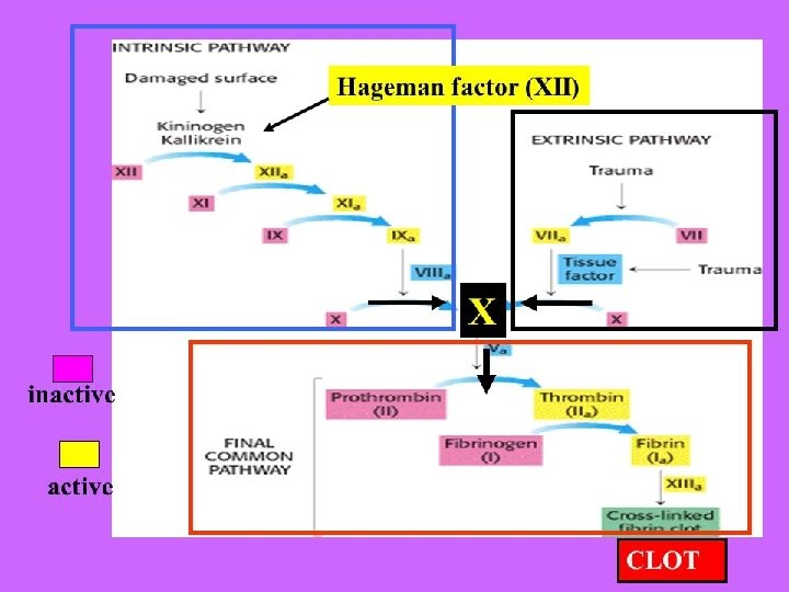


 3. The")


 Significance of plasmin system: The lysis")


 Small punctate hemorrhages occur throughout the body all body tissues. Purplish")


- Slides: 47
Topics of today lectures: Hemostasis • Meaning of hemostasis • Mechanisms of hemostasis - Vascular contraction - Platelets plug - Blood coagulation (clotting) - Structure and functions of platelets - Blood coagulation - Mechanisms - Clotting factors - Prevention of clot formation inside normal blood vessels. - Lysis of blood clots (Fibrinolysis)
Meaning of Hemostasis • It means prevention of blood loss from circulatory system. Also it concerns with maintaining of blood in the fluid state in the vascular compartment. • Hemostasis consists of series of events responsible for the rapid repair of any break in the vascular endothelium. • Abnormality in hemostasis system may result in Hemorrhagic disorder or intravascular clotting (Thrombosis)
Haemostasis Blood vessels The haemostatic response to vascular injury has three elements: 1. Vasoconstriction 2. Platelet aggregation 3. Formation of Clot Platelets Clotting factors
Vasoconstriction after cut in blood vessels • First response to damage or cut in blood vessels. It is localized response. • It occurs almost immediately after cut. • The degree of vascular constriction is directly proportional to severity of damage (i. e the more a vessel is traumatized the greater the degree of spasm). • The vasoconstriction may last for many minutes and may continue for hours. • It narrows the lumen of the vessel to minimize the loss of blood. • The aim of vasoconstriction to reduce or stop blood flow from ruptured vessel.
Vasoconstriction (cont. ) • Nervous reflexes - initiated by pain or other sensory impulses originate from the damaged area. It starts almost immediately after the cut. • Local myogenic spasm due to direct damage to vascular wall. The smooth muscle in the vessel wall contracts and the diameter of the blood vessels decreases. • Vascular constriction is also mediated by release of local humoral factors from traumatized tissue. Blood platelets release thromboxane A 2 and serotonin which are vasoconstrictors.
Second mechanism of hemostasis is formation of Platelets plug (platelets Aggregation)
Characteristics of Platelets • • • Known also as thrombocytes. Smallest of the circulating formed elements diameter 1 to 4 m. Are anuclear bodies produced in bone marrow from the cytoplasm of Megakaryocytes. • Normal concentration of platelets in circulation is 150000300000/ l. Spontaneous hemorrhaging occurs when platelet count gets below 10 x 109/L (10000/ l) • Have a life span of 8 -12 days. Old platelets are removed by macrophages (mostly in spleen). • 75% of platelets are in circulation and 25% in spleen - no. of platelets in circulation is increased after splenectomy • Production is regulated by thrombopoietin
Platelets have many of the functional characteristics of whole cells • Contain contractile proteins – actin, myosin, thrombosthenin. These proteins enable platelets to contract. • Endoplasmic reticulum, Golgi apparatus – protein synthesis, calcium storage • Mitochondria – ATP, ADP formation • Enzyme systems for synthesis of prostaglandins
Platelets characteristics (cont. ) Platelets produce : • Clotting factors like Fibrin Stabilizing Factor (clotting factor no. XIII) and von Willebrand factor. • Platelet derived growth factor (PDGF) - causes vascular endothelial cells, smooth muscles and fibroblasts to grow and repair damaged vessel wall and stimulates wound healing. • • Serotonin Prostaglandines ADP Thromboxane A 2
Platelets Plasma Membrane • Has Glycoprotein coat that repulses normal endothelium while adhering to injured endothelium and any exposed collagen. • Contain receptors for collagen. • Is rich in phospholipids that activate many steps of clotting process.
FUNCTIONS OF PLATELETS • Serotonin released by platelets contributes to VASOCONSTRICTION observed immediately after a vascular injury. • Platelets aggregate to PLUG the vascular injury. • Platelets provide phospholipids which accelerate the process of CLOTTING. • Contractile proteins of the platelets bring about CLOT RETRACTION. • Platelets have a growth factor which stimulates mitosis in vascular wall leading to REPAIR of damaged vessels. • Platelets seem to be involved in all stages of HEMOSTASIS - from the initial reaction till the final repair.
Platelets production • Megakaryocytes, giant cells in the bone marrow, form platelets by pinching off bits of cytoplasm and extruding them into the circulation • Colony stimulating factors regulate the production of Megakaryocytes • Thrombopoietin, a circulating protein factor (produced in the Liver and Kidney) facilitates maturation of Megakaryocytes. Provides a feedback control for maintaining a normal platelet count. megakaryocytes Platelets released from megakaryocytes
Role of platelets in hemostasis • Platelets contribution in hemostasis consists of many steps. – Interact with injured vessel wall (adhesion) – Interact with each other (mass of platelets) – Produce platelets plug • Platelet plug is – Fragile – Can easily be dislodged from the vessel wall ** The platelet plug is reinforced by fibrin fibers (clot).
1. Platelet Adhesion • Damage to endothelium exposes blood to the subepithelial tissue matrix (collagen) • Injured endothelium releases a plasma cofactor protein (von Willebrand Factor) which attaches to the platelet membrane glycoprotein receptor causing adhesion. (Congenital absence of v. WF causes bleeding disorder)
2. Formation of Platelet plug • Contact with damaged endothelium/collagen changes in platelets shape. • Swelling, more irregular shape with multiple pseudopodia, forceful contraction (due to calcium mediated contraction of actin and myosin) and degranulation with release of multiple active factors • Become sticky and adhere to collagen via von Willebrand factor • Secrete large quantities of ADP and form thromboxane A 2 from arachidonic acid. • The release of large amounts of ADP combined with thromboxane A 2 amplifies the initial aggregation of platelets (positive feedback ) into a large platelet mass formation of the platelet plug.
FORMATION OF PLATELETS PLUG
The third mechanism of hemostasis Mechanism of Blood Clotting
Balance between anticoagulants and procoagulants • Normally blood in blood vessel kept in fluid state because of dominancy of anticoagulants factors. • When blood vessel is traumatized, locally the procoagulants are activated and clot develops clot procoagulants Fluid state anticoagulants
Blood coagulation in ruptured BV • Starts quickly -20 seconds in severe bleeding, and 1 -2 minutes in minor trauma. • Interaction between substances released from traumatized blood vessel wall, platelets and clotting factors.
Fate of blood clot • Blood clot formed in the body follows one of these two courses: 1. invasion of the clot by fibroblasts starts few hours after clot formation. It converts clot in the wall of the blood vessel into fibrous tissue within 1 -2 weeks. *** platelets enhance this process through growth factor that they secrete. 2. Dissolving of the clot.
General mechanism of blood clotting ** Three essential steps: • When blood vessel is injured a cascade of complex chemical reactions results in formation of activated substances collectively called prothrombin activator. • Prothrombin activator converts prothrombin into Thrombin • Thrombin converts fibrinogen into Fibrin fibers
Prothrombin (clotting factor II) • Plasma protein of globulin type. • MW= 68000 • Produced by liver. It must be produced continuously because it is continuously consumed by the body. • Its production needs vit. K. • Plasma concentration 15 mg/dl. • When activated, it produces thrombin which acts on fibrinogen. • Prothrombin attaches to platelets on the site of injury.
Fibrinogen (clotting factor I) • • MW=340000 Plasma concentration 0. 1 -0. 7 g/dl Formed in the liver. Thrombin acts on fibrinogen producing fibrin monomers which polymerize into long fibrin fibers. • Fibrin fibers form the meshwork of the clot. • The meshwork of the fibrin fibers is strengthened by fibrin stabilizing factor (XIII).
Fibrinogen (cont. ) • 3 stages of conversion of fibrinogen to fibrin – Proteolysis • Thrombin cleavage of fibrinogen results in fibrin monomers – Polymerization • Spontaneous self-assembly into fibrin polymers – Stabilization • Introduction of covalent bonds into fibrin polymers by XIIIa
Formation of prothrombin activator (Clotting Cascade**) • • • Has two semi-independent pathways The intrinsic pathway has all of its components within the blood. It begins in the blood itself. The extrinsic pathway is triggered by extravascular tissue damage; this pathway is activated by exposure to a tissue factor (trauma to blood vessel wall and surrounding tissues) Both pathways result in activation of prothrombin (factor II) The intrinsic pathway is relatively slow (1 -6 minutes) The extrinsic pathway is quite fast (15 seconds) ** In Clotting Cascade, Each coagulation factor is converted to an active form by the previous factor in the cascade
CLOTTING FACTORS
EXTRINSIC PATHWAY Tissue factor (tissue thromboplastin) : Phospholipid from damaged tissues. Note: Prothrombin activator is a complex consists of Xa, Va, Ca++ and platelet phospholipid The extrinsic pathway is initiated when there is an injury to a blood vessel wall and release of tissue factor
INTRINSIC PATHWAY Starts when blood exposes to rough surface (collagen in blood vessel wall. Exposure of blood to collagen activates factor XII and causes release of phospholipids from platelets.
Contact with collagen
Important points in Blood clotting 1. All factors or procoagulants present in blood in INACTIVE forms. 2. INJURY to tissue, blood vessel wall, cells, platelets is essential for initiation of blood clotting. 3. Ca++ is required for all steps of intrinsic and extrinsic mechanisms EXCEPT for converting XIIa and Xla. 4. Clotting promotes more clot formation (vicious cycle - positive feed back). 5. Most of clotting factors are produced in liver. 6. Formation of clotting factors II, VII, IX and X in the liver needs presence of vitamin K.
Summary of sequence of events after vessel injury - Vasoconstriction- Controlled by vessel smooth muscle; enhanced by chemicals secreted by platelets – Platelet adhesion • Adhesion to exposed subendothelial connective tissue – Platelet aggregation • Interaction and adhesion of platelets to one another to form initial plug at injury site – Fibrin-platelet plug • Coagulation factors interact on platelet surface to produce fibrin; fibrin-platelet plug then forms at site of vessel injury – Fibrin stabilization • Fibrin clot must be stabilized by Factor-XIIIa
Why blood in the vessels does not normally clot? 1. Endothelial surface factors: a. The smoothness of intact endothelium of the blood vessels. This prevents activation of intrinsic pathway. b. a layer of glycocalyx on endothelium that repels the clotting factors. c. presence of protein called thrombomodulin which binds thrombin (removing thrombin). The complex of thrombinthrombomodulin activates protein C which inactivates the activated form of factors V and VIII. 2. The rapid rate of blood flow in the blood vessels, preventing stagnation of blood.
thrombin-thrombomodulin and protein C thrombin-thrombomodulin Tissue plasminogen activator
Why blood in the vessels does not normally clot ? (cont. ) 3. The presence of the natural anticoagulant substance, heparin. 4. Formation of an antithrombin (known as Antithrombin III) which removes free thrombin. The effectiveness of antithrombin III as anticoagulant is increased greatly by presence of heparin. The complex of heparin-antithrombin III can remove other activated clotting factors (XII, X, IX). *** HEPARIN: Is a negatively charged conjugated polysaccharide, produced by basophil and mast cells. It is used in medical practice to prevent intravascular clotting).
PREVENTION OF BLOOD CLOTTING IN THE NORMAL VASCULAR SYSTEM • Antithrombin action of Fibrin and antithrombin III. – – When a clot is forming 85 - 90% of thrombin formed from prothrombin becomes adsorbed to fibrin fibers. Antithrombin III combines with any thrombin which is not adsorbed to fibrin and blocks its effect on fibrinogen and inactivates the bound thrombin.
Lysis of blood clot • Blood contains protein called plasminogen which can be activated into proteolytic enzyme called PLASMIN. • Plasmin digest fibrin fibers (so causes lysis of the clot) it also digests fibrinogen, prothrombin, factors V, VIII and XII (It sometimes decreases the coagulability of the blood). Tissue plasminogen activator (t. PA) produced by injured tissues and endothelium. It coverts plasminogen which entrapped in the clot into plasmin which digest fibrin fibers.
Lysis of blood clots – Plasmin (cont. ) Significance of plasmin system: The lysis of blood clots allows slow clearing (over a period of several days) of extraneous clotted blood in tissues and sometimes allows reopening of clotted vessels. It helps to remove minute clots from the millions of tiny peripheral vessels. [this is the basis for using t-PA or streptokinase for treating intravascular clots in pulmonary or coronary artery]
LABORATORY EVALUATION OF HEMOSTASIS DISORDERS • Platelets disorders present as petechiae and bleeding into mucous membranes because of failure to form the platelets plug. • Patients with coagulation defects may develop deep spreading hematomas, bleeding into the joints and hematuria because of failure of blood clotting. • Platelets functions evaluation – Peripheral blood smear evaluation – bleeding time (N=1 -6 min) • Clotting factors evaluation – Clotting time (N=6 -10 min) – Prothrombin time (PT) –measures the extrinsic and common pathways (N=11 -13 sec). INR= Test / Normal – Activated partial thromboplastin time (APTT) –measure intrinsic pathway (N=30 -40 sec).
EXCESS BLEEDING 1 -Deficiency of vit K Cause: A- Dec Prothrombin B- Dec Factor VII, IX, and X
2 -Hmophilia Mostly in male, caused by abnormality or deficiency of (Factor VIII 85% A, and Factor IX 15%) Transmitted by female chromosome
3 -THROMBOCYTOPENIA (Idiopathic) Small punctate hemorrhages occur throughout the body all body tissues. Purplish blotches of the skin (Thrombocytopenic purpula)
Thromboembolic condition Thrombus: Abnormal clot develop in a blood vesell. Emboli : Break the clot (Thrombus) and flow with the blood. Emboli in the arteries can plug arteries or arterioles in Brain (CVA), Kid, or elswhere. Emboli in the venous system (Right Heart) cause pulmonary artery embolism
CAUSE OF THROMBOEMBOLIC CONDITION 1 -Rough endothelial surface : arteriosclerosis, infection, trauma 2 - Very slow flow : small quantities of thrombin and other procoagulants.
THROMBOLYSIS Streptokinase for coronary occlusion
ANTICOAGULANT 1 - Heparin IV 2 -Coumarins: Warfarin
Prevent blood coagulation outside the body 1 -Heparin 2 -Decrease concentration of Ca by using oxalate to cause precipitated of Ca oxalate from the plasma