TOPIC VESICULAR TRAFFICKING GENERAL INTRODUCTION Vesicles Vesicular trafficking


: Also called transitional ER Contain ER exit sites FUNCTIONS:")
: • Many types of cells export protein produced by")


 are of three types : Transitional vesicles :")


 • Biogenesis Tiny membrane-bound Vesicles Intracellular digestion Hydrolases")


 is the")


: • The ligand-loaded receptors diffuse into these coated-pits.")


- Slides: 39
TOPIC: VESICULAR TRAFFICKING
GENERAL INTRODUCTION: Vesicles Vesicular trafficking Two Main Pathways Communication pathway • • Release molecule Carries molecules Secretory pathway Trafficking SECRETORY PATHWAY: Sends Vesicles From Endoplasmic reticulum Organelles Send or Receive Messages Importing pathway Golgi Apparatus Plasma Membrane Extracellular space Plasma membrane IMPORTING PATHWAY (DEGRADATION PATHWAY): Plasma membrane Endocytic molecules Vesicles originates Which degrades By membrane invagination Becomes lysosomes Fuse with Endosomes
BRIEF INTRODUCTION OF ORGANELLES INVOLVED IN VESICULAR TRAFFICKING • ENDOPLASMIC RETICULUM: Endoplasmic reticulum Membranous synthesis Endoplasmic reticulum forms SHEET Transport organelle “ER Lumen” (ER cisternal space) 10% of entire cell volume. Its membrane allows molecules to selectively transferred between lumen and cytoplasm. It also provides channel between nucleus and the cytoplasm. Producing ER has a central role Processing “Biochemical compounds” Transporting Use inside and outside of the cell. Extension of nuclear envelope.
CONT…. . Its membrane is the side of production of all transmembrane proteins and lipids For most of cell organelles including ER itself Golgi apparatus Almost all of the proteins Lysosomes Endosomes Exit the cell Mitochondria Peroxisomes Secretory vesicles Plasma membrane. Those staying in lumen of ER, Golgi apparatus or lysosome Delivered from endoplasmic reticulum. ER resident proteins. Made up of specialized retention signal Identifies other proteins Made up of specific CHAPERON PROTEIN (BIP) Sequence of amino acid
CONT…… SMOOTH ENDOPLASMIC RETICULUM (FUNCTION): Also called transitional ER Contain ER exit sites FUNCTIONS: • Transport vesicles • Carrying newly synthesized proteins and lipids, including • Synthesis of lipids, • Metabolism of carbohydrates, • Detoxification of drugs an poisons. • Enzymes of SER are vital to the synthesis of lipids and steroids Example: Sex hormones of vertebrates and steroid hormones of adrenal glands • SER performing carbohydrates metabolism cells are abundant in liver cells.
CONT…… ROUGH ENDOPLASMIC RETICULUM (FUNCTION): • Many types of cells export protein produced by ribosomes attached to the rough ER. • The ribosome assemble amino acid into protein units which are carried into RER for further adjustments. • These proteins may be transmembrane proteins, which become embedded in the membrane of endoplasmic reticulum OR • Water soluble proteins which are able to pass through the membrane into lumen, then those retained proteins are folded into correct three dimensional conformation. • Chemicals such as carbohydrates or sugars are added, then ER either transport the completed protein called Secretory proteins
ENDOPLASMIC RETICULUM
GOLGI APPARATUS: Golgi apparatus Main function Apparatus Opposite end Stack of sacs Cisternae. Protein modification. Receives vesicles From ER Modified compounds leave Cis-face Trans-face Facing the plasma membrane Where most of the modifies are sent. • Vesicles sent off by ER containing proteins are further altered at Golgi apparatus and then prepared for secretion from the cell or transport to other parts of the cell. • • • Modification and synthesis of carbohydrate The Golgi apparatus removes and substitutes various sugar monomers. In addition to modifying proteins the Golgi also manufactures macromolecules itself. In plants Golgi apparatus produce pectin and other polysaccharides need by plant structure. Molecular identification labels or tags are added by Golgi enzymes to help in their transport. After this, Golgi apparatus sends off its products by budding vesicles from its Trans-face.
GOLGI APPARATUS
VACUOLES: These are membrane bound sacs larger then vesicles. In plant cells vacuoles cover 30 -90% of total cell volume.
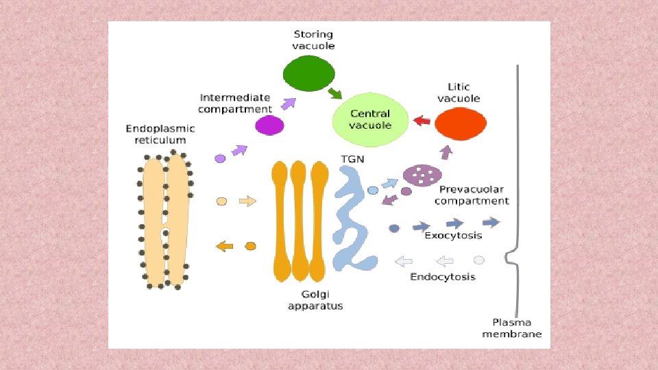
VESICLES: These are small membrane bound transport unit that can transfer molecules between different compartments. Endoplasmic reticulum Golgi apparatus Clathrin-coated 3 -Various locations COPI-coated COPII-coated • CLATHRIN-coated: Transport substances between Golgi apparatus and plasma membrane. • COPI-coated & COPII-coated: Frequently used between ER & Golgi apparatus. i. e. COPI coats vesicles transporting from cis-Golgi back to the RER and between Golgi compartments. This type of transport is called as Retrograde(Backward) transport. COPII coats vesicles transporting proteins from the RER to the cis-Golgi.
VESICLES MECHANISM OF TRANSPORT
ROLE OF CLATHRIN COPII
The vesicles (60 nm in diameter) are of three types : Transitional vesicles : These are small membrane limited vesicles which are thought to form as blebs from the transitional ER to migrate and converge to cis face of Golgi, where they coalesce to form new cisternae. Secretory vesicles: These are varied-sized membrane-limited vesicles which discharge from margins of cisternae of Golgi. They, often, occur between the maturing face of Golgi Clathrin-coated vesicles These are spherical protuberances, about 50 µm in diameter and with a rough surface. They are found at the periphery of the organelle, usually at the ends of single tubules, and are morphologically quite distinct from the secretory vesicles. The clathrin-coated vesicles are known to play a role in intra-cellular traffic of membranes and of secretory products, i. e. , between ER and Golgi, as well as, between GELR region and the endosomal and lysosomal compartments.
The GERL Region : To the trans face of Golgi Trans-reticular Golgi TGN (trans Golgi-network) OR GERL (=Golgi + smooth ER + lysosomal) Involved in the origin of Primary Lysosomes + Melanin granules TUBULES: In Golgi apparatus a complex array of associated vesicles and anastomosing tubules (30 to 50 nm diameter) surround the dictyosome and radiate from it. In fact, the peripheral area of dictyosome is fenestrated (lace-like) in structure.
• GERL REGION GOLGI APPARATUS ENDOPLASMIC RETICULUM LYSOSOMES
LYSOSOMES: • (Gr. lyso=digestive + soma=body) • Biogenesis Tiny membrane-bound Vesicles Intracellular digestion Hydrolases and Membrane proteins. • Both classes of proteins Synthesized in ER Endolysosome Transported To an intermediate compartment Through the Golgi apparatus Trans Golgi network transport vesicle (coated by clathrin protein) Lysosomal enzymes Glycoproteins M 6 P-receptors Recognized Containing N-linked oligosaccharides that are processed Mannose 6 -phosphate (M 6 P) (which are transmembrane proteins) In Trans Golgi network Packaged Budding clathrin-coated vesicles Which quickly lose their coats.
FUNCTIONS OF LYSOSOMES: The important functions of lysosomes are as follows: Digestion of large extracellular particles. The lysosomes digest the food contents of the phagosomes or pinosomes. Digestion of intracellular substances. During the starvation, the lysosomes digest the stored food contents, viz. , proteins, lipids and carbohydrates (glycogen) of the cytoplasm and supply to the cell necessary amount of energy. Autolysis In certain pathological conditions the lysosomes start to digest the various organelles of the cells and this process is known as autolysis or cellular autophagy. Extracellular digestion. The lysosomes of certain cells such as sperms discharge their enzymes outside the cell during the process of fertilization. The lysosomal enzymes digest the limiting membranes of the ovum and form penetration.
LYSOSOME IN CELL
SECRETORY PATHWAY
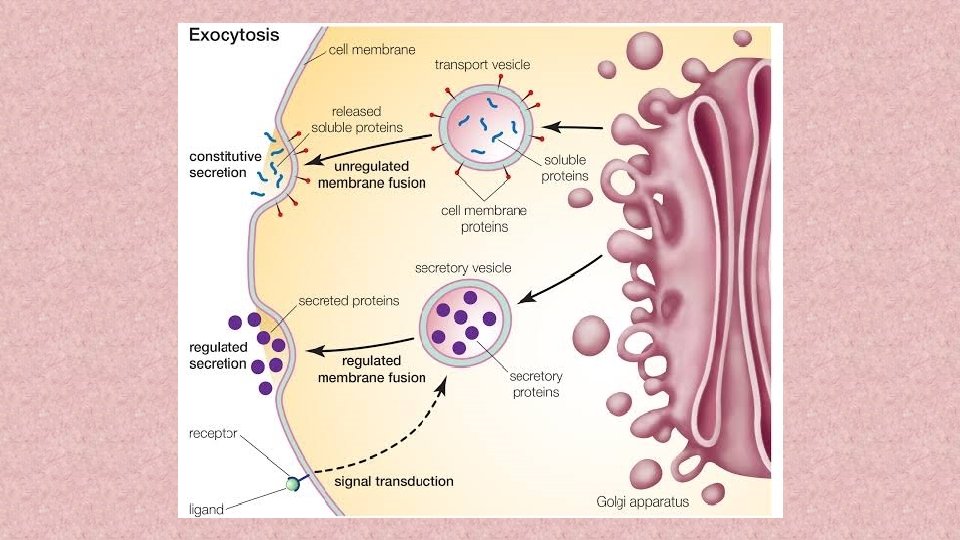
Cells routinely import and export large molecules across the plasma membrane. Macromolecules are secreted out from the cell by Exocytosis Macromolecules are ingested into the cell from outside through Phagocytosis and Endocytosis. EXOCYTOSIS: • It is also called Emeiocytosis and Cell vomiting. • In all eukaryotic cells, secretory vesicles are continually carrying new plasma membrane and cellular secretions such as proteins, lipids and carbohydrates from the Golgi apparatus to the plasma membrane or to cell exterior by the process of exocytosis. • The proteins to be secreted are synthesized on the rough endoplasmic reticulum (RER). • They pass into the lumen of the ER, glycosidated and are transported to the Golgi apparatus by ER-derived transport vesicles. • In the Golgi apparatus the proteins are: Modified, Concentrated, Further Glycosidated, Sorted and Finally packaged into Vesicles that pinch off from trans Golgi tubules and migrate to Plasma membrane to fuse with it and release the secretion to cell’s exterior. Small molecules From the cytosol Vesicles. Where they are complexed to specific macromolecules (e. g. a network of proteoglycans, in case of histamine)
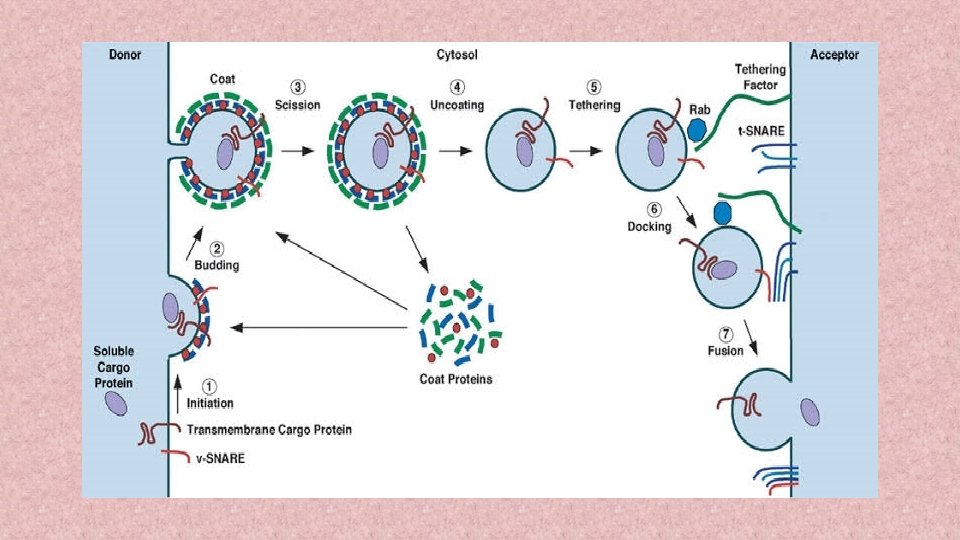
STEPS IN VESICULAR TRAFFICKING There are five steps to exocytosis: • Vesicle trafficking: In this first step the vesicle containing the waste product or chemical transmitter is transported through the cytoplasm towards the part of the cell from which it will be eliminated. • Vesicle tethering: As vesicle approaches the cell membrane, it is secured and pulled towards the part of the cell from which it will be eliminated. • Vesicle docking: In this step the vesicle comes in contact with the cell membrane, where it begins to chemically and physically merge with the proteins in the cell membrane. • Vesicle priming: In those cells where chemical transmitters are being released, this step involves the chemical preparations for the last step of exocytosis. • Vesicle fusion: In this last step, the proteins forming the walls of the vesicles merge with the cell membrane and reach, pushing the vesicle contents (waste products or chemical transmitters) out of the cell. This step is the primary mechanism for the increase in the size of the cell’s plasma membrane.
IMPORTING PATHWAY
ENDOCYTOSIS: In endocytosis, small regions of the plasma membrane fold inwards or invaginate, until it has formed new intracellular membrane limited vesicles. In eukaryotes, the following two types of endocytosis can occur : pinocytosis and receptormediated endocytosis. PHAGOCYTOSIS: (Gr. , phagein=to eat, kytos=cell or hollow vessel) The process of ingestion of large-sized solid substances by the cell is known as phagocytosis. Occurrence of phagocytosis: • The process of phagocytosis occurs in most protozoans and certain cells of multicellular organisms. • In multicellular organisms such as mammals, the phagocytosis occurs very actively in granular leucocytes and in the cells of mesoblastic origin. • The cells of the mesoblastic origin are collectively known as the cells of macrophagic or reticuloendothelial system. • The cells of macrophagic system are histiocytes of the connective tissue, the reticular cells of the hemopoietic organs (bone marrow, lymph nodes and spleen) and the endothelial cells which form the lining of capillary sinusoid of the liver, adrenal gland hypophysis. • The cells of macrophagic system can ingest bacteria, Protozoa, cell debris or even colloidal particles by the process of phagocytosis.
Process of phagocytosis. • In phagocytosis, first the target particle is bound, to the specific receptors on the cell’s surface Plasma membrane expands along the surface of the particle and eventually engulfs (phagolysosome) Fuse with the pre-existing lysosomes Adsorption Formed Vesicles Phagosomes Migrate to the interior of the cell The food is digested by the hydrolytic enzymes (acid hydrolase) of the lysosomes and the digested food is ultimately diffused to the surrounding cytoplasm. The undigested food is expelled from the plasma membrane by the process Ephagy or Egestion. Kinds of phagocytosis. • Ultraphagocytosis or Colloidopexy: The process in which plasma membrane ingests smaller colloidal particles is known as colloidopexy or ultraphagocytosis, e. g. leucocytes and the macrophagic cells of mammals. • Chromopexy: When the cell ingests colloidal chromogen particles phagocytotically the process is known as chromopexy, e. g. , some mesoblastic cells.
1. Pinocytosis. : • Pinocytosis (Gr. pinein = to drink ‘cell drinking’) is the non-specific uptake of small droplets of extracellular fluid by endocytic vesicles or pinosomes having diameter of about 0. 1 µm to 0. 2 µm. • Any material dissolved in the extracellular fluid is internalized in proportion to its concentration in the fluid. • The process of pinocytosis was first of all observed by Edward in Amoeba and by Lewis (1931) in the cultured cells. • The light microscopy has shown that in Amoeba tiny pinocytic channels are continually being formed at the cell surface by invagination of the plasma membrane. • From the inner end of each channel small vacuoles or pinosomes are pinched off, and these move towards the center of the cell, where they fuse with primary lysosomes, to form food vacuoles. • Ultimately, ingested contents are digested, small breakdown products such as sugars and amino acids diffuse to cytosol.
Micropinocytosis: • Electron microscopic observations have been made on the pinocytotic process at sub-cellular or sub-microscopic level in the cells. • The pinocytosis which occurs at submicroscopic level is known as micropinocytosis.
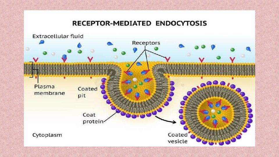
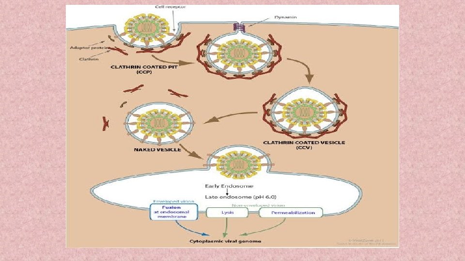
1. Receptor-mediated endocytosis. In this type of endocytosis, a specific receptor on the surface of the plasma membrane “recognizes” an extracellular macromolecule and binds with it. The substance bound with the receptor is called the ligand. Examples of ligands may include: Viruses, Small proteins, Vitamin B 12, Cholesterol containing LDL ( low density lipoprotein), Oligosaccharide etc. The region of plasma membrane containing the receptor-ligand complex undergoes endocytosis. The whole process of receptor mediated endocytosis, includes the following events : • Interaction of ligands and cell surface receptors • Formation of coated-pits and coated-vesicles • Functions of coated pits (clathrin dependent) • Fusion of endocytic vesicle and endosome
Functions of coated pits (clathrin dependent): • The ligand-loaded receptors diffuse into these coated-pits. • A coated-pit may accommodate about 1000 receptors of assorted variety. • In fact, coated-pits serve as molecular filters and selective concentrating devices, since, they tend to collect certain receptors and leave others. • They increase the efficiency of internalization of a particular ligand more than 1000 -fold and also carry minor components of extracellular fluid. • The life-time of each coated-pit is quite short—within a minute or so of being formed, it invaginates into the cell and pinches off to form the coated-vesicles. • The coat of coated pits and coated vesicles is made up of protein, called clathrin and certain other proteins. A molecule of clathrin is composed of three large polypeptide chains and three smaller polypeptide chains, all of which together form a three-legged structure, called triskelion. • A number of triskelions assemble into a basket-like network of hexagons and pentagons on the cytoplasmic surface of the membranes.
Endosome or Receptosome. • Recently it has been found that in the cells exists a complex set of heterogeneous membrane bound tubes and vesicles, called endosome, which extends from the periphery of the cell to the perinuclear region, where it lies quite close to Golgi apparatus. Thus, endosomes may be of two types: • Peripheral endosomes just beneath the plasma membrane. • Perinuclear or Internal endosomes. • These perinuclear endosomes are converted into endolysosomes and then into lysosomes due to following three activities: • The fusion of transport vesicles from the Golgi apparatus. • Transport vesicles capture a cargo of molecules, e. g. , proteins, from the lumen of one compartment as they pinch off from its membrane and then discharge that cargo into another compartment as they fuse with it. Thus, in such vesicular transport, the transported proteins do not cross any membrane and they are transferred from lumen to lumen. • Continuous membrane retrieval. • Increased acidification.
Membrane fusion during exocytosis and endocytosis. Both exocytosis and endocytosis involve the fusion of initially separate regions of lipid bilayer and occur in at least two steps : first the two bilayers come into close apposition, it is called bilayer adherence, and then they fuse together (This is called bilayer joining). Both of these steps are believed to be mediated by some types of specific proteins, called fusogenic proteins. MODIFICATIONS NECESSARY FOR TRANSPORT Invaginations Microvilli Basement Membrane Tight Junctions (Zonula Occludens) Basal lamia Reticular layer Desmosomes
DIAGRAMMATIC MECHANISM OF VESICULAR TRAFFICKING
THANK YOU