TOPIC Cell migration Cell migration Cell migration is


 have important functions in cell")


![� Cells adhesion occurs from the interactions between cell-adhesion molecules (CAMs), [2] transmembrane proteins](https://slidetodoc.com/presentation_image_h2/3ce97a5ba07adbfc0bb586dd06f5f33c/image-23.jpg "� Cells adhesion occurs from the interactions between cell-adhesion molecules (CAMs), [2] transmembrane proteins")


- Slides: 29
TOPIC : Cell migration
Cell migration �Cell migration is the directed movement of a single cell or a group of cells in response to chemical and/or. It is a fundamental cellular process that occurs throughout life, starting during embryonic development and continuing until death, and at times it can contribute to pathogenic states in disease. �mechanical signals.
� In a developing embryo, cell migration is the driving factor for various morphogenetic events. For instance, during gastrulation in very early embryos, groups of cells migrate as sheets to form the three germ layers. Subsequently, cells from the germ layers migrate to various target locations, where they specialize into distinct cell populations that make up various tissues or organs in the embryo.
� In adult organisms, cell migration occurs during vital cellular processes such as tissue renewal and repair, wherein old or damaged cells are replaced by the migration of newly formed cells from the underlying tissue layers. Such events are essential to maintain tissue integrity and homeostasis. Cell migration also plays a role in mediating immune responses during infections, in which phagocytic cells such as neutrophils circulating in the bloodstream migrate to the infected tissues and destroy the invading pathogens.
Cell migration as cyclic process �The migration of a single cell or a group of cells is regarded as a cyclic process, which involves the polarization of cells in response to migratory signals, the extension of filo podial or lamelli podial protrusions, the formation of adhesions between the cell and the underlying matrix, and the pushing of the cells over the adhesions as a result of traction forces generated by the adhesions.
Polarization of migrating cells: �In cell migration, polarity refers to the front-rear polarity - that is, the molecular and functional differences between the front (closest to the direction of migration) and rear (the opposite to the front) of the cell.
Extension of protrusions: �A polarized cell starts putting forth actinbased protrusions at its leading edge, such as lamellipodia or filopodia. Lamellipodia are formed as branched, dendritic networks of actin filaments, and therefore able to push along a broader stretch of the membrane. Filopodia, on the other hand, are formed as parallel bundles of actin filaments, and have roles mainly in sensing the physical properties of the extracellular environment
How and Why Cells Move: � Cell movement is a necessary function in organisms. Without the ability to move, cells could not grow and divide or migrate to areas where they are needed. The cytoskeleton is the component of the cell that makes cell movement possible. This network of fibers is spread throughout the cell's cytoplasm and holds organelles in their proper place. Cytoskeleton fibers also move cells from one location to another in a fashion that resembles crawling
Steps of Cell Movement � Cell adhesion molecules on cell surfaces hold cells in place to prevent undirected migration. Adhesion molecules hold cells to other cells, cells to the extracellular matrix (ECM) and the ECM to the cytoskeleton. The extracellular matrix is a network of proteins, carbohydrates and fluids that surround cells. The ECM helps to position cells in tissues, transport communication signals between cells and reposition cells during cell migration. Cell movement is prompted by chemical or physical signals that are detected by proteins found on cell membranes. Once these signals are detected and received, the cell begins to move.
� In the first phase, the cell detaches from the extracellular matrix at its foremost position and extends forward. � In the second phase, the detached portion of the cell moves forward and re-attaches at a new forward position. The rear portion of the cell also detaches from the extracellular matrix. � In the third phase, the cell is pulled forward to a new position by the motor protein myosin. Myosin utilizes the energy derived from ATP to move along actin filaments, causing cytoskeleton fibers to slide along one another. This action causes the entire cell to move forward.
Movement Within Cells: �Not all cell movement involves the repositioning of a cell from one place to another. Movement also occurs within cells. Vesicle transportation, organelle migration, and chromosome movement during mitosis are examples of types of internal cell movement.
� Vesicle transportation involves the movement of molecules and other substances into and out of a cell. These substances are enclosed within vesicles for transportation. Endocytosis, pinocytosis, and exocytosis are examples of vesicle transportation processes. In phagocytosis, a type of endocytosis, foreign substances and unwanted material are engulfed and destroyed by white blood cells. The targeted matter, such as a bacterium, is internalized, enclosed within a vesicle, and degraded by enzymes.
�Organelle migration and chromosome movement occur during cell division. This movement ensures that each replicated cell receives the appropriate complement of chromosomes and organelles. Intracellular movement is made possible by motor proteins, which travel along cytoskeleton fibers. As the motor proteins move along microtubules, they carry organelles and vesicles with them.
Cilia and Flagella: � Some cells possess cellular appendage-like protrusions called cilia and flagella. These cell structures are formed from specialized groupings of microtubules that slide against one another allowing them to move and bend. Compared to flagella, cilia are much shorter and more numerous. Cilia move in a wavelike motion. Flagella are longer and have more of a whip-like movement. Cilia and flagella are found in both plant cells and animal cells.
� Sperm cells are examples of body cells with a single flagellum. The flagellum propels the sperm cell toward the female oocyte for fertilization. Cilia are found within areas of the body such as the lungs and respiratory system, parts of the digestive tract, as well as in the female reproductive tract. Cilia extend from the epithelium lining the lumen of these body system tracts. These hair-like threads move in a sweeping motion to direct the flow of cells or debris. For example, cilia in the respiratory tract help to propel mucus, pollen, dust, and other substances away from the lungs.
Molecular Process 0 f mogration �There are two main theories for how the cell advances its front edge: the cytoskeletal model and membrane flow model. It is possible that both underlying processes contribute to cell extension.
Cytoskeletal model � Leading edge: � Experimentation has shown that there is rapid actin polymerisation at the cell's front edge. ] This observation has led to the hypothesis that formation of actin filaments "push" the leading edge forward and is the main motile force for advancing the cell's front edge. In addition, cytoskeletal elements are able to interact extensively and intimately with a cell's plasma membrane.
� Trailing edge: � Other cytoskeletal components (like microtubules) have important functions in cell migration. It has been found that microtubules act as “struts” that counteract the contractile forces that are needed for trailing edge retraction during cell movement. When microtubules in the trailing edge of cell are dynamic, they are able to remodel to allow retraction. When dynamics are suppressed, microtubules cannot remodel and, therefore, oppose the contractile forces.
� The morphology of cells with suppressed microtubule dynamics indicate that cells can extend the front edge (polarized in the direction of movement), but have difficulty retracting their trailing edge. On the other hand, high drug concentrations, or microtubule mutations that depolymerize the microtubules, can restore cell migration but there is a loss of directionality. It can be concluded that microtubules act both to restrain cell movement and to establish directionality
Membrane flow model �Studies have also shown that the front of the migration is the site at which the membrane is returned to the cell surface from internal membrane pools at the end of the endocytic cycle. This has led to the hypothesis that extension of the leading edge occurs primarily by addition of membrane at the front of the cell
� If so, the actin filaments that form at the front might stabilize the added membrane so that a structured extension, or lamella, is formed rather than a bubble-like structure (or bleb) at its front. [15] For a cell to move, it is necessary to bring a fresh supply of "feet" (proteins called integrins, which attach a cell to the surface on which it is crawling) to the front. It is likely that these feet are endocytosed toward the rear of the cell and brought to the cell's front by exocytosis, to be reused to form new attachments to the substrate.
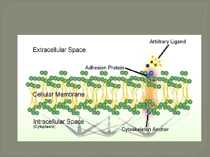
Cell adhesion � Cell adhesion is the process by which cells interact and attach to neighbouring cells through specialised molecules of the cell surface. This process can occur either through direct contact between cell surfaces or indirect interaction, where cells attach to surrounding extracellular matrix, a gel-like structure containing molecules released by cells into spaces between them
� Cells adhesion occurs from the interactions between cell-adhesion molecules (CAMs), [2] transmembrane proteins located on the cell surface. Cell adhesion links cells in different ways and can be involved in signal transduction for cells to detect and respond to changes in the surroundings. [1][3] Other cellular processes regulated by cell adhesion include cell migration and tissue development in multicellular organisms. [4]
General mechanism � � Cell–cell junctions can occur in different forms. In anchoring junctions between cells such as adherens junctions and desmosomes, the main CAMs present are the cadherins. This family of CAMs are membrane proteins that mediate cell–cell adhesion through its extracellular domains and require extracellular Ca 2+ ions to function correctly. [2] Cadherins forms homophilic attachment between themselves, which results in cells of a similar type sticking together and can lead to selective cell adhesion, allowing vertebrate cells to assemble into organised tissues. [1] Cadherins are essential for cell–cell adhesion and cell signalling in multicellular animals and can be separated into two types: classical cadherins and non-classical cadherins. [2]
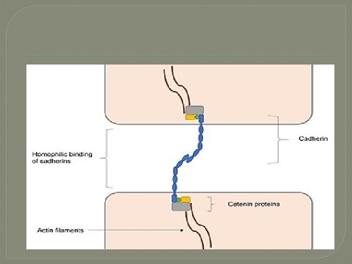
Adherens junctions � Adherens junctions mainly function to maintain the shape of tissues and to hold cells together. In adherens junctions, cadherins between neighbouring cells interact through their extracellular domains, which share a conserved calcium-sensitive region in their extracellular domains. When this region comes into contact with Ca 2+ ions, extracellular domains of cadherins undergo a conformational change from the inactive flexible conformation to a more rigid conformation in order to undergo homophilic binding. Intracellular domains of cadherins are also highly conserved, as they bind to proteins called catenins, forming catenin-cadherin complexe
� These protein complexes link cadherins to actin filaments. This association with actin filaments is essential for adherens junctions to stabilise cell– cell adhesion. [10] [11] [12] Interactions with actin filaments can also promote clustering of cadherins, which are involved in the assembly of adherens junctions. This is since cadherin clusters promote actin filament polymerisation, which in turn promotes the assembly of adherens junctions by binding to the cadherin–catenin complexes that then form at the junction.
THA NK YOU