Topic 6 Human Physiology Organization of the body

Topic 6 Human Physiology

Organization of the body • The human body is composed of cells that are organized into tissues, tissues organized into organs, and organs organized into organ systems. • Anatomy is the study of identifying structures. • Physiology is the study of how the organs and tissues of the body function.

6. 1 Digestive System • Digestion is an enzyme facilitated chemical process. • The Order of Events: • Ingestion Eating • Digestion Breaking food down • Absorption Food molecules into blood/lymph • Transport circulatory system delivers to body

Digestion • • Proteins Amino Acids Lipids Glycerol and fatty acids Carbohydrates monosaccharides Nucleic acids Nucleotides • This is accomplished thru hydrolysis reactions

Enzymes in Digestion • Alimentary Canal – The whole passage along which food passes through the body from mouth to anus. • Enzymes are added to food as it travels the alimentary canal. • Each enzyme is specific for a food type. • The human body temp (370 C) provides a high enough activation energy for digestive reactions to occur.

Anatomy of Human Digestive System • A long tube called the Alimentary canal plus the pancreas and liver which are connected to the alimentary canal by ducts. • Begins with mouth, ends with anus. • All solids and liquids are either absorbed by the body or excreted as feces.

Alimentary canal • Muscular tube where food is moved along by smooth muscles. • The contractions of the muscles and the movement of the food is called peristalsis. • Peristalsis in the stomach is called “churning”.

Roll of the pancreas • The pancreas produces many molecules necessary for normal function of the body. • Two hormones – insulin and glucagon, help regulate glucose levels. • Three enzymes used in digestion • Lipase – used to metabolize lipids • Amylase – used to metabolize carbohydrates • Endopeptidase – used to metabolize protein • As a group, these three enzymes are called pancreatic juice

Pancreas and Liver • Pancreatic duct carries juices to the beginning of the small intestine • Liver produces bile which aids in lipid digestion. Bile is stored in the gallbladder and is carried to the small intestine by the bile duct.

Table of Enzymes

Digestion of starch begins in the mouth • The enzyme salivary amylase begins breaking down (hydrolyzing) starch in the saliva of the mouth and converting it into maltose. • The activity of this enzyme ends at the stomach due to its low p. H environment.

Digestion of starch • Most of the starch arrives at the beginning of the small intestine still undigested. • The pancreas secretes pancreatic amylase into the beginning of the small intestine, called the duodenum. • The p. H of the small intestine is slightly basic which is perfect for this enzyme. • Starch continues to be hydrolyzed to maltose.

Maltase • The small intestine itself produces the enzyme maltase, which further converts the maltose into glucose. • Maltase remains attached to the cells of the small intestine.

Mucosa • The lining of the small intestine is called the mucosa • The mucosa contains finger like projections called villi which allow for an increased surface area. • The villi absorb the nutrients from the lumen. • Within the villi are blood capillaries as well as a lacteal (part of the lymph system)

absorption • Most nutrients are absorbed into the capillaries of the blood stream while fatty acids are usually absorbed into a lacteal.

Ways epithelial cells absorb nutrients

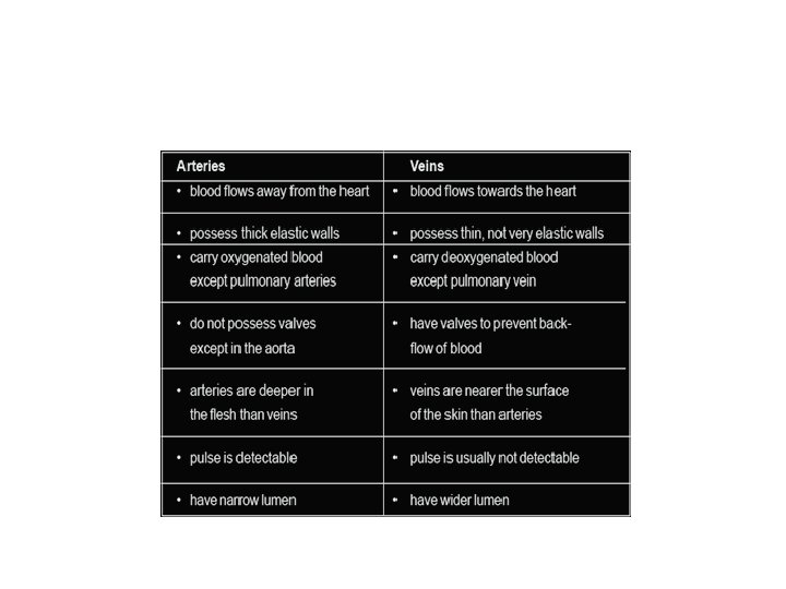
6. 2 Blood System • Arteries, capillaries and veins • Arteries are blood vessels carrying blood away from the heart. • Veins are blood vessels carrying blood towards the heart. • Capillaries are very small, often one cell thick, where the exchanges between the blood and the tissues of the body happen.

Arteries • Have a thick layer of muscle that can be used to change the diameter of the lumen. • Have a layer of elastic tissue which stretches and contracts to allow for the pressure created by the heart pumping. • By the time the blood enters the capillaries, most of the pressure is gone.

Capillaries • Capillaries are only one cell thick to assist in easier diffusion into and out of the blood. • Capillaries are the site of the exchanges which take place between the blood and the tissues of the body. • Blood cells move through the capillaries in a single file line, much slower than they did through the arterioles.

Veins • Veins receive blood at low pressure from the capillaries. • Because the blood is moving slower, veins are thinner walled and have a larger lumen than arteries. • Many veins have one way valves to assist in the movement of blood back towards the heart.

Structure vs Function

Heart

Heart • The heart is a side by side pump • Each side has a chamber that incoming blood slowly enters, and a chamber the blood exits from under pressure. • The sequence of movement exiting the heart is: • Large artery, smaller arteries, arterioles, capillary bed, venules, smaller veins, large vein

Right Side of the Heart • Receives deoxygenated blood from the body. • Sends blood to the lungs to get oxygen and release CO 2 • Called Pulmonary circulation • Capillaries where exchanges take place are in the lungs • Oxygenated blood then returns to the heart.

Left Side of the Heart • • Receives oxygenated blood from the lungs. Sends blood out to the body Called Systemic circulation. We will begin one full trip around the body at this point.

Systemic Route • Oxygenated blood leaves the left side of the heart through a major artery called the Aorta. • The Aorta branches, taking blood to almost every part of the body. • Eventually, the oxygenated blood will arrive at capillary beds where exchanges take place.

Systemic Route • From the capillary beds of the body, deoxygenated blood travels back to the heart through branches of a major vein called the Vena Cava and arrives at the right side of the heart.

Pulmonary Route • Deoxygenated blood is pumped out of the right side of the heart through the pulmonary artery to the lungs. • Exchanges in capillary beds takes place. • Oxygenated blood returns to the left side of the heart through the pulmonary vein.

Anatomy of the Heart

Control of Heart Rate • The heart is made of Cardiac Muscle which is different than other types of muscle. • Cardiac muscle can spontaneously contract without input from the nervous system, called Myogenic muscle contraction. • The heart needs to have the rate and timing of the contractions controlled.
 • Found in the wall of the right atrium. •")
Sinoatrial Node (SA node) • Found in the wall of the right atrium. • Acts as a natural pacemaker by sending out an electrical signal to cause the Atria to contract. • Also in the right Atrium is a mass of muscle tissue called Atrioventricular Node (AV node). • When the AV node receives the electrical signal from the SA node, it delays it. 1 seconds then sends out the signal to the ventricles.

Control of the SA Node • The Medulla monitors the CO 2 levels in your blood and can send a message through the cardiac nerve to have the SA node increase its rate of impulse. • When the medulla senses the CO 2 levels are dropping, it sends another message, this time through the vagus nerve, to slow back down.

Control of the SA Node • The SA node can also be influenced by certain chemicals, like epinephrine (adrenaline). • During stress or excitement, the adrenal gland secretes epinephrine into the bloodstream, causing the SA node to increase its rate of impulse.

Blood Pressure • Diastole is the term used to describe when a heart chamber is not contracting. • Systole is the term used to describe when a heart chamber is contracting. • Heart valves open and close based on the pressure on either side of the valve. • The sound of you heart beating is caused by the valves when the snap closed.

Atherosclerosis • A slow build-up of material in the arteries called plaque. • Plaque is composed of lipids, cholesterol, cell debris and calcium. • As arteries slowly build plaque, they become less flexible. • The amount of plaque depends on genetics, eating habits and several other factors. • http: //medmovie. com/library_id/7556/topic/ cvml_0070 a/

Heart Attack • Coronary arteries supply the heart itself with oxygen rich blood. • They branch directly off of the Aorta. • Because the heart never stops beating, it requires a lot of oxygen. • If any of the three major coronary arteries gets blocked, part of the heart doesn’t receive oxygen. • Plaque can cause this. Called coronary thrombosis, myocardial infarction, or heart attack. • http: //medmovie. com/library_id/7556/topic/cvml_00 71 a/

6. 3 Defense against Infectious Disease • Pathogen – any living organism or virus that is capable of causing a disease. • Pathogens include viruses, bacteria, protozoa, fungi and worms. • Most of the pathogens we are exposed to never get into our bodies.

Primary Defense • Ways of making it difficult for pathogens to enter the body • Skin – contains two primary layers, dermis and epidermis. • The underneath layer is dermis and it is alive, the top layer is the epidermis and it is mainly dead cells. • This top layer of epidermis is an excellent barrier against most pathogens.

Primary Defense • Entry points that are not covered by skin are covered by mucus membranes. • Mucus traps incoming pathogens to prevent them from reaching cells. • Some mucus membranes are covered with cilia which are hair-like projections that move trapped pathogens out of the body.

Locations of mucus membranes • Trachea – the tube that carries air to and from the lungs • Nasal passages – tubes that allow air to enter the nose and then the trachea • Urethra – a tube that carries urine from the bladder to the outside • Vagina – the reproductive tract leading from the uterus to the outside

Blood clotting • When the skin is damaged, exposing an opening for pathogens, blood will clot. • This prevents further blood loss and it plugs the opening to prevent pathogens from gaining access. • In the blood plasma there are different plasma proteins with different functions. • Two of the proteins are involves in clotting of the blood.

Prothrombin and Fibrinogen • Proteins always in the plasma but “inactive”. • Also in the plasma are cell fragments called platelets. • Platelets form in bone marrow and break apart into several fragments, they have no nucleus and are very short lived (8 -10 days)

Clotting Process • If blood vessel is damaged, the cells release chemicals that cause platelets to adhere to damaged area. • The damaged cells and the platelets release chemicals called clotting factors that convert the prothrombin in the blood to thrombin. • Thrombin is an enzyme that catalyzes fibrinogen to fibrin, which is insoluble and helps to form a plug.

Immune Response • When a pathogen does get into the body, it causes an immune response. • If it’s the first time the particular pathogen has been in the body, it’s called a primary immune response, if not the first time, it’s called a secondary immune response. • Primary response can take more than a week to be successful, secondary response is very quick with little symptoms. “immunity”
 • A type of leucocyte (white blood cell) that is involved")
Phagocytes (non-specific response) • A type of leucocyte (white blood cell) that is involved early in the immune response is a macrophage. • Macrophages are large and can change shape to easily squeeze in and out of the blood and engulf pathogens by phagocytosis. • Macrophages can recognize a cell as being “self” or “not self” based on cell membrane surface proteins. • This is a non specific response. • https: //www. youtube. com/watch? v=FPO_9 F 9 BJv k

Specific Response • Antibodies are proteins produced by the body in response to a specific pathogen. • Each antibody is different because each pathogen is different. • Pathogens are cells with membranes that contain proteins, or viruses with a protein coat called a capsid. • These foreign proteins are called antigens

Antibodies • Antibodies are Y-shaped proteins. • At the ends of the forks are binding sites at attach to the antigen. • Antibodies are produced by a type of leucocyte called a plasma cell. • There are many different types of plasma cells, and each type can only produce one type of antibody. • http: //highered. mheducation. com/sites/0072507 470/student_view 0/chapter 22/animation__the_i mmune_response. html

Primary Immune Response 1. A specific pathogen is identified 2. A specific plasma cell is identified to produce an antibody against the pathogen 3. The specific plasma cell begins cloning itself to make many 4. Plasma cells begin making antibodies 5. Antibodies circulate in blood until they find pathogen 6. In various ways, antibodies eliminate pathogen 7. Some plasma cells remain in blood to provide immunity against secondary infection. (Memory Cells) 8. Memory plasma cells respond quickly if same pathogen encountered again. (Secondary Immune Response)

HIV • HIV is the abbreviation for a virus called the Human Immunodeficiency Virus. • Like all pathogens, it has a specific type of cell it likes to infect • HIV attacks a type of lymphocyte in our immune system. • The immune system slowly loses the ability to make antibodies.
 is the name of the disease")
AIDS • AIDS ( Acquired Immune Deficiency Syndrome) is the name of the disease caused by HIV. • Symptoms don’t show for years after the initial HIV infection. • With the immune system not working properly, the infected person becomes infected with multiple infections.

AIDS • No cure, but medicines exist that can prolong the time period between infection and symptoms. • Transmitted most commonly through unprotected sex and sharing needles. • Sometimes transmitted from HIV+ mother to child during pregnancy or breastfeeding. • Initially spread by blood transfusions.

Antibiotics • Bacteria are prokaryotic organisms, humans are eukaryotic. • Antibiotics are chemicals that take advantage of the differences and block some of the biochemistry of bacteria. ( certain reaction, cell wall production etc) • Viruses have no metabolic reactions so antibiotics don’t work on them.

Antibiotic Resistance • Because of genetic variation, high numbers and very fast reproduction times, a bacterium with a genetic variant causing it to not be affected is possible. • That bacterium can then produce large numbers of bacteria resistant to a particular antibiotic. • Odds are increases by long-term use and overuse. (S. aureus has become MRSA)

6. 4 Gas Exchange • Our lungs work with our heart and blood vessels to ensure that body cells are well supplied with oxygen and are able to give up carbon dioxide. • Oxygen is needed for cellular aerobic respiration and the production of ATP

Ventilation • The process of filling the lungs with air and then expelling the air. • The purpose of which is to allow diffusion of gasses in the lungs. • The site of this diffusion within the lung are small sacs called alveoli. • Oxygen in the alveoli diffuses into the bloodstream, while CO 2 in the bloodstream diffuses into the alveoli.

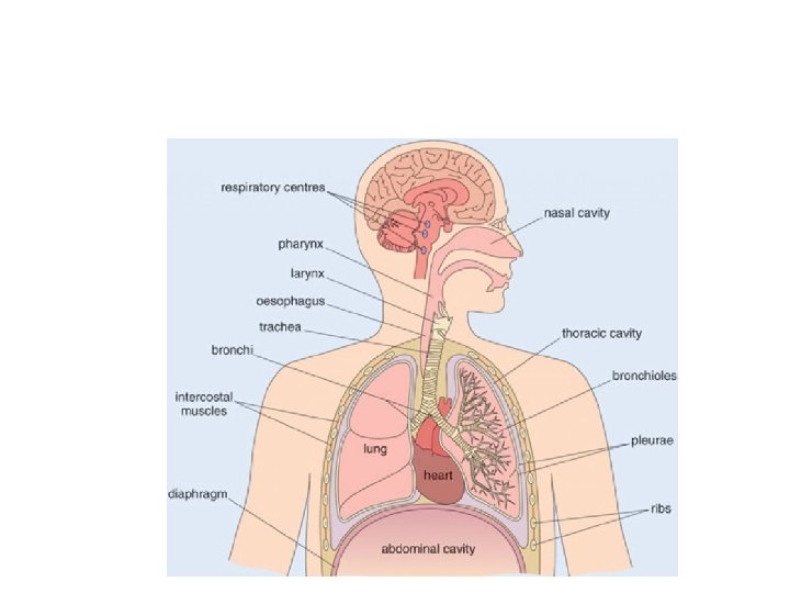
Mechanism of Ventilation • The tissue that makes up our lungs is not muscular so the lungs are not capable of purposeful movement. • The mechanism is based on the inverse relationship between pressure and volume. • The lungs are located in a cavity called the thorax or thoracic cavity. • Pressure differences between this cavity and the outside are the cause of air movement.
 • The bottom of the thoracic cavity is a muscle called")
Inspiration (Breathing In) • The bottom of the thoracic cavity is a muscle called the diaphragm. • The ribs are covered with the external and internal intercostal muscles. • When the diaphragm and external intercostals contract, it increases the volume of the cavity which makes the pressure in the cavity and the lungs drop. • The pressure in the lungs is now lower (a vacuum)than the outside of the body, so air rushes in to equalize the pressure.
 • The diaphragm and intercostal muscles relax • The volume of")
Expiration (Breathing Out) • The diaphragm and intercostal muscles relax • The volume of the cavity gets smaller, raising the pressure. • The pressure is now greater than the outside air. • Air moves out to equalize pressure.

Gas Exchange in the Alveoli • • • Air first enters your mouth or nasal passages. Then it enters your trachea Then the left and right bronchi Then smaller and smaller branches of bronchi Then very small bronchioles Then it enters small sacs in the lung called alveoli

Alveoli structure

• Alveoli are found in clusters at the ends off the bronchioles • There approximately 300 million in each lung. • They are surrounded by capillary beds. • Blood entering these capillaries is coming from the right ventricle of the heart via the pulmonary arteries.

• Carbon dioxide diffuses out of the blood into the alveoli and oxygen diffuses from the alveoli into the blood. • Only a two cell trip due to alveoli and capillary each being one cell thick

Pneumocytes • The single cell layer of the alveoli are made of two different types of cells called pneumocytes. • Type 1 pneumocytes – very thin with a large surface area for diffusion. If damaged, these cells are incapable of mitosis for replacement. • Type 2 pneumocytes – Cube shaped, not good for diffusion but secrete a solution that acts as a surfactant. Keeps the alveoli from sticking closed. • Can undergo mitosis to make both types of cells

Emphysema • A disease where the alveoli are destroyed. • Leading cause is tobacco smoking • One of the diseases collectively called COPD, chronic obstructive pulmonary disease. • Chronic (slow) disease that turns alveoli into large, irregular structures with holes in them. • Reduces the surface area for diffusion causing shortness of breath. • Other causes include: Marijuana smoke, smog, coal dust.
.")
Lung Cancer • Begins in the lungs but is very prone to spreading (metastasizing). • Common sites of spreading include brain, bones, liver and adrenal glands. • Caused by carcinogenic substances that cause the lungs cell to mutate. • Most common source of carcinogens is cigarette smoke, but other substances like asbestos can also cause cancer. • Early diagnosis is vital due to high mortality rate.

Cancer Rates • Recent data supports a direct correlation between countries that have decreased the number of people who smoke and a decrease in lung cancer. And visa versa

6. 5 Neurons and Synapses • The brain and spinal cord comprise the central nervous system (CNS). • The CNS receives sensory info from receptors and processes it. • If a response is needed, the CNS initiates a motor response. • Sensory neurons bring info to the CNS, motor neurons carry response from CNS to muscles.

Peripheral Nervous System • Sensory and motor neurons are peripheral nerves. • A neuron is an individual cell, many neurons working together is a nerve. • Neurons and nerves carry electrical impulses around the body. • Nerves are a group of neurons covered by a protected sheath.

• Spinal nerves come directly from the spinal cord and are mixed, containing sensory and motor neurons. There are 31 pairs of spinal nerves. • Cranial nerves come from the brainstem, there are 12 pairs. Example: optic nerve

Neurons • Transmit electrical impulses • Varying lengths, one neuron runs from lower spinal cord to the toes (1 meter long single cell)

Parts of a neuron • • Dendrites Cell body Axon At the end of the axon are synaptic terminal buttons that release chemicals called neurotransmitters which help the electrical impulse continue to the next neuron or muscle.

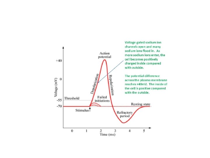
Nerve Impulse • Travels from the dendrites and body down the axon to the terminal buttons. • Actually in a neuron, not a nerve. • The change in potential (voltage) across the neuron membrane.

Resting Potential • Not currently sending an impulse but ready to. • Referred to as being polarized • Resting Potential is created by sodium potassium pumps, pumping Na+ ions out of the cell and P+ ions into the cell. • 3 Na+ out/ 2 K+ in requires 1 ATP • Once a potential of approximately 70 millivolts is reached,

Depolarization • Depolarization is sending an impulse or signal. • Called an action potential • Involves the movement of ions into and out of the neuron, not along the neuron. Think of a wave at a stadium or what happens when dominos fall. • A sodium channel opens and allows the accumulated sodium ions to rush into the neuron, changing a local area to positive. • Called depolarization • This depolarization causes the nearby sodium channel to open, letting more sodium ions in.

• Becomes a self propagating wave. • Once it begins at the dendrite end of the neuron, it runs to the synaptic terminal end. • There is a minimum electrical potential or threshold that must be reached to begin an action potential. • Begins with a specialized receptor neuron that converts a physical stimulus into the initial action potential.

Repolarization: return to resting potential • After the action potential has passed an area, potassium channels open allowing potassium to leave the cell. • Now sodium potassium pump moves them into position to re-establish resting potential.

Saltatory Conduction • Myelinated neurons have an axon containing Schwann cells which wrap around the axon. • Schwann cells are spaced evenly with spaces between called nodes of Ranvier. • Action potential can jump from node to node allowing the signal to travel faster as well as use less energy.

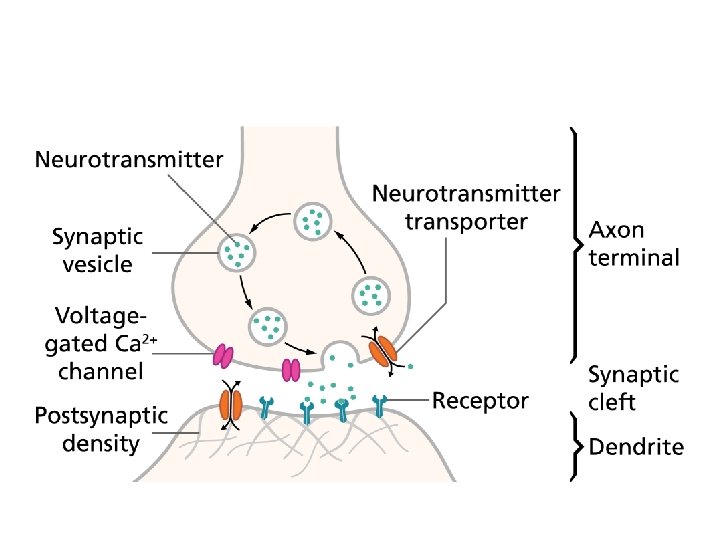
Synapses • Communication from one neuron to another is done chemically. • Synapse is the gap between the terminal end of one and the dendrite of another. • The chemical is called a neurotransmitter and is released from the terminal buttons of a neuron. • Allows the continuation of an impulse when it is received by the dendrite of another neuron.

• Terminal buttons contain vesicles filled with neurotransmitters. There are many, but a very common one is acetylcholine. • When an action potential reaches a terminal button, the following occurs: 1. Action potential causes calcium ions to diffuse into buttons. 2. Vesicles of neurotransmitter fuse with plasma membrane and release neurotransmitter.

3. Neurotransmitter diffuses across synaptic gap 4. Neurotransmitter binds with receptor protein on postsynaptic neuron membrane. 5. Ion channel opens allowing sodium to diffuse into the neuron. 6. This initiates an action potential. 7. The neurotransmitter degrades due to enzymes and is released by receptor protein. 8. The sodium channel closes. 9. Neurotransmitter fragments diffuse back across gap to be reused

Insecticides • Neonicotinoid insecticides similar to nicotine. • Binds to postsynaptic receptors preventing acetylcholine from binding. • They also don’t break down. • Leads to paralysis of insect.

6. 6 Hormones, homeostasis, and reproduction • Homeostasis: The maintaining of internal variables within certain limits. • Examples: blood p. H, blood CO 2 conc. , blood glucose conc. , body temp. , water balance. • All of these variables have a range that is considered normal. • If they get too high or low, the body will do something to try to bring them back into range. This is called a negative feedback loop.

• The nervous and endocrine systems work together to maintain homeostasis. • The endocrine system consists of glands that produce various hormones that travel the body in the bloodstream. • Examples: pituitary gland, pineal gland, hypothalamus, thyroid gland, pancreas, ovaries, testes.

Hormones and their functions • Each hormone has a specific gland the secretes it into the bloodstream. • Hot all body cells are influenced by any one hormone. • The cells that are influenced are called the hormones target cells or tissues.

Thyroxin • Thyroxin – produced by the thyroid gland found in two forms called T 3 and T 4. • Created from an amino acid and iodine. • T 3 has 3 iodine atoms and T 4 has 4 iodine atoms. • Target cells are most cells in the body. • In the cells, T 4 is converted to T 3. • T 3 enters the nucleus where it increases the rate of transcription, increasing the cells metabolism and heat. • Too much thyroxin is hyperthyroidism, too little is hypothyroidism.

Leptin • • • Produced by fat tissue. The more fat tissue, the more leptin produced. Target cells are in the hypothalamus. Leptin lowers your appetite. Too skinny, low leptin, increased appetite Too much fat, high leptin, decreased appetite.

Melatonin • Produced by pineal gland in the brain. • Pineal gland helps maintain your internal clock or circadian rhythm. • Low melatonin production during the day, highest at night. • If you alter amount of light and sleep, rhythum is changed (jet lag)

Insulin/glucagon • Produced by the pancreas • Help to control blood glucose levels. • Cells are constantly using glucose for cellular respiration so are lowering blood glucose levels. • Glucose is entering the bloodstream via the villi of the small intestine as a result of digestion. • Because we eat at certain times, there is a natural fluctuation of blood glucose levels. It still must be maintained within certain levels.

Human Reproduction • Human reproduction is necessary to continue the species but it also ensures genetic variability. • Hormones play an important roll in the creation of male vs female, as well as the function of the system. • The structures of the male and female reproductive system are adapted for the production of gametes, and the female system also provides a suitable environment for embryo growth until birth.

Male anatomy
 View")
Side (sagittal) View

Front view

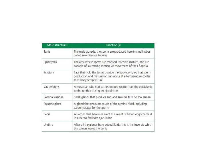
Structure and Function

Male vs female • Depends on the presence of a Y chromosome • We are the same until about 8 weeks • If no Y, the hormones coded for by the X chromosome create estrogen and progesterone, causing female development • Y chromosome codes for Testosterone causing male development. • The same embryonic tissue is involved, homologous structures

Puberty • The same hormones responsible for determination of sex in utero are made in higher amounts. • Causes secondary sex characteristics • Increases estrogen and progesterone in females cause: enlargement of breasts, pubic and arm hair, widening of hips • QUESTION: WHAT PURPOSE IS SURVED?

PUBERTY • Testosterone increase in males causes: facial, pubic, arm, chest hair, large larynx with deeper voice, increased muscle mass, enlargement of the penis. • SAME QUESTION

Menstrual Cycle • A hormone cycle that begins at puberty • Lasts on average, 28 days • Purpose – time the release of an egg for possible fertilization and implantation into uterus. • Implanting needs to happen when endometrium is thick with blood vessels. • If no implantation, endometrium breaks down and leads to bleeding (menstruation)

FSH and LH • Hypothalamus regulates the menstrual cycle by producing gonadotropin-releasing hormone. • Acts on the pituitary gland to release Folliclestimulating hormone (FSH) and Luteinizing hormone (LH). • The target for these 2 hormones is the ovaries.

Effects of FSH and LH • Causes follicle cells to release Estrogen. The target cells of estrogen are the endometrium of the uterus, causing a build up of blood vessels. Estrogen also has positive feedback loop to pituitary to release more FSH and LH. • FSH and LH also cause follicle cells and oocytes to form a Graafian follicle, and eventually cause ovulation or release of the egg.

• After ovulation, the follicle reorganizes into a corpus luteum and begins producing Progesterone. • Corpus Luteum will produce progesterone for 1012 days. It maintains the thick endometrium during this time, awaiting the implantation of the egg. • Progesterone, along with estrogen also provide a negative feedback loop to the hypothalamus to drop FSH and LH so another egg won’t be produced.

• If there isn’t a pregnancy, after 10 -12 days the progesterone and estrogen levels start to fall, causing the vascular endometrium to begin to break down, causing bleeding and the beginning of menstruation. • This drop in estrogen and progesterone triggers the hypothalamus to secrete Gn. RH and begin another cycle.

In vitro fertilization • Natural fertilization usually occurs in the fallopian tubes 24 -48 hours after ovulation. • It takes several days to travel down the tube and reach the uterus. It has divided to become approximately 100 cells and is called a blastocyst. • Things that can go wrong: • Low sperm count, impotence, non ovulation, blocked fallopian tubes

Steps of IVF 1. Females take drugs that shut down he normal cycle and use LH to produce many Graafian follicles. Called superovulation 2. Several eggs are harvested surgically 3. Sperm is collected form male 4. Eggs are mixed with the sperm 5. 1 -3 fertilized eggs are implanted into the female’s uterus. 6. Unused embryos can be frozen and used later if the procedure doesn’t work
- Slides: 108