Topic 1 6 Genetic information is copied and


 Mitosis in plant cells occurs only")


 – PMAT • Chromosomes arrange themselves at the middle or equator of")
 - PMAT • Anaphase is a very rapid stage – the fastest")
 - PMAT • Fastest stage in Mitosis. • The spindle fibres contract")
 - PMAT • Final stage of mitosis. The chromatids have now reached")


- Slides: 47
Topic 1. 6 Genetic information is copied and passed on to daughter cells (cell division)
Assessed outcome – CB pg 117
Assessed outcome – CB pg 118
Cell Division Cell division is the process where a parent cell divides into two daughter cells. There are two types of cell division: Mitosis occurs in body cells (somatic cells). Meiosis occurs in the sex organs and produces gametes (sex cells). Sperm Ovum (egg) Meiosis (meiotic division) produces sex cells or gametes, sperm and ovum (above). The examination of a root tip of an onion plant (left) shows a proportion of the cells are undergoing mitosis (some indicated with arrows).
Eukaryote DNA • In eukaryotes such as plants and animals, the majority of DNA is found in the nucleus and is called nuclear DNA. • But remember the mitochondria and chloroplasts also contain their own DNA.
Video A good video explaining the key terms chromatin, chromosomes and chromatids: • https: //www. khanacademy. org/science/high-school-biology/hsreproduction-and-cell-division/hs-chromosome-structure-andnumbers/v/chromosomes-chromatids-chromatin-etc
Chromatin • In a cell, DNA does not usually exist by itself, but instead associates with specialised proteins that organize it and give it structure. • In eukaryotes, these proteins include the histones, a group of basic (positively charged) proteins that form “bobbins” around which negatively charged DNA can wrap. • In addition to organizing DNA and making it more compact, histones play an important role in determining which genes are active. • So, DNA + histone structural proteins = chromatin.
Chromatin • For most of the life of the cell, chromatin is decondensed, meaning that it exists in long, thin strings that look like squiggles under the microscope. • In this state, the DNA can be accessed relatively easily by cellular machinery (such as transcription protein enzymes that read and copy DNA), which is important in allowing the cell to grow and function. Chromatin
Chromatin Just before a cell divides the chromatin condenses. Condensation takes place when the cell is about to divide. When chromatin condenses, the eukaryotic DNA is not just one long string, instead it becomes shorter and fatter so you can see it as separate, linear pieces called chromosomes. Bacteria also have chromosomes, but their chromosomes are typically circular. DNA replication Chromatin condenses to form chromosomes
Chromosomes Each species has its own characteristic number of chromosomes. For instance, humans, have 46 chromosomes in a typical body cell. Like many species of animals and plants, humans are diploid (2 n), meaning that most of their chromosomes come in matched sets known as homologous pairs. The 46 chromosomes of a human cell are organized into 23 pairs, and the two members of each pair are said to be homologues of one another (with the slight exception of the X and Y chromosomes).
Chromosomes Human sperm and eggs, which have only one homologous chromosome from each pair, are said to be haploid (n). When a sperm and egg fuse, their genetic material combines to form one complete, diploid set of chromosomes. So, for each homologous pair of chromosomes in your genome, one of the homologues comes from your mum and the other from your dad. (Genome = the DNA found in the nucleus)
Chromosomes The two chromosomes in a homologous pair are very similar to one another and have the same size and shape. Most importantly, they carry the same type of genetic information: that is, they have the same genes in the same locations. However, they don't necessarily have the same versions of genes. That's because you may have inherited two different gene versions from your mum and your dad.
Chromosomes and Chromatids • As the cell prepares to divide, it must make a copy of each of its chromosomes. The two copies of a chromosome are called sister chromatids. The sister chromatids are identical to one another (and are attached to each other by proteins called cohesins – term not needed for exam) • The attachment between sister chromatids is tightest at the centromere, a region of DNA that is important for their separation during later stages of cell division. • As long as the sister chromatids are connected at the centromere, they are still considered to be one chromosome. However, as soon as they are pulled apart during cell division, each is considered a separate chromosome. chromatin condensation
NB. The genes are located in rows on the chromosome Homo – same Logous - relation A pair of homologous chromosomes (homologous pairs)
Homologous chromosomes
Mitosis and the Cell Cycle – pg 120 In Mitosis a cell will divide once to produce two daughter cells that are genetically identical to the parent cell. They have the diploid (2 n) number of chromosomes (GCSE). Dividing cells undergo a regular pattern of events known as the cell cycle (IPMAT). This is a continuous process but for ease of teaching it is subdivided into different stages for one complete cell division. Metaphase Mitosis inesis Prophase cytok Interphase (‘resting stage’) Mitosis (nuclear division – 4 stages (PMAT) Anaphase Telophase Interphase Cytokinesis (division of the cytoplasm)
The Cell Cycle The cell cycle can be thought of as the life cycle of a cell. In other words, it is the series of growth and development steps a cell undergoes between its “birth”—formation by the division of a mother cell—and reproduction—division to make two new daughter cells. Stages of the cell cycle To divide, a cell must complete several important tasks: it must grow, copy its genetic material (DNA), and physically split into two daughter cells. Cells perform these tasks in an organized, predictable series of steps that make up the cell cycle. The cell cycle is a cycle, rather than a linear pathway, because at the end of each go-round, the two daughter cells can start the exact same process over again from the beginning.
The Cell Cycle In eukaryotic cells, or cells with a nucleus, the stages of the cell cycle are divided into two major phases: interphase and the mitotic (M) phase. • During interphase (G₁, S, and G₂ phases): the cell grows and makes a copy of its DNA. • During the mitotic (M) phase: the cell separates its DNA into two sets and cytokinesis (C) divides its cytoplasm, forming two new cells. DNA
Let’s enter the cell cycle just as a cell forms, by division of its mother cell. What must this newborn cell do next if it wants to go on and divide itself? It must prepare itself for division and this happens in three steps: G 1 - Growth 1 - the cell increases in size because it makes copies of organelles and the molecular building blocks it will need in later steps. (DO NOT say the cell grows – it increases in size) In S phase – Synthesis - the cell replicates the DNA in its nucleus. The chromosome DNA number stays the same but the DNA content doubles. In G 2 - Growth 2 - the cell increases in size even more, synthesising organelles, proteins, ribosomal material and ATP. The cell begins to reorganise its contents in preparation for mitosis. G 2 ends when mitosis begins. G 1, S and G 2 together are known as interphase. The prefix inter- means between, reflecting that interphase takes place between one mitotic (M) phase and the next.
Interphase CB pg 121 Interphase is not a part of mitosis, but plays an essential role in the cell cycle. It is often called the resting phase but the cell is NOT resting! During interphase the following occurs: Replication of DNA (semi-conservative). Replication of organelles which have their own DNA – mitochondria and chloroplasts. Making new organelles (replication is not acceptable here, only organelles with DNA can be replicated). Synthesis of ribosomal material. Synthesis of ATP. Synthesis of proteins. Increase in cell size (not growth).
Interphase CB pg 121 Top tip - The image below shows a cell during interphase. The DNA is found as chromatin - the chromosomes have not condensed yet and the chromatids are not visible. The nucleolus and nuclear membrane are clearly visible. Top tip - At any given time most cells (80%) will be in interphase as it is the longest part of the cell cycle. In plants mitosis only takes place in the meristems – tip of the root, tip of the shoot, buds and tree rings. See next slide DNA
Plant Root Tip (diagram not explicit on spec) Mitosis in plant cells occurs only in regions of meristematic tissue located at the root tip, shoot tip and buds. In contrast, mitosis can occur throughout the body of a growing animal. Zone of specialization Root tip growing in this direction Zone of elongation Zone of cell division Meristematic tissue (area of cell division) Root cap
After Interphase comes the Mitotic Phase where the cell divides its copied DNA and cytoplasm to make two new cells. The Mitotic Phase has two distinct division-related processes: mitosis and cytokinesis DNA
Mitosis In mitosis, the nuclear DNA of the cell condenses into visible chromosomes and is pulled apart by the mitotic spindle, a specialized structure made out of microtubules. Mitosis takes place in four stages (PMAT): prophase, metaphase, anaphase and telophase. DNA
Cytokinesis In cytokinesis, the cytoplasm of the cell is split in two, making two new cells. Cytokinesis usually begins just as mitosis is ending, with a little overlap. Importantly, cytokinesis takes place differently in animal and plant cells. DNA
Mitotis – PMAT …. .
Prophase - PMAT • First stage of mitosis. • Longest stage of mitosis. • Chromatin condenses (becomes shorter and thicker) forming chromosomes. Chromatids joined by the centromere become visible. • In animal cells the centrioles move to opposite poles of the cell. Protein microtubules form from each centriole and the spindle develops, extending from pole to pole. Nuclear membrane disintegrates • Towards the end of prophase the nuclear membrane and the nucleolus disintegrate. Pairs of chromatids can clearly be seen lying free in the cytoplasm. centrioles
Metaphase (middle) – PMAT • Chromosomes arrange themselves at the middle or equator of the cell. • Spindle fibres attach to the chromosomes at the centromere.
Anaphase (Apart) - PMAT • Anaphase is a very rapid stage – the fastest in Mitosis. • The spindle fibres contract and this splits the centromere, pulling the (sister) chromatids apart to opposite poles of the cell. The chromatids separate and are pulled to opposite poles of the cell; the centromeres lead the way.
Anaphase (Apart) - PMAT • Fastest stage in Mitosis. • The spindle fibres contract and this splits the centromere, pulling the (sister) chromatids apart to opposite poles of the cell. The chromatids separate and are pulled to opposite poles of the cell; the centromeres lead the way.
Telophase (Two) - PMAT • Final stage of mitosis. The chromatids have now reached the poles of the cells and are referred to as chromosomes again. • The chromosomes decondense and lengthen back into chromatin. • The spindle breaks down. • The nucleolus reappears and the nuclear membrane reforms around each group of chromosomes.
Cytokinesis CB pg 123 Cytokinesis is the division of the cytoplasm. It is different in plants and animals cells. • In animal cells cytokinesis occurs by the parent cell membrane pinching the cell into two from the outside inwards, forming a cleavage furrow. • In plant cells the cell wall prevents a cleavage furrow forming. Instead, a cell plate forms across the equator of the parent cell from the centre outwards until the cell is divided in two. After cytokinesis the two cells are now separate cells in their own right. Nucleus Cell plate
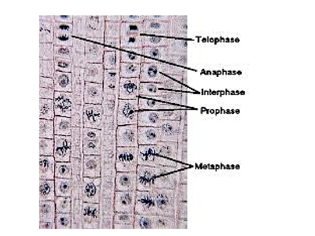
Images of cells undergoing Mitosis – pg 124 Interphase Telophase Prophase Metaphase Anaphase
Images of cells undergoing Mitosis – pg 124
Alternative video: https: //www. youtube. com/watch? v=xsr. H 050 wn. IA
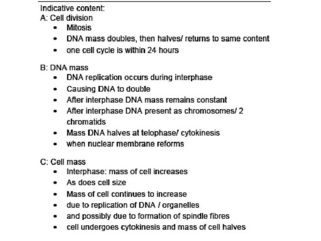
Changes in DNA content and the importance of mitosis – CB pg 125 The number of chromosomes remains constant throughout the cell cycle, but the amount of DNA present in the cell changes. Look at the cell cycle and the table below: • The DNA content doubles in the S phase when the DNA is replicated to ensure the new daughter cells have correct DNA content after cytokinesis. • The DNA content halves during cytokinesis (C) when the cytoplasm and cell splits into two
Changes in DNA content – pg 166 At the end of mitosis two genetically identical daughter cells are produced; genetically identical or clones of the parent cell. The graph below shows ONE complete cell cycle (error in your book – says two!): S G 2 Cyto Interphase Mitosis G 1 Look at the graph above: The time periods A and B represent interphase. Time period C is mitosis and Time period D is cytokinesis. During time period B on the graph, the DNA content of the cell has doubled due to replication. During time period D the DNA content is halved after cytokinesis. You must be able to interpret a graph like this in the exam.
June 2017 WJEC U 1
The importance of mitosis – pg 126 Mitosis is essential for growth, the repair of tissues and the replacement of dead or worn out cells. Asexual reproduction takes place by mitosis. Offspring produced asexually are genetically identical to the parent. An advantage of asexual reproduction is the ability to increase in numbers quickly to take advantage of an ideal environment. The disadvantage is the lack of genetic variation, leading to an inability to adapt if the environment changes. Top tip – Remember mitosis maintains the diploid chromosome number. The daughter cells are genetically identical to the parent cell. Each parent cell produces two new daughter cells.
Calculating the mitotic index – pg 127/8 The mitotic index is the percentage of cells in mitosis. For any sample it can be calculated using the equation above.
Calculating the number of cells at a particular stage – pg 128 You will also be expected to calculate the proportion of cells at a particular stage. You should have prepared onion or garlic root tip squashes in order to calculate these for yourself. An example is given below: If a preparation of a root tip meristem has 40 cells with 36 in interphase the calculation is as follows … Proportion of cell cycle spent in interphase = 36 ÷ 40 x 100 = 90% Therefore the proportion of the cell cycle spent in mitosis and cytokinesis = (100 – 90) = 10% You may be asked to estimate the time taken by a stage of the cell cycle. If 90% of the cell cycle is spent in interphase, and the cell cycle takes 24 hours: Time spent in interphase = 90 ÷ 100 x 24 = 21. 6 hours
Calculating the length of a stage in the cell cycle Let's say takes a particular cell type 24 hours to complete a cell cycle. Count the number of cells from a slide that are in a particular stage e. g. there are 20 cells out of 200 in prophase. Therefore 20 x 100 = 10% 200 10% of 24 hours = 2. 4 hours This means 2. 4 hours of the cell cycle is spent in prophase Calculate the length of time spent during telophase for a cell with a cycle that lasts 80 hours. A slide for these cells had 240 cells and 40 of them were seen to be in telophase.
Calculating the length of a stage in the cell cycle 40 x 100 = 16. 67% 240 80 x 16. 67 = 13. 34 hours spent in telophase out of 80 hours 100 Calculate the length of time spent during telophase for a cell with a cycle that lasts 80 hours. A slide for these cells had 240 cells and 40 of them were seen to be in telophase.
Mitosis and cancer – pg 129 Cancers are the result of uncontrolled mitosis. Cancerous cells divide repeatedly, out of control, with the formation of a tumour. A tumour is an irregular mass of cells; tumours prevent the normal function of body organs. Cancers are thought to be initiated when mutations (changes) occur in the genes that control cell division.
The importance of Mitosis – pg 130 Daughter cells from Mitosis are genetically identical this leads to genetic stability. Mitosis is associated with the growth and repair of somatic cells in the body. Mitosis provides for asexual reproduction e. g. bacteria. Comparing Mitosis in plants and animal cells