Tooth Development Odontogenesis Dentition Primary dentition develops during
")


 – cuboidal shape –")


- Slides: 29
Tooth Development (Odontogenesis)
Dentition • Primary dentition – develops during prenatal period – 20 teeth • Permanent dentition – develops as the jaw grows and matures – 32 teeth • period in between – during the preteen years – mixed dentition period
THERE ARE MULTIPLE STAGES IN TOOTH DEVELOPMENT – but three major ones actually • initiation stage – 6 th to 7 th week • bud stage – 8 th week • cap stage – 9 th to 10 th weeks • bell stage – 11 th to 12 th weeks • apposition stage – vaires per tooth • maturation stage – varies per tooth
Tooth formation • first signs of formation – day 11 • thickening of the epithelium where tooth formation will occur on the 1 st branchial arch • signals – earliest mesenchymal markers for tooth formation are the Lim-homeobox genes (Lhx-6 and Lhx 7) – expressed as early as day 9 in the neural crest cells of the tooth region • positions of the teeth are controlled by signals from the oral epithelium – role of FGF-8 and Pax-9 – determine the position of the tooth germs • more than 90 genes have been identified in the oral epithelium, dental epithelium and dental mesenchyme!! so exact signaling mechanisms remain unclear
Tooth formation: Initial stages -involves the physiologic process of induction -induction of ectodermal tissues by the developing mesenchyme -mechanisms remain unknown -at the 6 th week the stomatodeum is lined with ectoderm – outer portion is the oral epithelium -this gives rise to the primary epithelial bands -also is a developing mesenchyme which contains neural crest cells that have migrated to the area -a basement membrane separates the developing oral epithelium and mesenchyme Primary epithelial bands: Horseshoe-shaped bands that appear approximately around the 37 th day of development, one for each jaw. -there are two subdivisions: vestibular lamina and dental lamina -the dental lamina develops a series of epithelial outgrowths - grow deep into the mesenchyme -develops in the future spot for the dental arches -will form the midline for these arches -arches then form posteriorly from this point -the ingrowths represent the future sites for each deciduous tooth -the vestibular lamina cells rapidly enlarge and then degenerate – forms a cleft that becomes the vestibule of the oral cavity The initiation of tooth formation starts around the 37 th day of gestation.
Bud Stage • • • marked by the incursion of epithelium into the mesenchyme period of extensive proliferation and growth of the dental lamina forms into buds or oral masses that penetrate into the mesenchyme each tooth bud is surrounded by the mesenchyme buds + mesenchyme develop into the tooth germ and the associated tissues of the tooth this developing tooth forms from both the ectoderm and mesenchyme and from neural crest cells that have migrated into the mesenchyme 1. Tooth bud 2. Oral epithelium 3. Mesenchyme
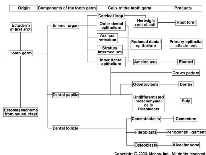
Cap Stage • • • characterized by continuation of the ingrowth of the oral epithelium into the mesenchyme. tooth bud of the dental lamina proliferates unequally in different parts of the bud – forms a cap shaped tissue attached to the remaining dental lamina – looks like a cap sitting on a ball of condensing mesenchyme occurs for the primary dentition (during the fetal period) this stage marks the beginning of histodifferentiation (differentiation of tissues) the tooth germ also begins to take on form – start of morphodifferentiation a depression forms in the deepest part of each tooth bud and forms the cap or enamel organ (or dental organ) – produces the future enamel (ectodermal origin) below this cap is a condensing mass of mesenchyme – dental papilla – produces the future dentin and pulp tissue (mesenchymal origin) the basement membrane separating the dental organ and the dental papilla becomes the future site for the dentinoenamel junction (DEJ) remaining mesenchyme surrounds the dental/enamel organ and condenses to form the dental sac or the dental follicle
Cap stage • -together the enamel organ + dental papilla + dental follicle is considered the developing tooth germ or tooth primordium • -these primordium will be housed in the developing dental arches and will develop into the primary dentition
Bell Stage • • • Continuation of histodifferentiation and morphodifferentiation cap shape then assumes a more bell-like shape differentiation produces four types of cells within the enamel/dental organ – – • • 1. inner enamel epithelium 2. outer enamel epithelium 3. stellate reticulum 4. stratum intermedium during the bell stage the dental lamina is separated from the dental organ the dental papilla undergoes differentiation and produces two types of cells – 1. outer cells of the DP – forms the dentin-secreting cells (odontoblasts) – 2. central cells of the DP – forms the primordium of the pulp • dental sac increases its collagen content and differentiates at a later stage than the EO and DP
Differentiation of the Enamel/Dental organ • outer enamel epithelium (OEE) – cuboidal shape – – • – – – short, columnar cells differentiates into the enamel secreting cells = ameloblasts separated from the dental papilla below it by a basement membrane also cells accumulate large amounts of glycogen may also be called the inner dental epithelium the IEE and OEE are continuous region where they connect – curved rim of the EO = cervical loop stellate reticulum – – • OEE inner enamel epithelium (IEE) – – • protective barrier during enamel production may also be called the outer dental epithelium very little cytoplasm cells are separated from the dental follicle by a basement membrane star-shaped cells in many layers center of the enamel organ forms a network = reticulum supports production of enamel stratum intermedium – – – inner layer of compressed flat to cuboidal cells very high levels of the enzyme alkaline phosphatase supports production of enamel IEE cervical loop
Bell Stage -the cells in the center of the enamel organ begin to synthesize and secrete GAGs -this pulls water into the EO -increasing amount of fluid in the EO forces the central cells apart -however, they remain connected via cellular processes which makes them star shaped = stellate ret. B = inner dental epithelium (inner enamel epithelium)
Bell stage – early crown formation • the dental papilla is separated from the enamel organ by a basement membrane • immediately below this BM is a region called the acellular zone • this is where the first enamel proteins will be laid down • the dental lamina begins to break up into discrete islands of epithelial cells (epithelial pearls)– separates the oral epithelium from the developing tooth • these pearls may form cysts and delay eruption or they may develop into supernumerary teeth • the IEE completes its folding and you can begin to identify the shape of the future crown pattern
Tooth development so far
Cap and Bell stages & Permanent teeth • • • during the cap stage the development of the permanent dentition begins – anterior teeth the primordia for these teeth appears as an extension off the developing dental lamina penetrates into the mesenchyme lingual to the primary primordium its site of origin is called the succesional dental lamina these permanent teeth are called succedaneous teeth (anterior teeth and the premolars) – teeth that form with the primary tooth buds (primary predecessors) – permanent molars are nonsuccedaneous - they are formed by posterior proliferation of the dental lamina.
Appositional stage • secretion of enamel, dentin and cementum • these tissues are initially secreted as a matrix that is partially calcified – serves as a framework for later calcification • time period varies • multiple inductions occur between the ectodermal tissues of the enamel organ and the mesenchymal tissues of the dental papillae and dental sac – these inductions are crucial for the production of enamel, dentin and cementum – these interactions are mediated by the basement membrane found in between these ectodermal and mesenchymal tissues
Maturation stage • characterized by the completion of calcification
• Ameloblasts and Odontoblasts ameloblasts – the cells of the IEE assume a more columnar shape or they elongate – differentiate into pre-ameloblasts – this differentiation is characterized by the repolarization of these PAs – movement of the nucleus away from the basement membrane – this repolarization is critical to the differentiation of the PAs – continued differentiation and maturation results in the formation of ameloblasts – the pre-ABs induce the cells of the dental papilla to differentiate also • odontoblasts – differentiation by the mesenchyme of the dental papilla – occurs after differentiation of pre-ABs begins – results because the pre-ABs induce differentiation of the mesenchymal cells also – also undergo repolarization – mirror image of the pre-ABs (see Figure 6 -12 and 6 -13) – after differentiation – the ODs then start dentinogenesis • • • begin to deposit predentin on the side of their basement membrane – forms a layer immediately below the BM and above the cells (figure 6 -13) therefore dentin formation begins before enamel synthesis explains why dentin is thicker than enamel
• • At 1 the epithelium is separated from the dental papilla by an acellular zone. At 2 the cells of the inner dental epithelium have elongated, and the acellular zone begins to be eliminated as odontoblasts differentiate from ectomesenchymal cells in the tooth pulp. At 3 the odontoblasts retreat toward the center of the pulp, leaving behind formed dentin. At 4 the cells of the inner dental epithelium, now ameloblasts, begin to migrate outward and leave behind formed enamel. before dentin forms – cells of the EO receive blood supply from vessels of the dental lamina as dentin forms, it cuts of this papillary source of blood/nutrients this causes a drastic reduction in the amount of nutrients that reach the EO but the ABs require extensive nutrients to form enamel – stellate reticulum collapses and invagination of the OEE – this brings in blood supply from peripheral vessels found outside the tooth
Dentinoenamel junction • • • after OD differentiation and the initiation of dentinogenesis – the BM between the pre-ABs and ODs disintegrates this allows direct contact between the pre-ABs and ODs – results in the completion of pre-AB differentiation to mature ABs then begin amelogenesis – apposition of enamel matrix replaces the disintegrating BM each ameloblast forms a tapered portion that faces the disintegrating BM - called a tome or Tome’s process upon contact of the enamel matrix and dentin – the disintegrating BM begins to mineralize – forms the dentinoenamel junction or DEJ the ODs form cellular process as they retreat toward the dental papilla (figure 6 -14) that penetrate the forming predentin = dentinal tubules mineralization of the developing dentin and enamel is distinct for each type of tissue the cell bodies of the ODs remain in the pulp tissue the cell bodies of the ABs participate in tooth eruption and will disappear shortly after
Timetable for tooth development • • • Entire primary dentition initiated between 6 and 8 weeks of embryonic development. Successional permanent teeth initiated between 20 th week in utero and 10 th month after birth permanent molars between 20 th week in utero (first molar) and 5 th year of life (third molar)
ROOT FORMATION • • • takes place as the crown is completely shaped and the tooth begins to erupt therefore the tooth forms from the “top down” – i. e. crown to root formation is through the formation of a cervical loop the CL is the most cervical portion of the enamel/dental organ – two layers consisting of IEE and OEE the CL begins to grow down into the dental sac – elongates and moves away from the dental papilla it forms a Hertwig's root sheath rim of the sheath = epithelial diaphragm – encloses the developing primary alveolar foramen also grows down to encompass all but the basal portion of the pulp this sheath shapes the root and induces dentin formation in the root area by the ODs of the dental papilla – • • – ensures that it is continuous with the crown dentin this sheath lacks the stellate reticulum and stratum intermedium is capable of differentiating into ODs BUT NOT ABs A, The root is beginning to form as an extension of the inner and outer dental epithelia in the cervical loop region (circles) which form a bilayered structure called Hertwig’s epithelial root sheath. B. formation of dentin by odontoblasts above the root sheath
Root Dentin • The root of the tooth is composed by dentin and cementum • dentin forms when the outer cells of the dental papilla are induced to differentiation into ODs • similar to what occurs at the crown area • influenced by the IEE of Hertwig’s root sheath • the ODs then undergo dentinogenesis and secrete predentin • after dentin formation – the BM disintegrates along with the Hertwig’s sheath
Cementum and Pulp formation • cementogenesis in the root area also occurs upon degradation of the H. root sheath • the degradation allows contact of the dental sac cells with the dentin surface – induces the formation of cementoblast cells • the CBs cover the root dentin and undergo cementogenesis – laying down cementoid • the CBs do NOT leave cellular processes within the cementum but many CBs become entrapped in the forming cementum • these entrapped CBs are called cementocytes • only upon mineralization of the cementoid can it be called cementum • the region of contact between cementum and root dentin = dentinocemental junction or DCJ • while the cementum is forming - the central cells of the dental papilla form the pulp (Chapter 13)
Periodontal ligament • • • the surrounding tissues of the tooth also develop as the crown and root form the mesenchyme of the dental sac condenses to form the periodontal ligament forms adjacent to the new cementum involves synthesis of collagen and bundling into fibers ends of these fibers insert into the outer layer of cementum and surrounding alveolar bone the cells of the disintegrating H. root sheath develop into discrete islands of epithelial cells – – become epithelial rests of Malassez (figure 6 -22) cells become located in the mature periodontal ligament no known function they can be identified in the periodontal ligament and are responsible for the development of radicular cysts.
Multirooted teeth • anterior teeth, premolars and molars all begin as a single root – root trunk • root of the posterior teeth divides from the trunk into the correct number of root branches • differential growth of the H. root sheath results in the division of the root trunk into two or three roots
Primary tooth eruption • • eruption takes place in chronological order involves active eruption – vertical movement of the tooth as opposed to passive eruption – receding of the gingiva how it occurs is not understood – root growth, hormonal action, contractile collagen, vascular pressure? ? • several stages – after enamel apposition into the crown area of each primary and permanent tooth, the ABs place an acellular dental cuticle on the new enamel’s surface – as the tooth erupts it is still covered with a layer of amelobasts and the remaining layers of the enamel organ – these layers become compressed as the tooth moves – forming a reduced enamel epithelium (REE) or reduced dental epithelium – the REE fuses with the oral epithelium lining the oral cavity (figure 6 -28) – the REE produces enzymes which disintegrates the central portion of this fused tissue – results in an epithelial tunnel through which the tooth erupts – this disintegration results in an inflammatory response – interpreted as the teething response – as the tooth erupts, the portion of the epithelium covering the crown pulls back and exposes the
Primary tooth eruption -the epithelium remains attached to the tooth at the cervical portion -i. e. as the tooth pierces the oral epithelium, the cells of the reduced dental epithelium and the oral epithelium form the initial junctional epithelium (thin dotted line) -this will eventually form the dentogingival junction
Permanent tooth eruption • as the succedaneous permanent teeth develop below the primary teeth, the primary tooth is exfoliated • eruption is lingual to the roots of the primary teeth – exception is the maxillary incisors which move to a more facial position as they erupt • eruption process is the same for the primary teeth – formation of an epithelium lined tunnel for eruption • the process is also similar for the nonsuccedaneous teeth except no primary tooth is shed