TOOTH dens dentis odus odonotos Teeth Dentes arcus

 • arcus dentalis superior (maxillaris) – ellipse • arcus dentalis inferior (mandibularis)")
 neck (cervix) root (radix) pulp (pulpa)")
 • lingualis (lower teeth) palatinalis (upper")
 = dentoalveolar joint • located in bony alveolus")
 •")

")


 – 1 -4 quadrants (from")
 • enamel – enamelum (substantia")
")


 • mucosa attached to periosteum • stratified nonkeratinizing squamous epithelium • papilla")
 • linea mucogingivalis • gingiva alveolaris (pars affixa gingivae) – pink, stippled,")
 • oral ectoderm • mesoderm • cells of neural crest ectomesenchyme")
 appears – thickening of stomodeum")
 – local thickening of epithelium,")
 – outer")
 – derived from mesenchyme cells")
 – from inner enamel epithelium")
 =")


- Slides: 39
TOOTH dens, dentis odus, odonotos
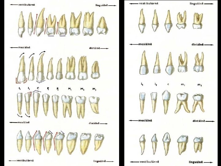
Teeth (Dentes) • arcus dentalis superior (maxillaris) – ellipse • arcus dentalis inferior (mandibularis) – parabola • permanent teeth (dentes permanentes) – 32 • deciduous teeth (dentes decidui) – 20 dens incisivus (= incisor tooth) 8/8 dens caninus (= canine tooth) 4/4 dens premolaris (= premolar tooth) 8/0 dens molaris (= molar tooth) 12/8
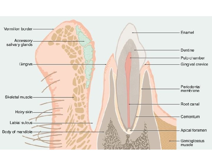
Teeth – parts • • crown (corona) neck (cervix) root (radix) pulp (pulpa)
Surfaces and directions • occlusalis • vestibularis (buccalis/labialis) • lingualis (lower teeth) palatinalis (upper teeth) • mesialis • distalis
Teeth – fixation • gomphosis (socket) = dentoalveolar joint • located in bony alveolus dentalis of jaw • periodontium • parodontium = all structures around tooth
Peridontium • between tooth and dental alveolus (fixed to the bone of alveolus) • collagen fibers (serve as alveolar periosteum) • fixation apparatus of tooth = fibers of various directions • penetrates into cement • rapid change of connective tissue, plasticity – orthodontics • atrophy in lack of proteins and vitamin C → scurvy (= skorbut)
Periodontium
Scurvy (scorbut)
Macroscopy of tooth and its fixation
Denture as a whole • • mordex = denture orthodental position – teeth vertically occlusion (occlusio) 80 % psalidodontia (scissors-like occlusion) = norm – progenia = lower jaw longer (lower teeth in front of upper ones) – stegodontia = roof-like occlusion – prognathia = upper jaw longer (upper teeth in front of lower ones) – opisthodontia = lower teeth too far behind upper ones – hiatodontia (= mordex apertus)
Dental chart / scheme • crossed with letters – tooth number designed with lower INDEX – lowercase = decicuous – UPPERCASE = PERMANENT • crossed with numbers – Roman numerals = decicuous – Arabic numerals = PERMANENT
Dental chart / scheme of deciduous teeth Dental chart / scheme of permanent teeth
Dental chart / scheme • binumeral (Féderation Dentaire Internationale) – 1 -4 quadrants (from right side above clock-wise) = PERMANENT – 5 -8 (similar) = deciduous • numeral (American Dental Association) – numerals 1 -32 (from right upper third mollar clockwise) = PERMANENT – letters A-T (similar from right upper second molar) = deciduous
Teeth – structure • dentine – dentinum (substantia eburnea) • enamel – enamelum (substantia adamantina) • cement – cementum (substantia ossea) • pulp – pulpa – loose connective tissue, vessels, nerves
Enamel • hardest tissue of body • organic part – secreted by ameloblasts (enameloblastus) – glycoproteins (amelogenins, enamelins) • anorganic part 95% – hydroxyapatite • arranged vertically in prisms (rods) • in between interprismatic substance
Fluoridation • fluorine is sired under the enamel surface, posteruptivelly from saliva and tooth-paste • re-covers defects • fluorapatite is more resistant to acids (Ph 4, 5) and is produced more quickly than hydroxyapatite (p. H 5, 5) • the more fluorapatite is in enamel, the more resistant to dental caries (tooth decay) • supplement: tooth-paste, salt, at dentist
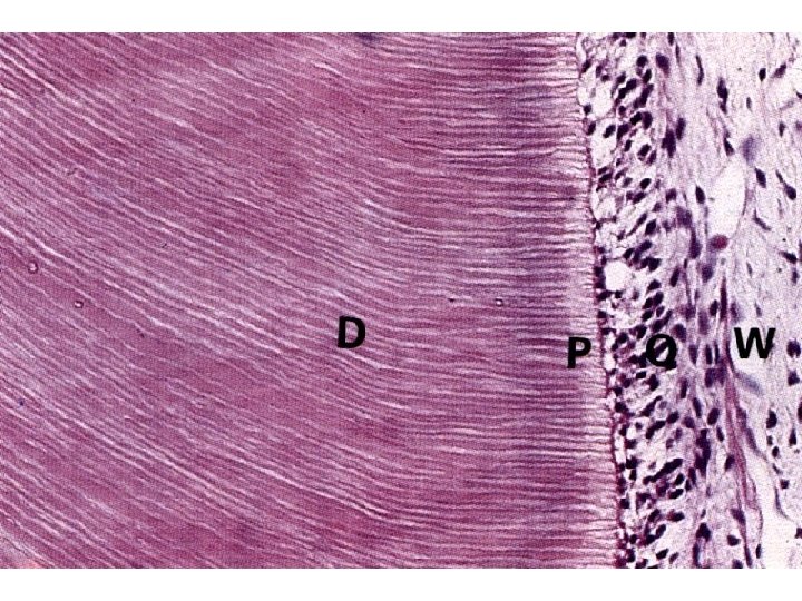
Dentine • calcified connective tissue • organic part – collagen I, proteoglycans – secreted by odontoblasts (dentinoblastus) • located on internal surface of dentine • Tomes fibers (fibrae dentinales) • anorganic part – hydroxyapatite • non-calcified dentine • predentine • close to enamel and cement
Pulp • loose connective tissue – fibroblasts – immune cells soustavy • vessels • nerve fibers (senstitive to pain)
Cement • thin layer at neck thick layer at root • fibrilar type of bone • cellular part – cementocytes
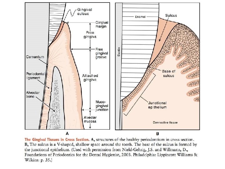
Gum (Gingiva) • mucosa attached to periosteum • stratified nonkeratinizing squamous epithelium • papilla gingivalis • no glands • no submucosa • gingivodental junction
Gum (Gingiva) • linea mucogingivalis • gingiva alveolaris (pars affixa gingivae) – pink, stippled, keratizing • „free gingival groove“ • gingiva marginalis (pars libera gingivae) – shiny, red, nonkeratinizing • sulcus gingivalis • junctio dentogingivalis • epithelium junctionale
Teeth development (Odontogenesis) • oral ectoderm • mesoderm • cells of neural crest ectomesenchyme • enamel is derived from ectoderm • other tissues are derived from ectomesenchyme
Teeth development • Week 6: lamina dentalis (dental lamina) appears – thickening of stomodeum epithelium • in each lamina 10 proliferation centers – dental buds
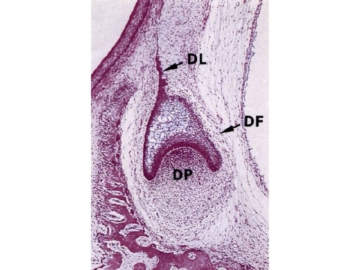
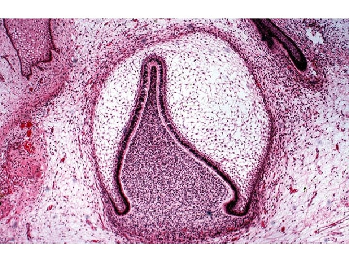
Stages of tooth development • dental bud (status gemmalis) – local thickening of epithelium, 10 in each jaw • dental cap (status galearis) – ectodermal part → enamel organ (organum enameleum) – invagination of mesenchyme → dental papilla
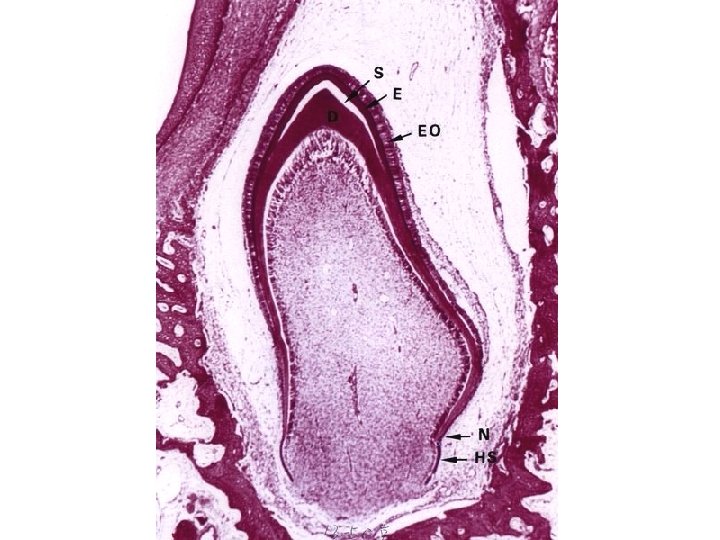
Stages of tooth development • dental cap → dental bell (status campanalis) – outer dental epithelium – enamel reticulum – inner dental epithelium – dental papilla → dental pulp – dental sac → cement, periodontal ligaments
Stages of tooth development – bell • odontoblasts (dentinoblasti) – derived from mesenchyme cells at inner enamel organ – produce (pre)dentine – Tomes fibers (fibrae dentinales) • cytoplasmatic processes left within dentine
Stages of tooth development – bell • ameloblasts (enameloblasti) – from inner enamel epithelium – basal surface becomes secretory – production of enamel
Stages of tooth development – bell • epithelial root sheath (vagina epithelialis radicis) = Hertwig sheath – transition between outer and inner enamel epithelium – ingrowth into mesenchyme and induction of root formation
Tooth eruption • decidual teeth: 6 th – 24 th month • enamel organ disrupted during tooth eruption
Permanent teeth • develop similarily to decidual teeth • secondary dental lamina – located at lingual side of dental lamina – prolonged distally (molars) • eruption from 6 th year • (finished in 30 th – 40 th year)
Clinical note • tetracycline antibiotics are contraindicated in children up to 8 years of age, pregnant and nursing women – high affinity to newly produced enamel – brown-yellow color – enamel hypoplasia