Tools of the Scientist One of the most


- Slides: 33
Tools of the Scientist
• One of the most important tools is the compound microscope. • Used to see the microscopic world or objects too small for the naked eye. • Plant cells, animal cells and bacteria cells can be observed. • Used to diagnose cancer
The compound light microscope • The compound microscope has two sets of lenses, • Each magnifies the what you are looking at
• light to pass through a specimen on the stage to form an image. • It can make objects 1000 times its normal size. light
• The microscope must have two characteristics to be useful: 1. Magnification 2. Resolution
• Magnification: the enlarging of the image so it can be seen easier
How to determine magnification: • Multiply the eye piece by the objective. • For example if the eye piece is 10 X and the objective is 25 X, your total magnification is 250 X. • 10 x 25 =250 x 10 x 25 x
• What is the total magnification of the following microscopes? Ocular: 10 x Ocular: 15 x objective: 40 x objective: 100 x
2. Resolution is the ability to distinguish between two points. • Similar to digital cameras (mega pixels) or LED Televisions. • Good resolution makes images clearer to view. Good resolution Poor resolution
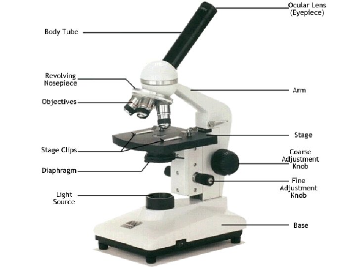
Parts of the microscope. 1. Ocular lens/eye piece: • Contains a magnifying lens, usually 10 x or 15 x.
2. Objective lens: • has three different magnifying lenses: A. Scanning lens: • magnifies image 4 X B. Low power: • magnifies 10 X C. High power: • magnifies 40 x
3. light source 4. diaphragm: regulates amount of light that passes up towards the eye piece.
5. Base: support microscope 6. Arm: supports body tube
7. Stage: supports slide to be observed. 8. Stage clips: Holds slide down on stage
9. Nosepiece: hold objectives, can be rotated. 10. Course Adjustment: moves body tube in order to focus the image.
11. Fine Adjustment: moves body tube slightly to sharpen image. 12. Body tube: keeps proper distance between eye piece and objectives.
• Images viewed under the light microscope are upside down and backwards • This is a compound F light microscope view of the letter F placed on a slide in its normal position. F
Other types of microscopes 1. Transmission electron microscope: • shines a beam of electrons through a thin specimen • Works like a compound light microscope but higher magnification.
mitochondria
2. Scanning Electron Microscope: • produces a 3 D image. • Runs a beam of electrons back and forth across the surface of the specimen. • Magnification of 500, 000 x
Image under scanning electron microscope
Sperm on egg
Red Blood Cell
1. Red blood cells under a compound light microscope 2. Red blood cell in a capillary under a transmission electron microscope 3. Red blood cells under a scanning electron microscope
Other important lab techniques 1. Chromatography 2. Gel electrophoresis
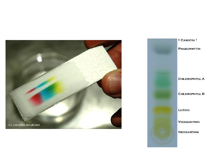
1. • Chromatography A lab technique used to separate mixtures. • Often done on paper • The chemicals separate based on density. • The solvent is absorbed up the paper, • separating the mixtures place on the paper by the scientist. Animation: http: //www 3. wooster. edu/chemis try/analytical/gc/default. html Solvent moves up paper
• How paper chromatography works. • Watch the water move up as it is absorbed by the paper. • Separates the chemicals that make up the black dot as it moves.
2. Electrophoresis • used to separate molecules such as DNA by size and charge • charged molecules like DNA are placed in an electric field, they migrate toward either the positive or negative pole according to their charge.
• Put DNA in the wells, • Plug it in • Separates based on size of molecules and charge • DNA has a negative charge, so its attracted to the positive side. • Smaller pieces move further • Every ones DNA is unique • Everyone's DNA separates in a unique pattern.
• Gel electrophoresis is one way to determine your baby’s momma • Also used to solve crimes Animation: http: //fotofox 17. deviantart. com/art/Gel-Electrophoresis-Animation-87035741
Electrophoresis pics The set up