TOOLS OF LIFE SCIENTISTS Class Notes Unit 1


 Transmission electron microscope – magnifies up to")
")
 MYELINSHEATHED NERVE CELL")


 flu virus (Center for Disease Control & Prevention)")
")
")


")


")


")


 Machines")


- Slides: 54
TOOLS OF LIFE SCIENTISTS Class Notes Unit 1 Lesson 2
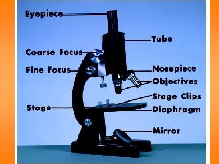
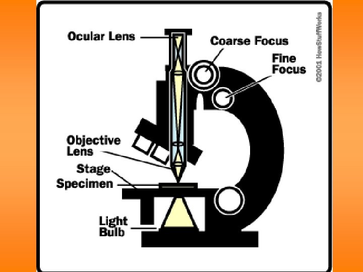
Compound Light Microscope
• Magnifies up to 1000 x • Tube w/ lenses – magnify the object • Stage – holds the specimen • Light – passes through the specimen
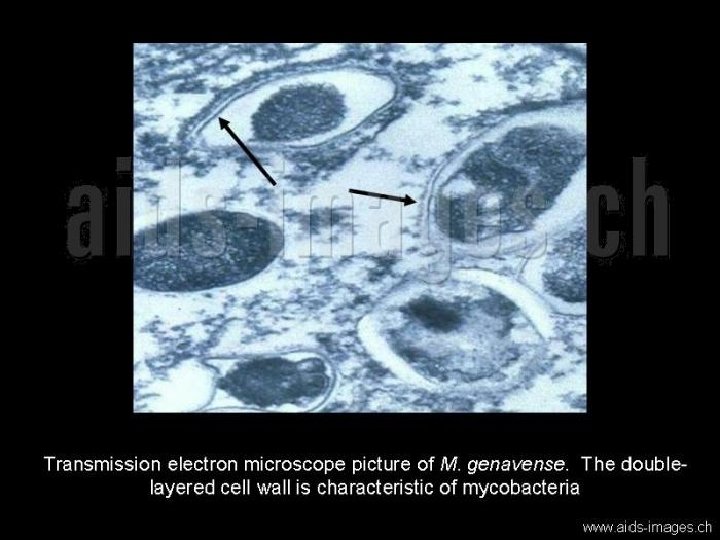
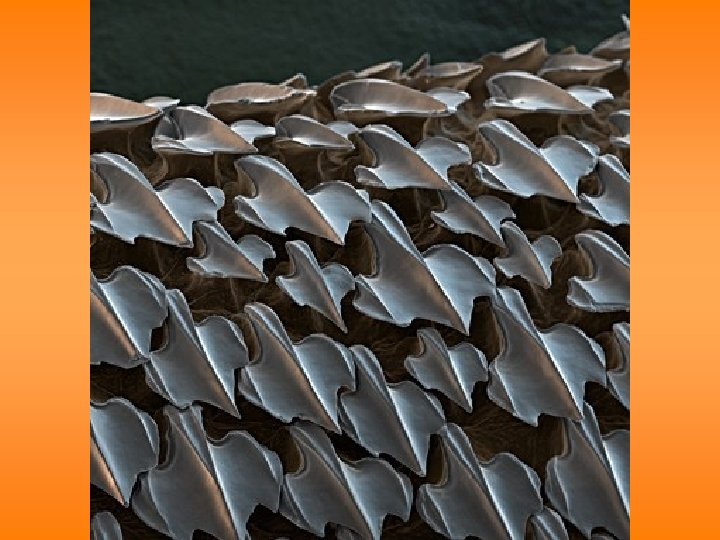
Electron Microscopes • Two types: • (1) Transmission electron microscope – magnifies up to 200, 000 x • e- pass through specimen • Flat (2 -D) image • (2) Scanning electron microscope – magnifies up to 100, 000 x • 3 -D image • e- bounce off surface of specimen • Preparation for viewing kills the specimen
Transmission Electron Microscope (TEM)
TEM images – flat (2 -D) MYELINSHEATHED NERVE CELL
BACTERIA
CELL ORGANELLES
ORGANELLES IN TOBACCO POLLEN GRAIN
H 1 N 1 (swine) flu virus (Center for Disease Control & Prevention)
Scanning Electron Microscope (SEM)
SEM images – 3 -Dimensional! ANTARCTIC MITE (1500 x)
DUST MITE
DUST MITE
FLEA
SNAIL RADULA
PENGUIN FEATHER (1500 x)
POLLEN
MICROBES
RED FIRE ANT
FLY FOOT
MOSQUITO (FALSE COLOR)
BAMBOO MITE
SPIDER
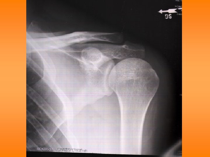
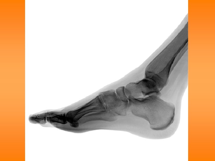
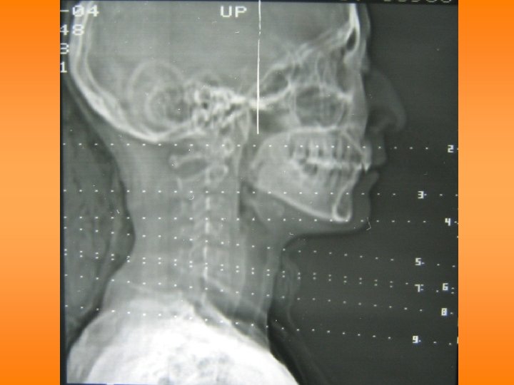
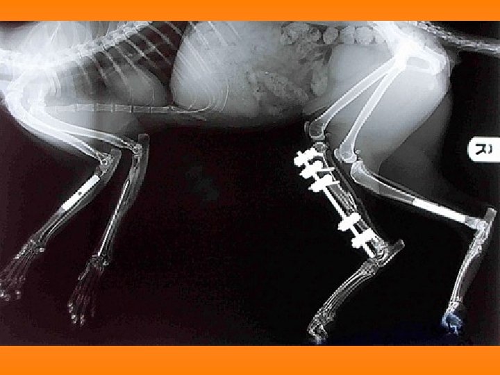
X-ray Machines • X-rays go through soft tissue • Blocked by bones, masses – leave shadows • Detect breaks, abnormalities, tumors, etc.
NORMAL LUNGS
NODULE IN LUNG
AORTIC RUPTURE (BLOOD IS CLOUDINESS ON LEFT SIDE)
A SWORD SWALLOWER’S X-RAY
WRIST REPAIR
OOPS!
OUCH
OUCH
PARTIAL HIP REPLACEMENTS
CAT Scan Machine
• Use X-rays • Clearer image than regular X-rays
CAT SCAN OF BRAIN
DO-ITYOURSELF CAT SCAN
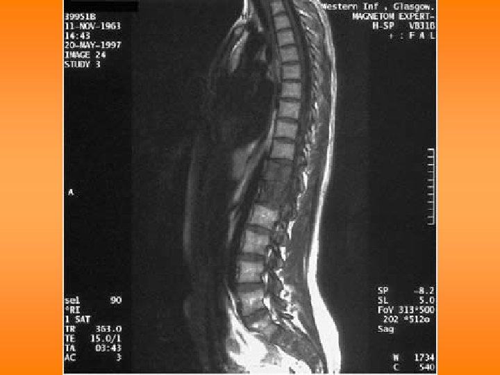
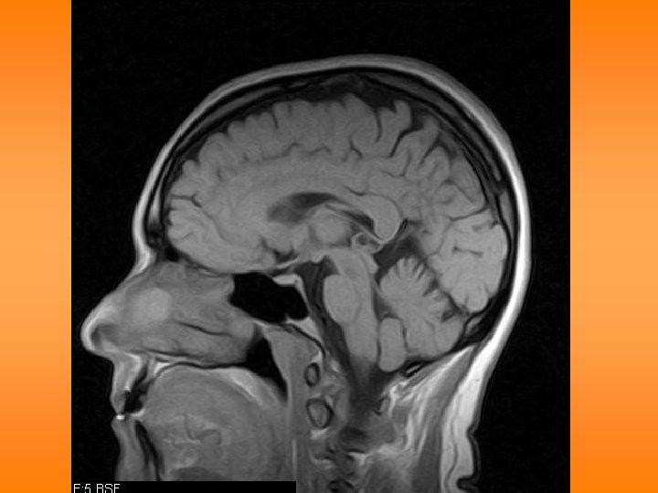
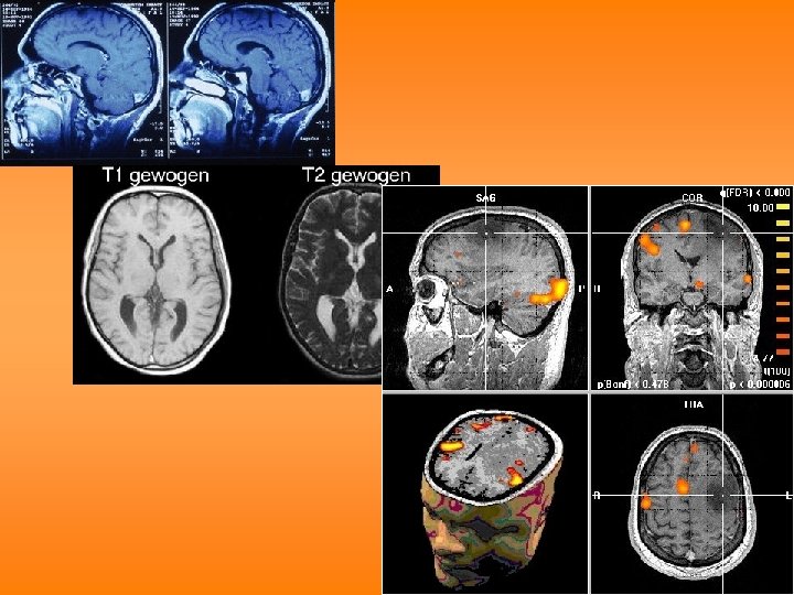
Magnetic Resonance Imaging (MRI) Machines
• Use magnetism, computers to make images
MRI Machine
THE END