
Tools II Voltammetric Detectors 1 2 3 4












- Slides: 92
Tools II Voltammetric Detectors 1. 2. 3. 4. Methods: Stripping, Metals DPV LC EC Rat Brains
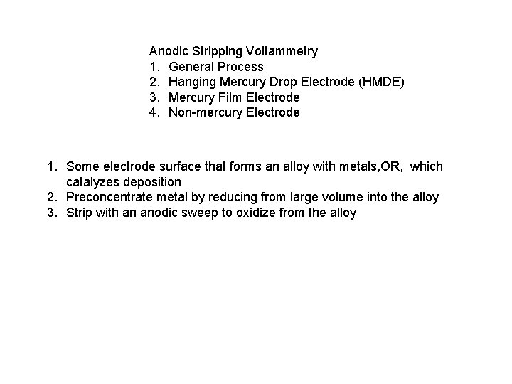
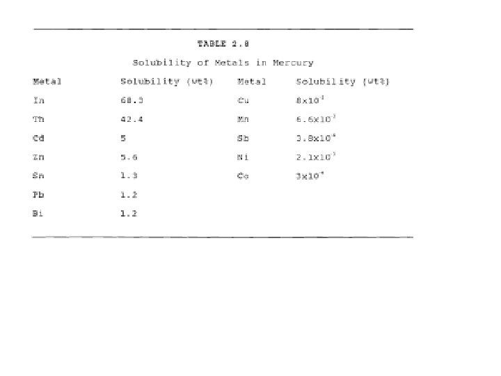
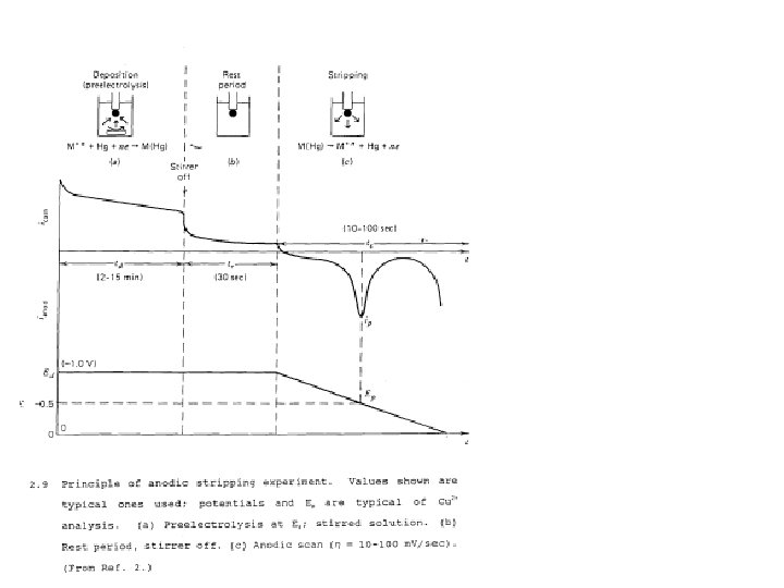
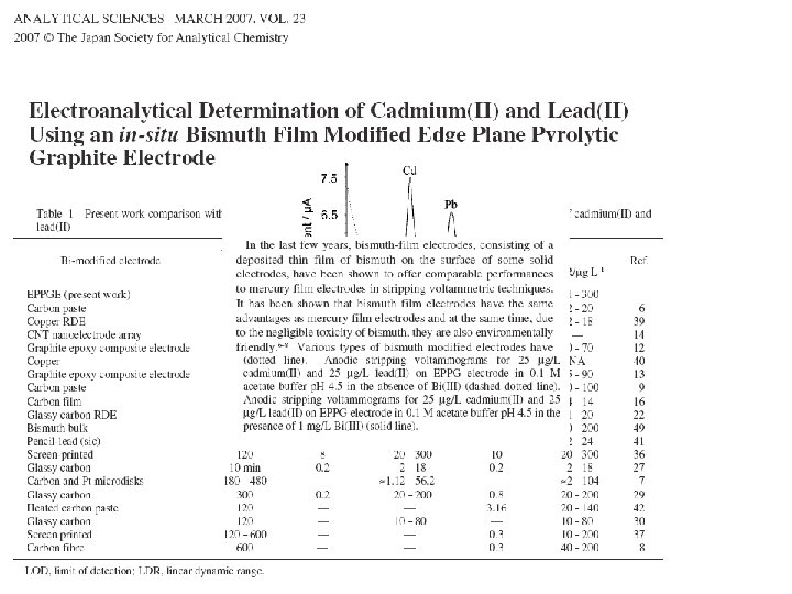
Anodic Stripping Voltammetry 1. General Process 2. Hanging Mercury Drop Electrode (HMDE) 3. Mercury Film Electrode 4. Non-mercury Electrode 1. Some electrode surface that forms an alloy with metals, OR, which catalyzes deposition 2. Preconcentrate metal by reducing from large volume into the alloy 3. Strip with an anodic sweep to oxidize from the alloy
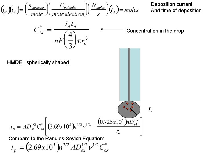
Deposition current And time of deposition Concentration in the drop HMDE, spherically shaped ro Compare to the Randles-Sevich Equation:
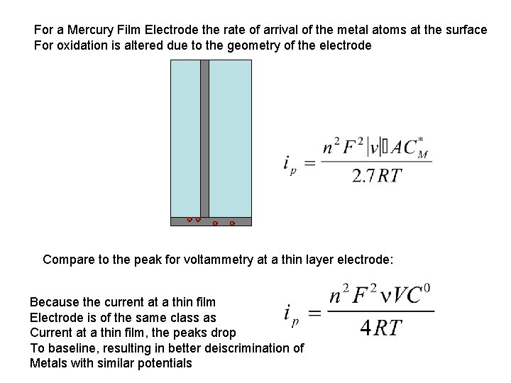
For a Mercury Film Electrode the rate of arrival of the metal atoms at the surface For oxidation is altered due to the geometry of the electrode Compare to the peak for voltammetry at a thin layer electrode: Because the current at a thin film Electrode is of the same class as Current at a thin film, the peaks drop To baseline, resulting in better deiscrimination of Metals with similar potentials
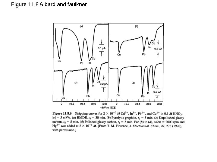
Figure 11. 8. 6 bard and faulkner
Tool II Voltammetric Detectors 1. 2. 3. 4. Methods: Stripping, DPV Metals LC EC Rat Brains
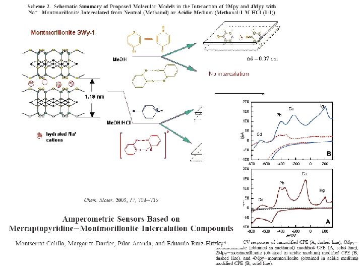
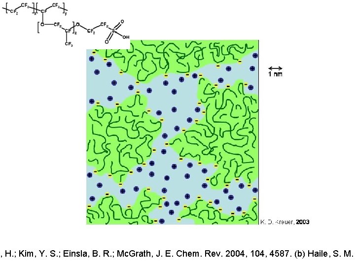
Goal to use the electrstatic attraction for accumpulation Similar goal in using Nafion – also anionic material easily manipulated
i, H. ; Kim, Y. S. ; Einsla, B. R. ; Mc. Grath, J. E. Chem. Rev. 2004, 104, 4587. (b) Haile, S. M.
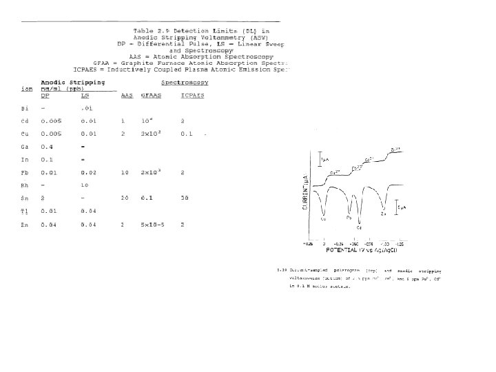
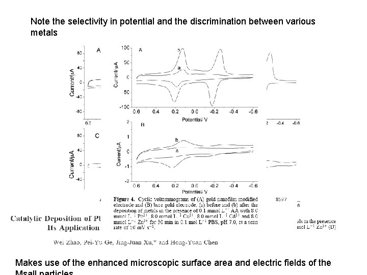
Note the selectivity in potential and the discrimination between various metals Makes use of the enhanced microscopic surface area and electric fields of the
JChem Ed 2007
Tool II Voltammetric Detectors 1. 2. 3. 4. Methods: Stripping, DPV Metals LC EC, LC EC MS Rat Brains
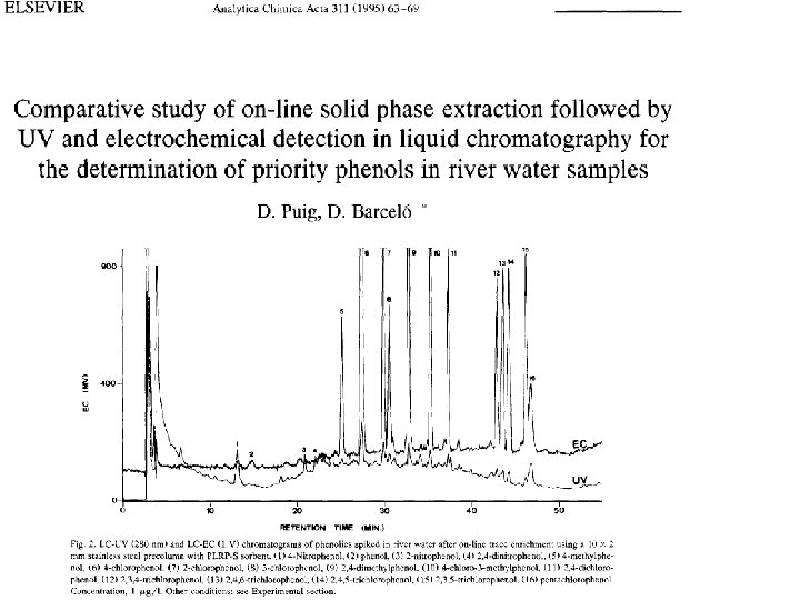
Many organics are easily oxidized Most solid metal electrodes do not oxidize in this range and so have low background current
Many others are easily reduced
All are candidates for voltammetric analysis
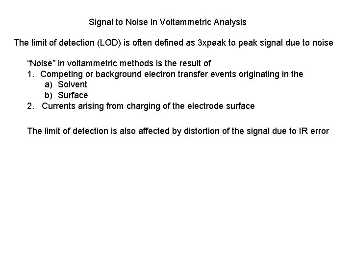
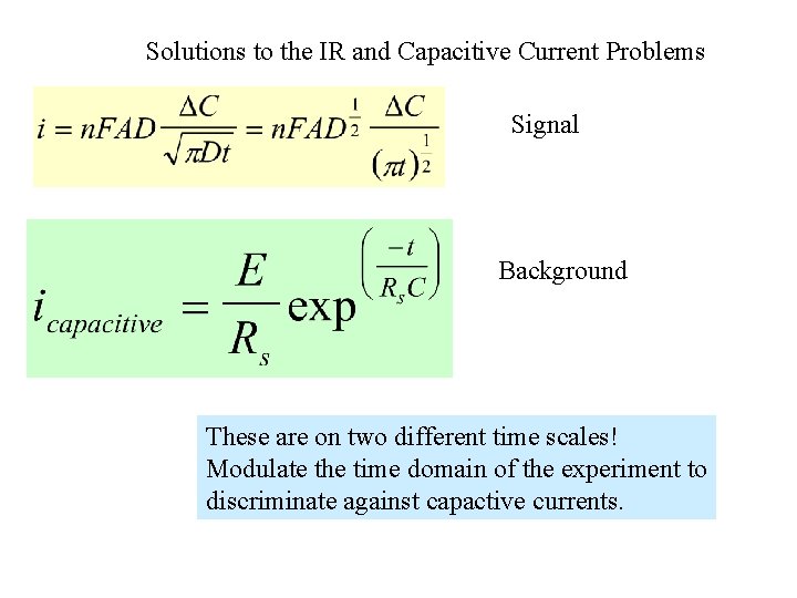
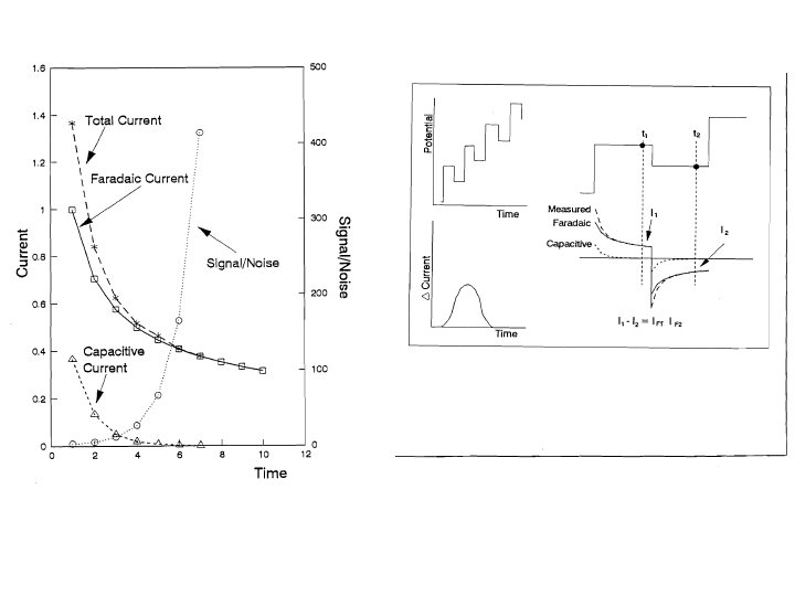
Signal to Noise in Voltammetric Analysis The limit of detection (LOD) is often defined as 3 xpeak to peak signal due to noise “Noise” in voltammetric methods is the result of 1. Competing or background electron transfer events originating in the a) Solvent b) Surface 2. Currents arising from charging of the electrode surface The limit of detection is also affected by distortion of the signal due to IR error
Solutions to the IR and Capacitive Current Problems Signal Background These are on two different time scales! Modulate the time domain of the experiment to discriminate against capactive currents.
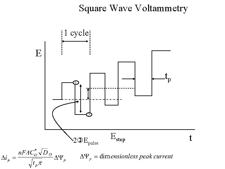
Square Wave Voltammetry 1 cycle E tp 1 2 2 Epulse Estep t
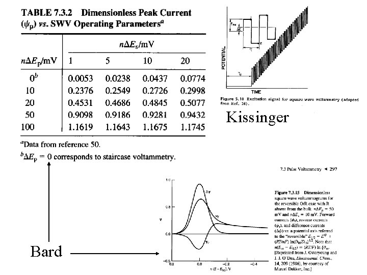
Kissinger Bard
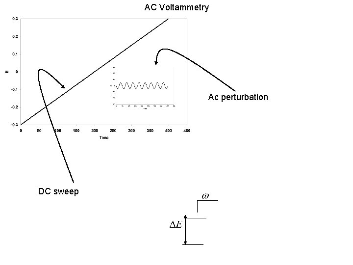
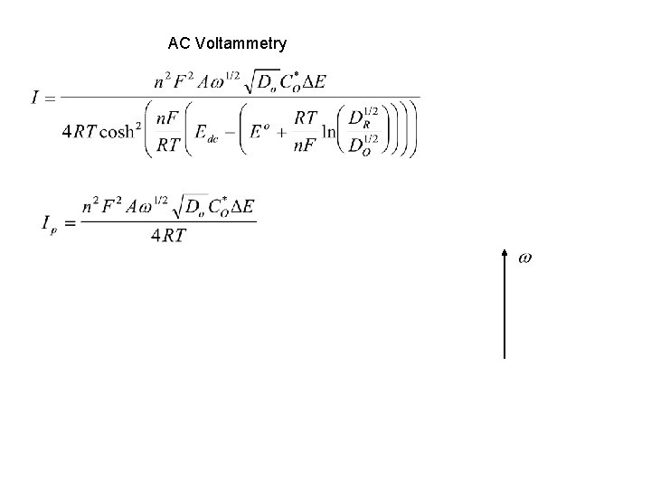
AC Voltammetry Ac perturbation DC sweep
AC Voltammetry
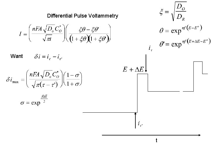
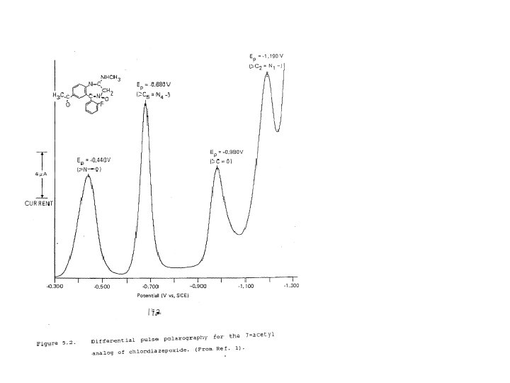
Differential Pulse Voltammetry Want t
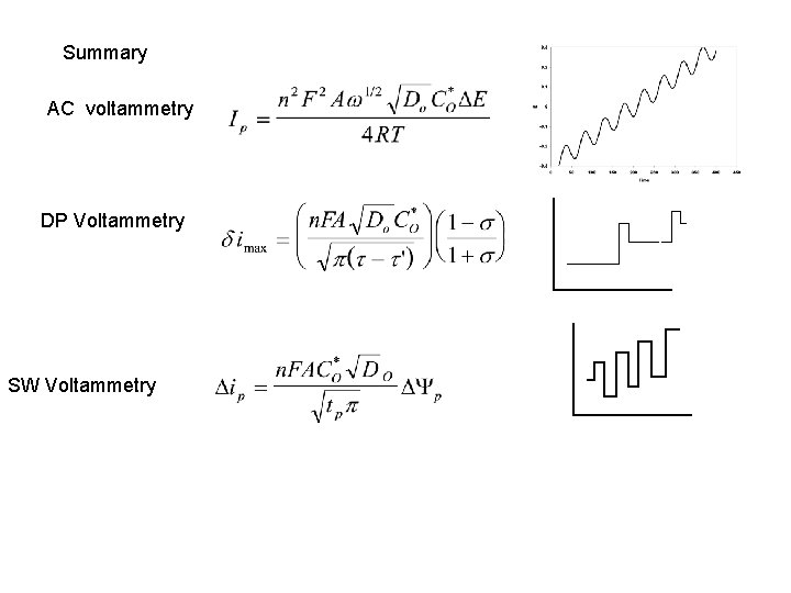
Summary AC voltammetry DP Voltammetry SW Voltammetry
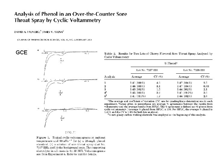
GCE
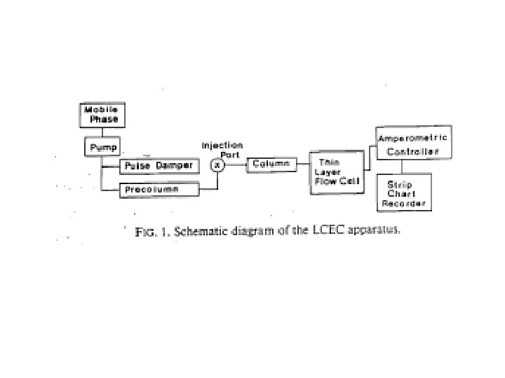
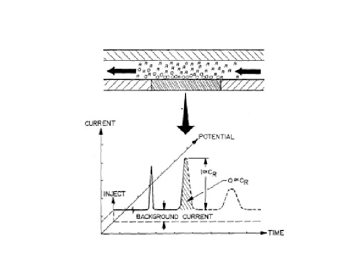
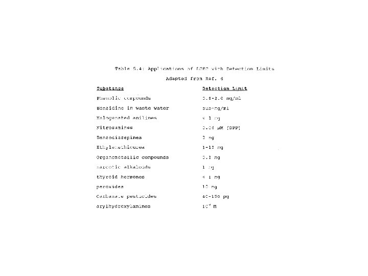
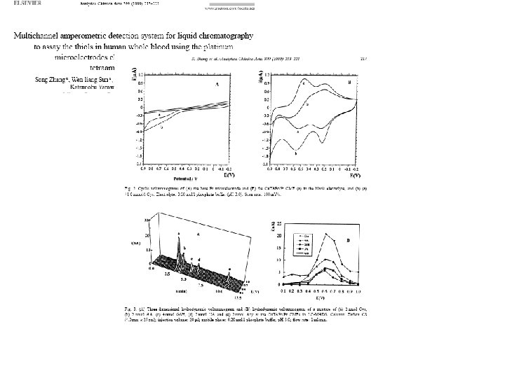
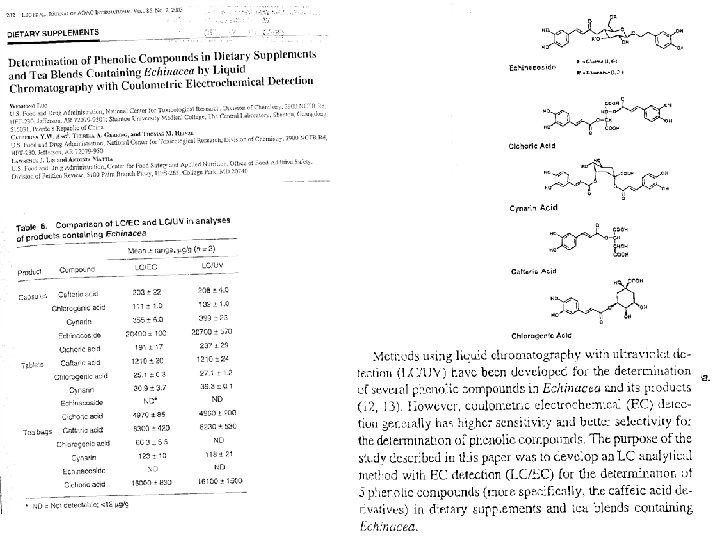
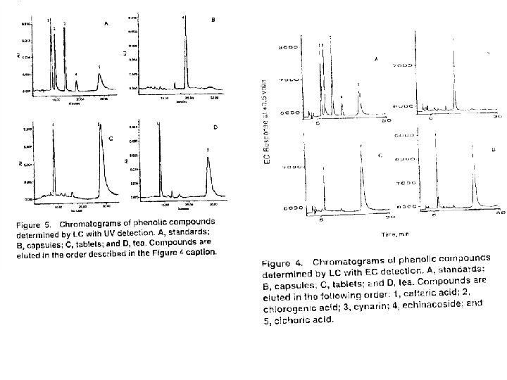
LC EC
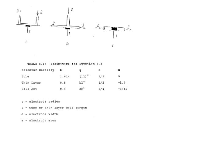
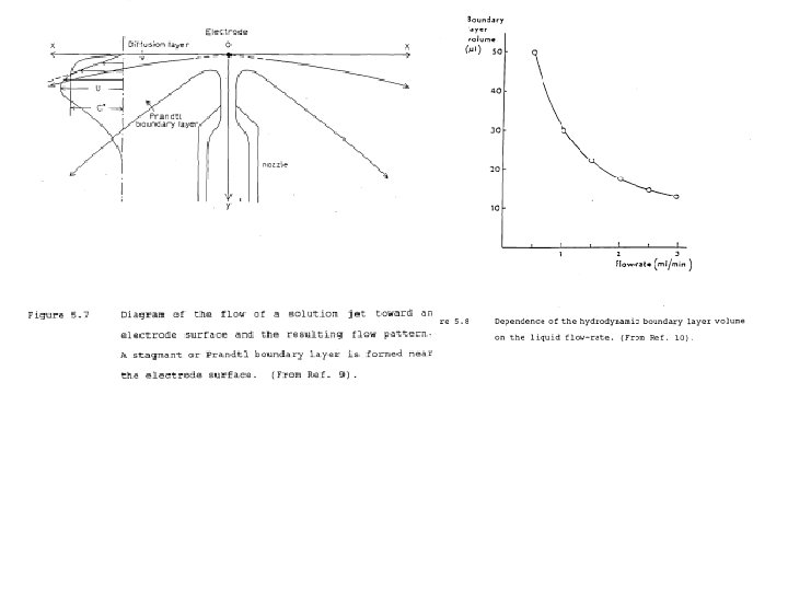
Control x. D at a fixed distance: Rotating Disk electrode, RDE Wall Jet Electrode, WJE
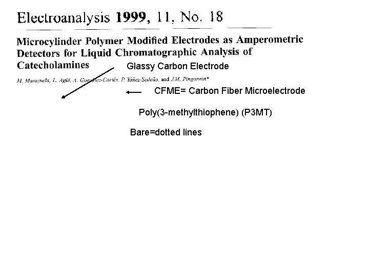
Glassy Carbon Electrode CFME= Carbon Fiber Microelectrode Poly(3 -methylthiophene) (P 3 MT) Bare=dotted lines
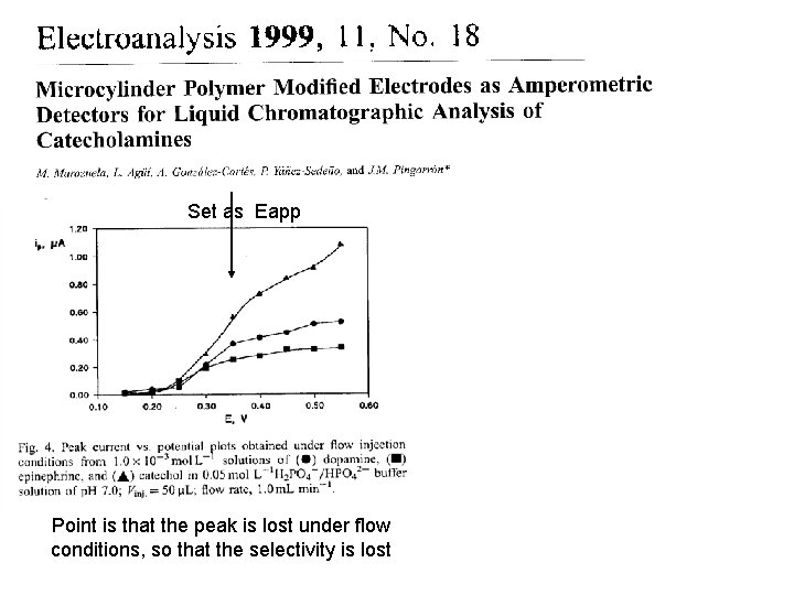
Set as Eapp Point is that the peak is lost under flow conditions, so that the selectivity is lost
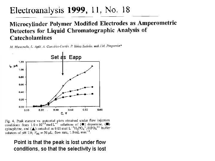
Set as Eapp Point is that the peak is lost under flow conditions, so that the selectivity is lost
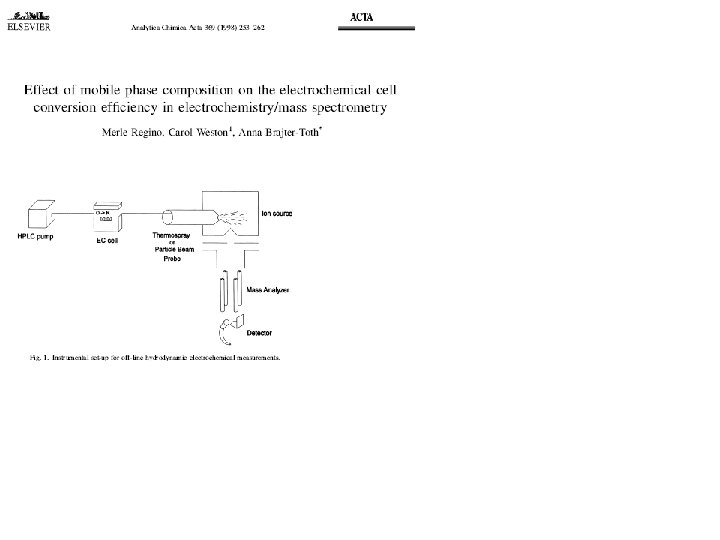
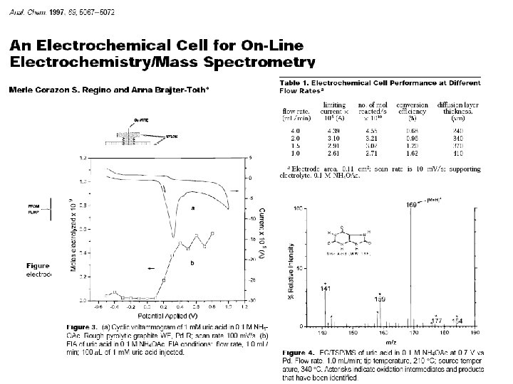
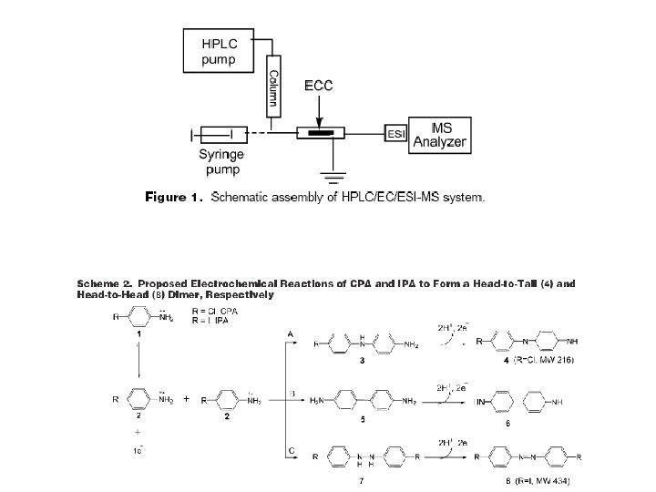
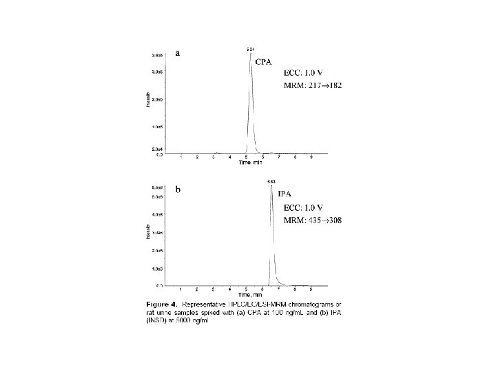
LC EC MS
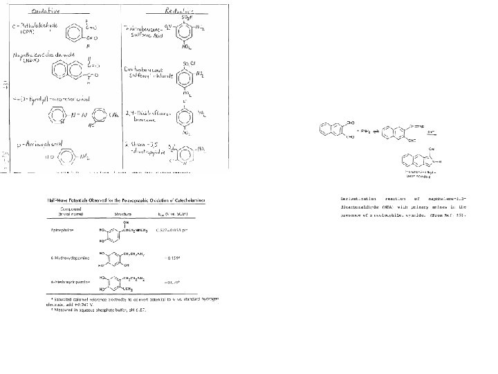
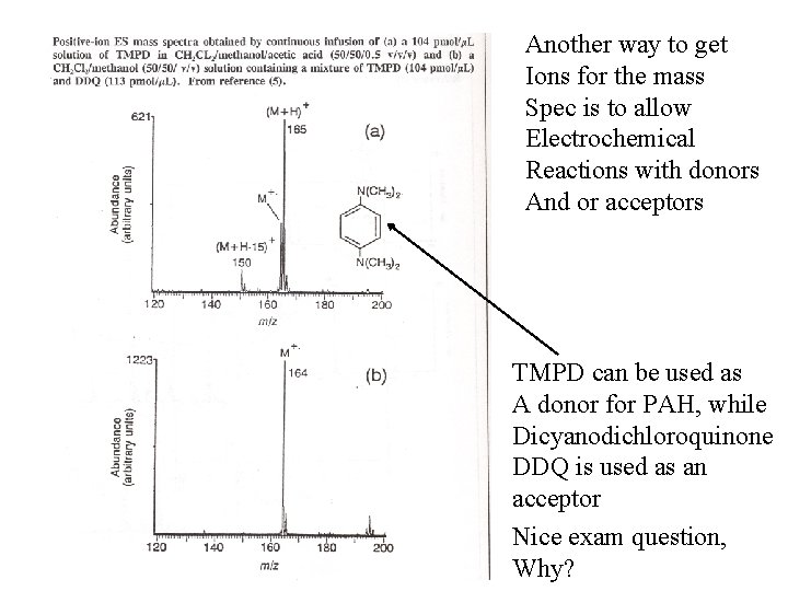
Some analytes are poorly suited to MS because of the difficulty in the ionization Pathway. P-chloroaniline (CPA) is one example Anticarcinogen sulofenur
Another way to get Ions for the mass Spec is to allow Electrochemical Reactions with donors And or acceptors TMPD can be used as A donor for PAH, while Dicyanodichloroquinone DDQ is used as an acceptor Nice exam question, Why?
Tool II Voltammetric Detectors 1. 2. 3. 4. Methods: Stripping, DPV Metals LC EC Rat Brains
An example of a unique system of electrochemistry which invokes 1. Concepts of microelectrodes 2. Suppression of capacitance 3. Blocking of surfaces to enhance selectivity 4. And the diquinone functionality just examined is The In vivo detection of dopamine in rat brains for nuerochemistry studies
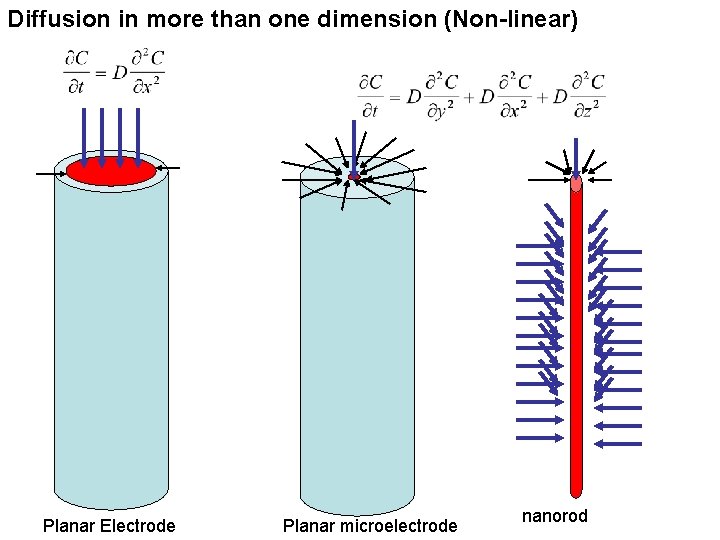
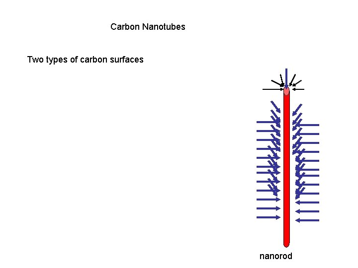
Diffusion in more than one dimension (Non-linear) Planar Electrode Planar microelectrode nanorod
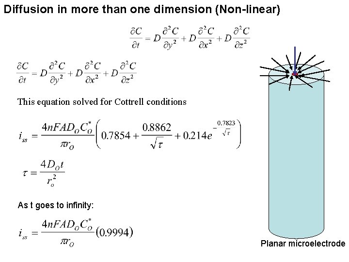
Diffusion in more than one dimension (Non-linear) This equation solved for Cottrell conditions As t goes to infinity: Planar microelectrode
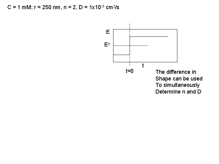
C = 1 m. M; r = 250 nm, n = 2, D = 1 x 10 -5 cm 2/s E Eo t=0 t The difference in Shape can be used To simultaneously Determine n and D
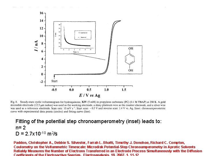
Fitting of the potential step chronoamperometry (inset) leads to: n= 2 D = 2. 7 x 10 -10 m 2/s Paddon, Christopher A. , Debbie S. Silvester, Farrah L. Bhatti, Timothy J. Donohoe, Richard C. Compton, Coulometry on the Voltammetric Timescale: Microdisk Potential-Step Chronoamperometry in Aprotic Solvents Reliably Measures the Number of Electrons Transferred in an Electrode Process Simultaneously with the Diffusion
Carbon Nanotubes Two types of carbon surfaces nanorod
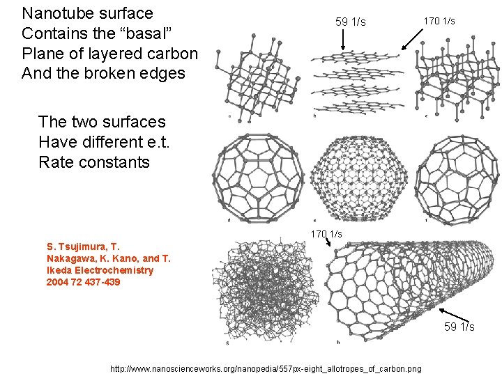
Nanotube surface Contains the “basal” Plane of layered carbon And the broken edges 59 1/s 170 1/s The two surfaces Have different e. t. Rate constants 170 1/s S. Tsujimura, T. Nakagawa, K. Kano, and T. Ikeda Electrochemistry 2004 72 437 -439 59 1/s http: //www. nanoscienceworks. org/nanopedia/557 px-eight_allotropes_of_carbon. png
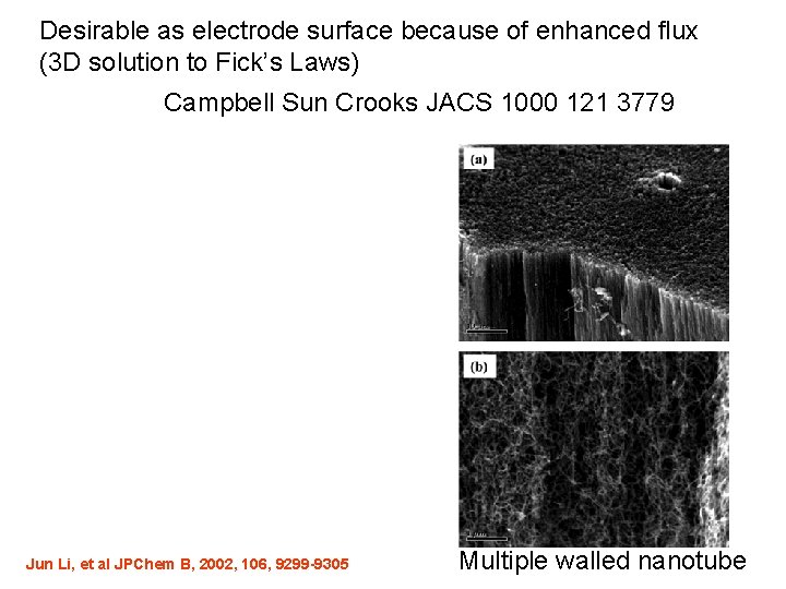
Desirable as electrode surface because of enhanced flux (3 D solution to Fick’s Laws) Campbell Sun Crooks JACS 1000 121 3779 Jun Li, et al JPChem B, 2002, 106, 9299 -9305 Multiple walled nanotube
Dopamine is a potent neurotransmitter and hormone in the brain. Can be supplied as medication that acts on the sympathetic nervous system producing effects such as increased heart rate and blood pressure. Deficits in dopamine are linked to behavioral diseases such as attentiondeficit hyperactivity disorder (ADHD). Abnormally high dopamine levels have been linked to psychosis, schizophrenia and cocaine addiction
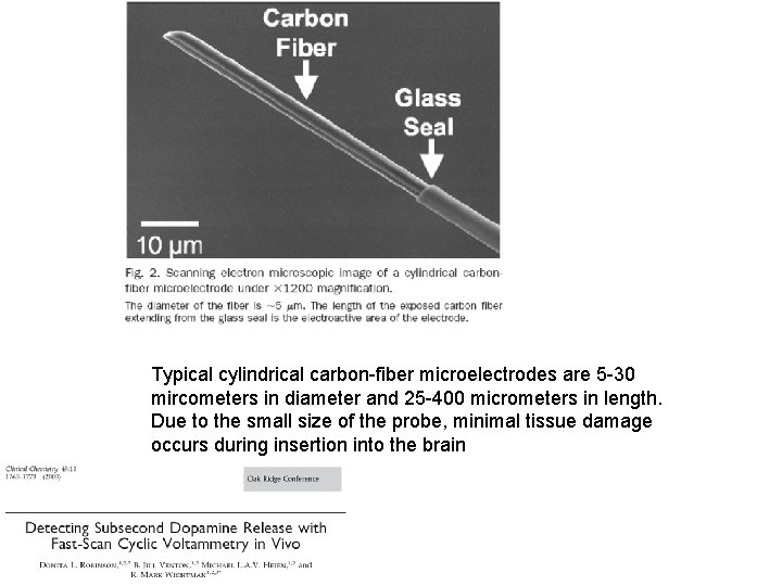
Typical cylindrical carbon-fiber microelectrodes are 5 -30 mircometers in diameter and 25 -400 micrometers in length. Due to the small size of the probe, minimal tissue damage occurs during insertion into the brain
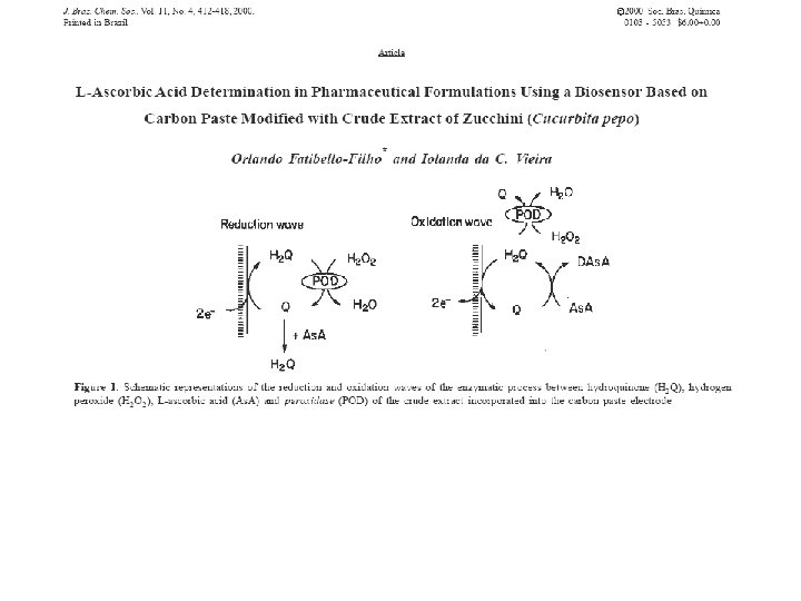
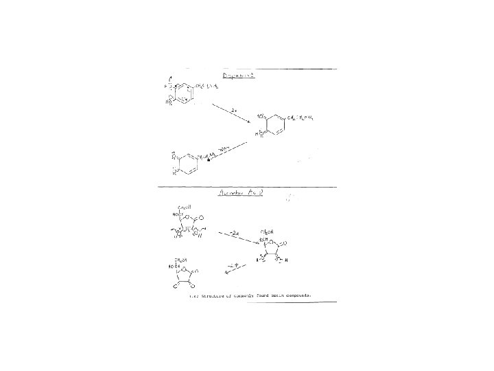
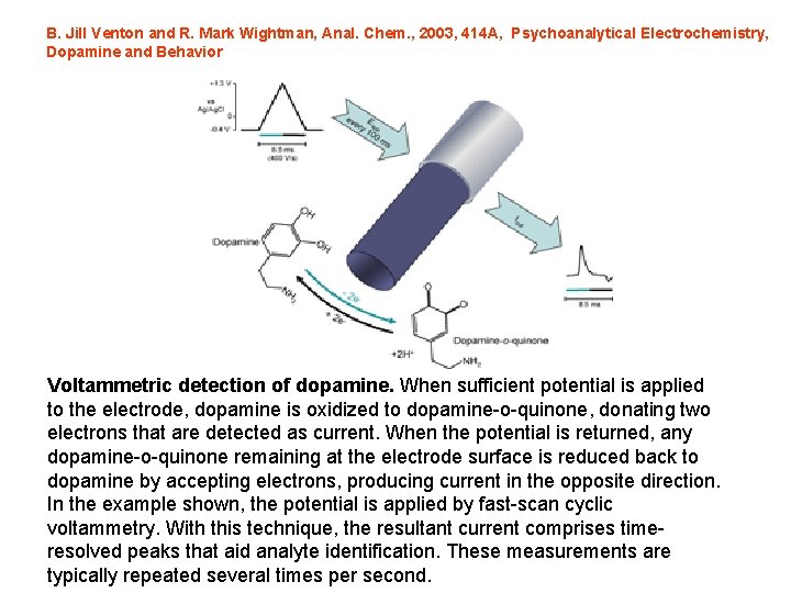
B. Jill Venton and R. Mark Wightman, Anal. Chem. , 2003, 414 A, Psychoanalytical Electrochemistry, Dopamine and Behavior Voltammetric detection of dopamine. When sufficient potential is applied to the electrode, dopamine is oxidized to dopamine-o-quinone, donating two electrons that are detected as current. When the potential is returned, any dopamine-o-quinone remaining at the electrode surface is reduced back to dopamine by accepting electrons, producing current in the opposite direction. In the example shown, the potential is applied by fast-scan cyclic voltammetry. With this technique, the resultant current comprises timeresolved peaks that aid analyte identification. These measurements are typically repeated several times per second.
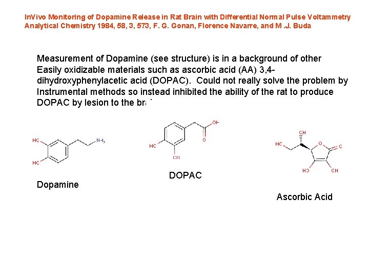
In. Vivo Monitoring of Dopamine Release in Rat Brain with Differential Normal Pulse Voltammetry Analytical Chemistry 1984, 58, 3, 573, F. G. Gonan, Florence Navarre, and M. J. Buda Measurement of Dopamine (see structure) is in a background of other Easily oxidizable materials such as ascorbic acid (AA) 3, 4 dihydroxyphenylacetic acid (DOPAC). Could not really solve the problem by Instrumental methods so instead inhibited the ability of the rat to produce DOPAC by lesion to the brain. Dopamine DOPAC Ascorbic Acid
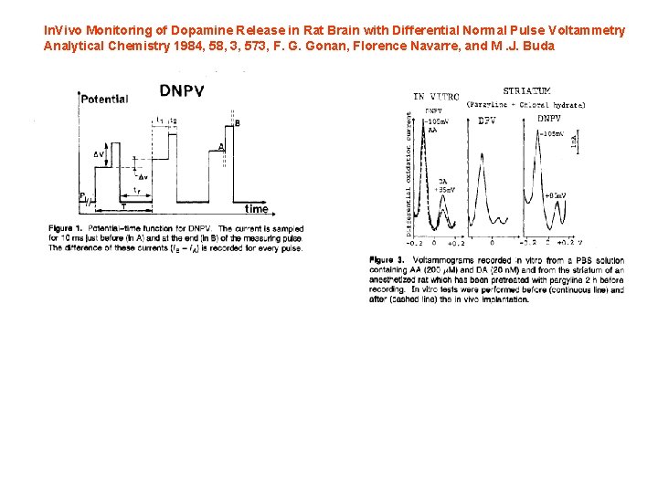
In. Vivo Monitoring of Dopamine Release in Rat Brain with Differential Normal Pulse Voltammetry Analytical Chemistry 1984, 58, 3, 573, F. G. Gonan, Florence Navarre, and M. J. Buda
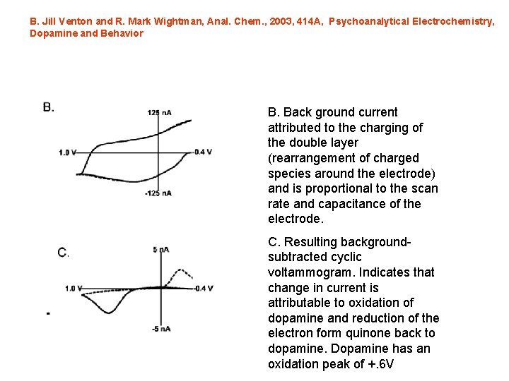
B. Jill Venton and R. Mark Wightman, Anal. Chem. , 2003, 414 A, Psychoanalytical Electrochemistry, Dopamine and Behavior B. Back ground current attributed to the charging of the double layer (rearrangement of charged species around the electrode) and is proportional to the scan rate and capacitance of the electrode. C. Resulting backgroundsubtracted cyclic voltammogram. Indicates that change in current is attributable to oxidation of dopamine and reduction of the electron form quinone back to dopamine. Dopamine has an oxidation peak of +. 6 V
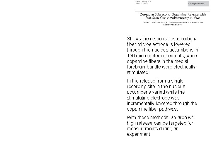
Shows the response as a carbonfiber microelectrode is lowered through the nucleus accumbens in 150 micrometer increments, while dopamine fibers in the medial forebrain bundle were electrically stimulated. In the release from a single recording site in the nucleus accumbens varied while the stimulating electrode was incrementally lowered through the dopamine fiber pathway. With these methods, an area w/ high release can be targeted for measurements during an experiment
CVs can be compared based on peak height, the relative ratio of oxidative and reductive peaks, as well as peak location and shape
Although using CV are a powerful tool for identifying electroactive compounds, some species such as norepinephrine and dopamine have nearly identical CV and cannot be differentiated by CV analysis alone. This is due to the structure similarity. To Identify dopamine changes in vivo, electrodes are placed in a known dopaminergic regions w/ low norepinephrine content
Reduce ascorbic acid background by coating the electrode with A blocking membrane which exlcudes anions Information about Nafion here
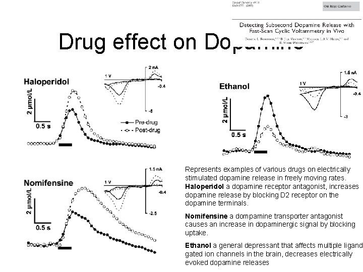
Drug effect on Dopamine Represents examples of various drugs on electrically stimulated dopamine release in freely moving rates. Haloperidol a dopamine receptor antagonist, increases dopamine release by blocking D 2 receptor on the dopamine terminals. Nomifensine a dompamine transporter antagonist causes an increase in dopaminergic signal by blocking uptake. Ethanol a general depressant that affects multiple ligandgated ion channels in the brain, decreases electrically evoked dopamine releases
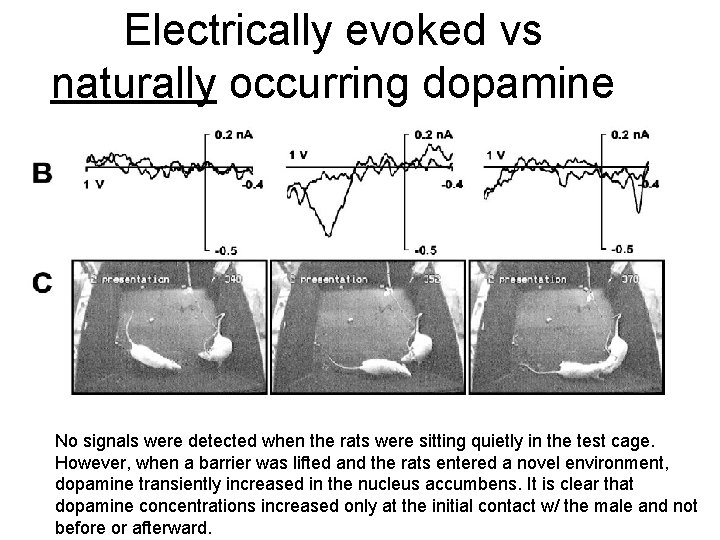
Electrically evoked vs naturally occurring dopamine No signals were detected when the rats were sitting quietly in the test cage. However, when a barrier was lifted and the rats entered a novel environment, dopamine transiently increased in the nucleus accumbens. It is clear that dopamine concentrations increased only at the initial contact w/ the male and not before or afterward.
Tools II Voltammetric Detectors 1. 2. 3. 4. Methods: Stripping, DPV Metals LC EC and LC EC MS Rat Brains END