
TONGUEI Presented by Dr Sushma Tomar Associate Professor
TONGUE-I Presented by : - Dr. Sushma Tomar Associate Professor Department of Anatomy
v Introduction v Presenting parts: • Root • Tip • Body v Muscles of Tongue v Arterial supply v Venous drainage v Lymphatic drainage v Nerve supply v Applied aspects Lesson Plan
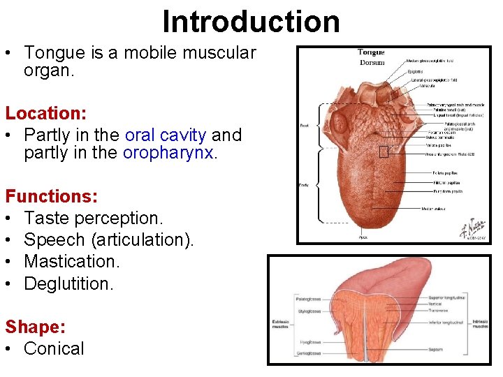
Introduction • Tongue is a mobile muscular organ. Location: • Partly in the oral cavity and partly in the oropharynx. Functions: • Taste perception. • Speech (articulation). • Mastication. • Deglutition. Shape: • Conical
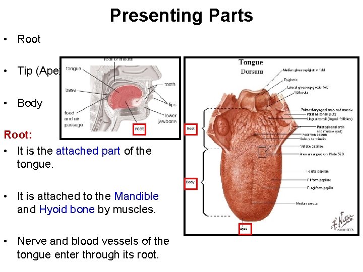
Presenting Parts • Root • Tip (Apex) • Body Root: • It is the attached part of the tongue. • It is attached to the Mandible and Hyoid bone by muscles. • Nerve and blood vessels of the tongue enter through its root.
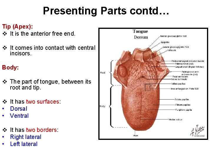
Presenting Parts contd… Tip (Apex): v It is the anterior free end. v It comes into contact with central incisors. Body: v The part of tongue, between its root and tip. v It has two surfaces: • Dorsal • Ventral v It has two borders: • Right lateral • Left lateral

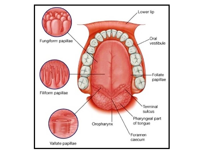
Dorsal Surface • Convex. Sulcus Terminalis: • An inverted Vshaped groove on the dorsum of the tongue. • Divides the dorsum of tongue into anterior 2/3 rd and posterior 1/3 rd. • Anterior 2/3 rd – Body of tongue [Oral part] rd
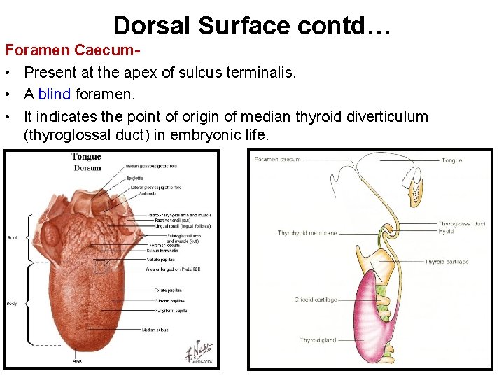
Dorsal Surface contd… Foramen Caecum • Present at the apex of sulcus terminalis. • A blind foramen. • It indicates the point of origin of median thyroid diverticulum (thyroglossal duct) in embryonic life.
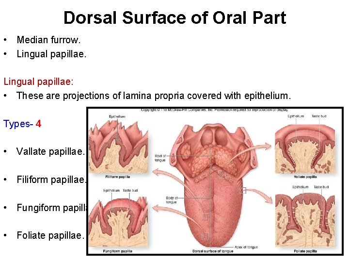
Dorsal Surface of Oral Part • Median furrow. • Lingual papillae: • These are projections of lamina propria covered with epithelium. Types- 4 • Vallate papillae. • Filiform papillae. • Fungiform papillae. • Foliate papillae.
![Vallate Papillae [Circumvallate Papillae] • Largest (1 -2 mm in diameter). Location- In front](http://slidetodoc.com/presentation_image_h2/06f48b3200274b568484130fe477342b/image-9.jpg "Vallate Papillae [Circumvallate Papillae] • Largest (1 -2 mm in diameter). Location- In front")
Vallate Papillae [Circumvallate Papillae] • Largest (1 -2 mm in diameter). Location- In front of sulcus terminalis in a ‘V’shaped row Shape- Truncated cone. • Each papilla is surrounded by a circular sulcus. • Circular sulcus is bounded on its periphery by a wall (vallum). • Ducts of several salivary glands open in the circular sulcus. • Taste buds are present in the papilla and its vallum.
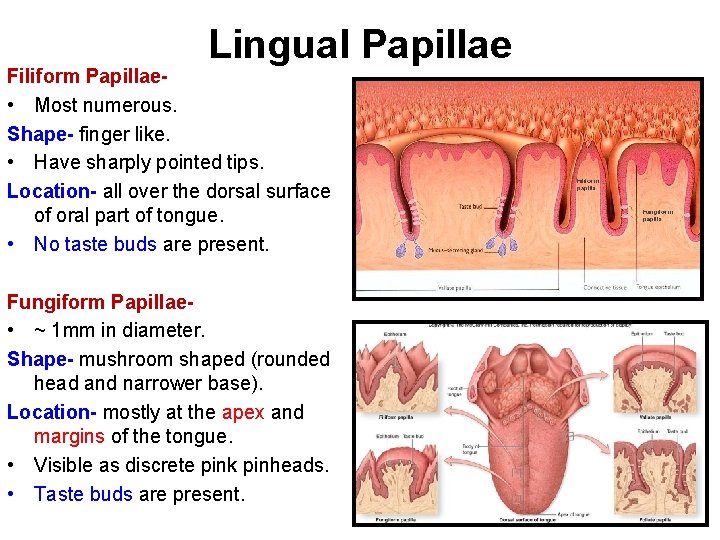
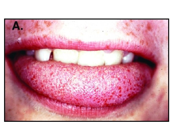
Lingual Papillae Filiform Papillae • Most numerous. Shape- finger like. • Have sharply pointed tips. Location- all over the dorsal surface of oral part of tongue. • No taste buds are present. Fungiform Papillae • ~ 1 mm in diameter. Shape- mushroom shaped (rounded head and narrower base). Location- mostly at the apex and margins of the tongue. • Visible as discrete pink pinheads. • Taste buds are present.
![• Dorsal Surface of Pharyngeal Part [Base of Tongue] This surface forms the](http://slidetodoc.com/presentation_image_h2/06f48b3200274b568484130fe477342b/image-13.jpg "• Dorsal Surface of Pharyngeal Part [Base of Tongue] This surface forms the")
• Dorsal Surface of Pharyngeal Part [Base of Tongue] This surface forms the anterior wall of oropharynx. • No lingual papillae. • A large number of lymphoid follicles are present in the submucosa [Lingual Tonsils]. • Mucosa of this surface is reflected onto the front of epiglottis as median glossoepiglottic fold. • Mucosa is reflected onto the lateral wall of pharynx as lateral glossoepiglottic folds. • The space on each side of median glossoepiglottic fold is termed ‘epigllotic vallecula’.
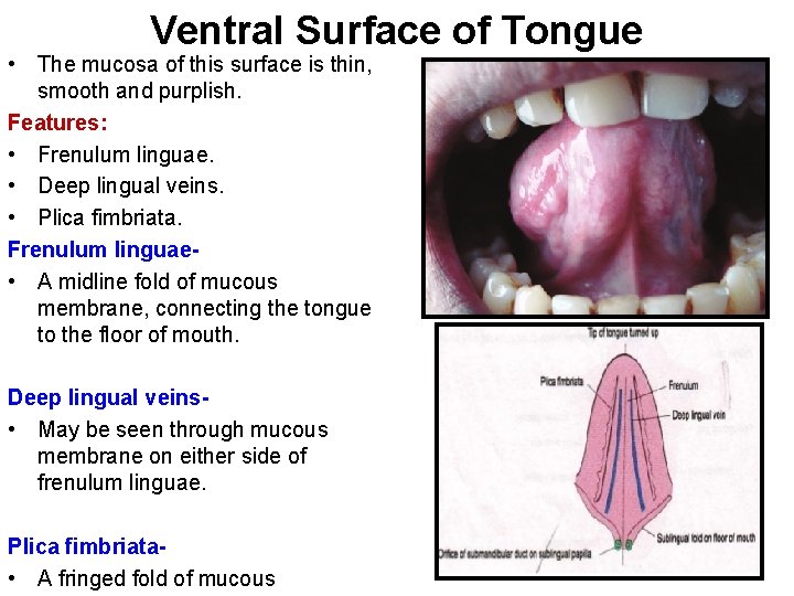
Ventral Surface of Tongue • The mucosa of this surface is thin, smooth and purplish. Features: • Frenulum linguae. • Deep lingual veins. • Plica fimbriata. Frenulum linguae • A midline fold of mucous membrane, connecting the tongue to the floor of mouth. Deep lingual veins • May be seen through mucous membrane on either side of frenulum linguae. Plica fimbriata • A fringed fold of mucous
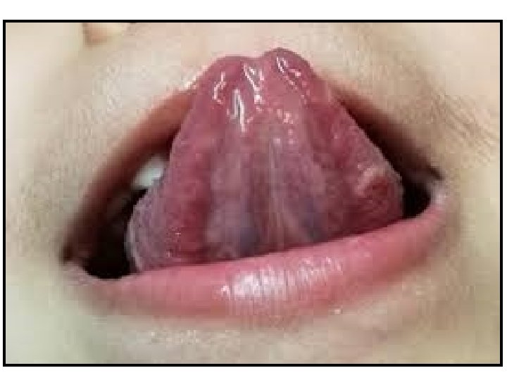
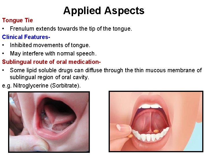
Applied Aspects Tongue Tie • Frenulum extends towards the tip of the tongue. Clinical Features • Inhibited movements of tongue. • May interfere with normal speech. Sublingual route of oral medication • Some lipid soluble drugs can diffuse through the thin mucous membrane of sublingual region of oral cavity. e. g. Nitroglycerine (Sorbitrate).
Muscles of Tongue • Extrinsic muscles. • Intrinsic muscles. Extrinsic Muscles • Take origin from structures outside the tongue. • Insert in the tongue. • Produce movements of tongue. • Change the shape of tongue. Intrinsic Muscles • Within the tongue. • Change the shape of tongue.
Muscles of Tongue contd… • Each half of tongue contains 4 extrinsic and 4 intrinsic muscles. Extrinsic Muscles • Genioglossus. • Hyoglossus. • Styloglossus. • Palatoglossus. Intrinsic Muscles • Superior longitudinal. • Inferior longitudinal. • Transverse. • Vertical.
![• • Genioglossus [Safety Muscle of A fan-shaped muscle. Tongue] Forms most of](http://slidetodoc.com/presentation_image_h2/06f48b3200274b568484130fe477342b/image-19.jpg "• • Genioglossus [Safety Muscle of A fan-shaped muscle. Tongue] Forms most of")
• • Genioglossus [Safety Muscle of A fan-shaped muscle. Tongue] Forms most of the bulk of tongue. Origin • Superior genial tubercle of mandible. Insertion • Fibers radiate backwards into the corresponding half of the tongue. Lower fibers – insert into the body of hyoid bone. Intermediate fibers • Pass beneath the anterior border of hyoglossus. • Extend upto stylohyoid ligament and middle constrictor of pharynx. Upper fibers • Turn upwards and forwards. • Extend up to the tip of tongue. Actions-
![Hyoglossus [Key Muscle of Suprahyoid Region] • A flat, quadrilateral muscle. Origin • Upper](http://slidetodoc.com/presentation_image_h2/06f48b3200274b568484130fe477342b/image-20.jpg "Hyoglossus [Key Muscle of Suprahyoid Region] • A flat, quadrilateral muscle. Origin • Upper")
Hyoglossus [Key Muscle of Suprahyoid Region] • A flat, quadrilateral muscle. Origin • Upper surface of greater cornu and adjacent part of body of hyoid bone. Insertion • Side of tongue between styloglossus laterally and inferior longitudinal muscle medially. Actions • Depression of Tongue (make the dorsal surface convex). • Retraction of Tongue.
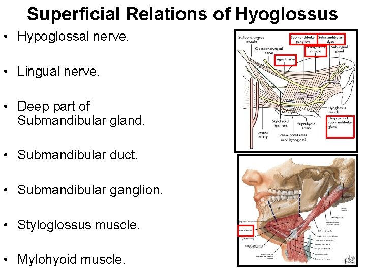
Superficial Relations of Hyoglossus • Hypoglossal nerve. • Lingual nerve. • Deep part of Submandibular gland. • Submandibular duct. • Submandibular ganglion. • Styloglossus muscle. • Mylohyoid muscle.
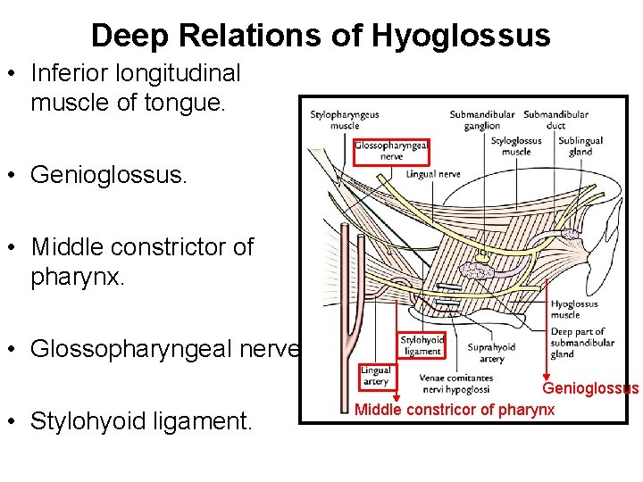
Deep Relations of Hyoglossus • Inferior longitudinal muscle of tongue. • Genioglossus. • Middle constrictor of pharynx. • Glossopharyngeal nerve. • Stylohyoid ligament. Genioglossus Middle constricor of pharynx
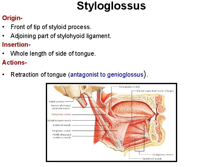
Styloglossus Origin • Front of tip of styloid process. • Adjoining part of stylohyoid ligament. Insertion • Whole length of side of tongue. Actions- • Retraction of tongue (antagonist to genioglossus).
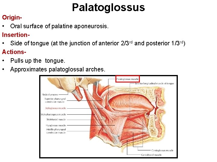
Palatoglossus Origin • Oral surface of palatine aponeurosis. Insertion • Side of tongue (at the junction of anterior 2/3 rd and posterior 1/3 rd) Actions • Pulls up the tongue. • Approximates palatoglossal arches.

- Slides: 25