Tokyo Guidelines 2018 initial management of acute biliary

Tokyo Guidelines 2018: initial management of acute biliary infection and flowchart for acute cholangitis

Dr gavidel journal club govaresh

Introduction Acute biliary infection, particularly acute cholangitis, may cause a rapid deterioration in condition due to sepsis, and prompt and appropriate treatment is therefore required.
")
Flowchart for the initial response to acute biliary infection The initial management (Fig. 1) of patients with suspected acute biliary infection starts with the measurement of vital signs to assess whether or not the situation is urgent. If the case is judged to be urgent, initial medical treatment should be started immediately including respiratory/circulatory management if required, without waiting for the definitive diagnosis. A detailed examination (consultation and physical examination) is then performed, after which blood tests and diagnostic imaging are performed; on the basis of the results, a definitive diagnosis is made following the diagnostic criteria for acute cholangitis and cholecystitis
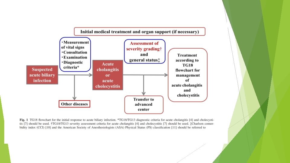

… Once the diagnosis has been confirmed, initial medical treatment should be started immediately, the severity should be assessed according to the severity grading criteria for acute cholangitis/cholecystitis, and the patient’s general status should be evaluated. The Charlson comorbidity index (CCI) (case series, CS) and the American Society of Anesthesiologists (ASA) Physical Status (PS) classification are useful for the evaluation of general status. Symptoms indicative of suspected acute biliary infection are fever, chills, abdominal pain, jaundice, nausea, vomiting, and disturbance of consciousness. If even one of these symptoms is present, acute biliary infection is suspected and it is necessary to proceed to diagnosis (CPG)

Management of patients with suspected acute biliary infection Vital signs include blood pressure, heart rate, respiration rate, temperature, urine volume, oxygen saturation (Sp. O 2), and consciousness level. In the physical examination, the evaluation and measurement of the patient’s state of consciousness goes without saying, and the presence or absence of yellowing of the palpebral conjunctiva, the location and severity of tenderness, and whether or not there any symptoms of peritoneal irritation must always be confirmed. The presence or absence of Murphy’s sign (compression of the right upper quadrant causes the patient to catch their breath due to pain when taking a deep breath), which is specific to acute cholecystitis, must always be confirmed.
 2)")
Tests required for the diagnosis of acute biliary infection Blood tests including: 1) 2) white blood cell count, platelet count, 3) C-reactive protein (CRP), 4) albumin, 5) alkaline phosphatase (ALP), 6) gamma-glutamyl transferase (GGT), 7) aspartate aminotransferase (AST), alanine aminotransferase (ALT), 8) bilirubin 9) Blood urea nitrogen (BUN), creatinine, 10) prothrombin time (PT), and PT-international normalized ratio (INR) are carried out for the purpose of diagnosis and severity grading, 11) and blood gas analysis should also be performed (CPG). If a high fever is present, blood culture should preferably be performed at this point.
 are useful")
In terms of diagnostic imaging, abdominal ultrasound and computed tomography (CT) are useful for the diagnosis of acute biliary infection, and at least one of these should be performed. Abdominal ultrasound in particular is minimally invasive, widely used, simple, and cheap, and should therefore be performed first in patients with suspected biliary infection, despite disadvantages including the fact that the results are easily affected by the operator’s skill and the patient’s condition (CPG). Inflammation in acute cholangitis is difficult to assess on diagnostic imaging, but it is possible to evaluate dilatation of the bile duct, or bile congestion due to occlusion/ stenosis of the bile duct or biliary calculus and its cause(CPG). The distinctive signs of acute cholecystitis on diagnostic imaging include enlargement of the gallbladder, gallbladder wall thickening, gallbladder calculi, fluid retention around the gallbladder, abscess around the gallbladder, and sludge debris in the gallbladder and the sonographic Murphy’s sign

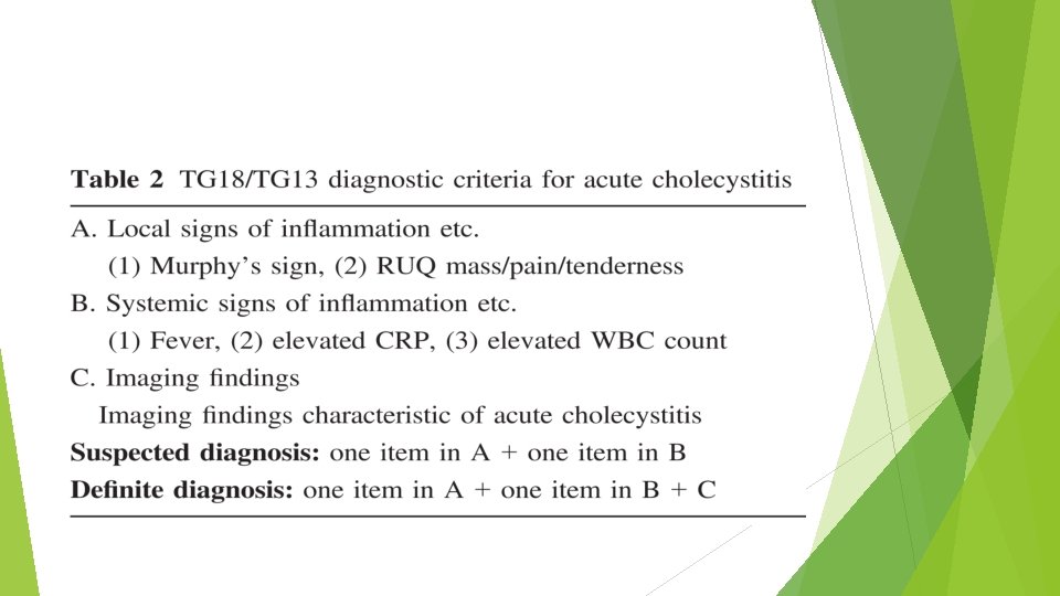
Diagnostic criteria for acute biliary infection

Initial treatment Once a definitive diagnosis of acute cholangitis or acute cholecystitis has been reached, initial treatment including the infusion of sufficient fluids and antibiotic and analgesic administration is started, with careful monitoring of blood pressure, heart rate, and urine volume. It goes without saying that if the patient is in a state of shock, initial treatment should be started without waiting for a definitive diagnosis. in principle patients should be fasted to enable immediate emergency drainage (CPG)

Despite the concern that analgesic administration may mask physical signs and cause a mistaken diagnosis, a randomized controlled trial (RCT) comparing intravenous morphine hydrochloride and an intravenous placebo for patients examined in the emergency room complaining of abdominal pain found no difference between them in the rate of diagnosis, and analgesics should therefore be administered proactively at an early stage. Opioid analgesics such as morphine hydrochloride and other similar types of drug (such as nonopioid analgesics and pentazocine) cause the sphincter of Oddi to contract, which may elevate biliary pressure, and must therefore be administered with caution. In the case of serious deterioration, such as the appearance of shock (hypotension), disturbance of consciousness, acute dyspnea, acute renal dysfunction, hepatic dysfunction, or disseminated intravascular coagulation (DIC) (reduced platelet count), emergency biliary drainage should be considered alongside appropriate organ support and respiratory/circulatory management (such as artificial ventilation, tracheal intubation, and the use of hypertensive agents) (CPG)

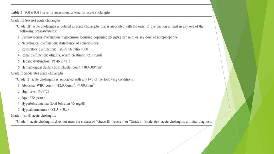
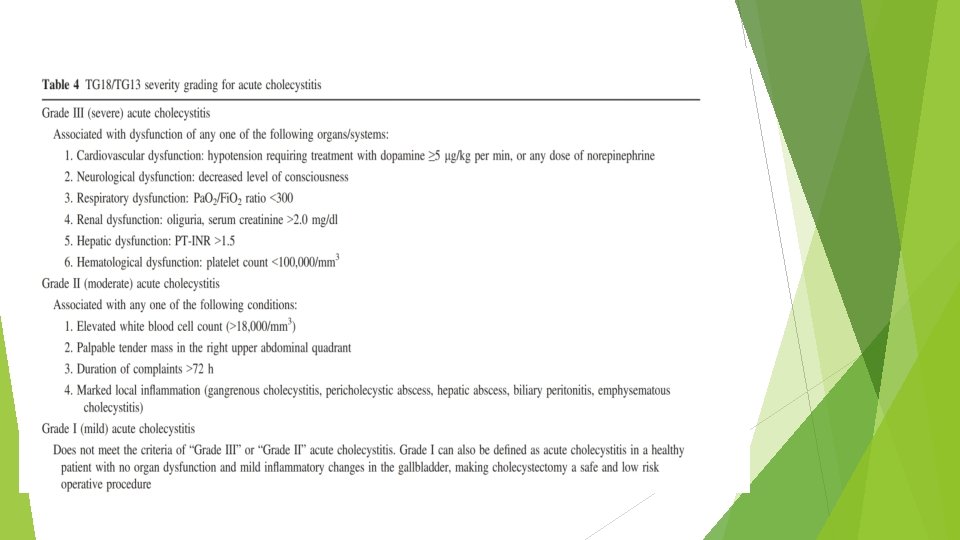
Severity assessment and general status evaluation Alongside initial treatment, severity assessment should be carried out using the TG 13/18 severity assessment criteria for acute cholangitis (Table 3) (CPG) or the TG 13/18 severity assessment criteria for acute cholecystitis (Table 4) (CPG), and the patient’s general status should also be evaluated using the CCI and the ASA-PS classification.

A multicenter joint study carried out in Japan and Taiwan found that for moderate cholangitis, mortality was significantly lower in 944 patients who underwent drainage within 24 h compared with 1, 081 patients who either underwent drainage after longer than 24 h or did not undergo drainage (1. 7% vs. 3. 4%, P = 0. 0172), but that there was no significant difference for mild or severe cholangitis. In the other observational study, which compared 130 patients with mild or moderate cholangitis who underwent drainage within 24 h and 82 who underwent drainage after longer than 24 h, although there was no significant difference in mortality which was zero in both groups, the mean duration of hospitalization was significantly shorter for patients who underwent drainage within 24 h (6. 8 days vs. 9. 2 days, P < 0. 01 (CS)
 (CS) and one RCT")
Three observational studies (one not use severity assessment criteria) (CS) and one RCT showed that single-stage lithotomy was safe and feasible in cases of mild or moderate acute cholangitis caused by choledocholithiasis. In the RCT, however, the rate of complications after endoscopic retrograde cholangiopancreatography (ERCP) was significantly higher than that after twostage lithotomy (6/35 = 17. 1% vs. 0/ 33 = 0%, P = 0. 025) (RCT) therefore, caution is required. Two studies have compared early and elective laparoscopic choledocholithotomy for patients with mild or moderate acute cholangitis associated with choledocolithiasis. Early laparoscopic choledocholithotomy was safe and there was no difference in the rate of complications,

Antibiotics for low risk community-acquired intra abdominal patient Including with mild to mod severity without risk factors for antibiotic resistance or treatment failure.

Antibiotics for low risk community-acquired intra abdominal patient

high risk community-acquired intra abdominal patient 1. Severe cholangitis 2. Recent travel to areas of the world with high resistance organisms 3. Known colonization with resistance organisms 4. Advanced age 5. Immunocompromising conditions 6. Major medical comorbidity
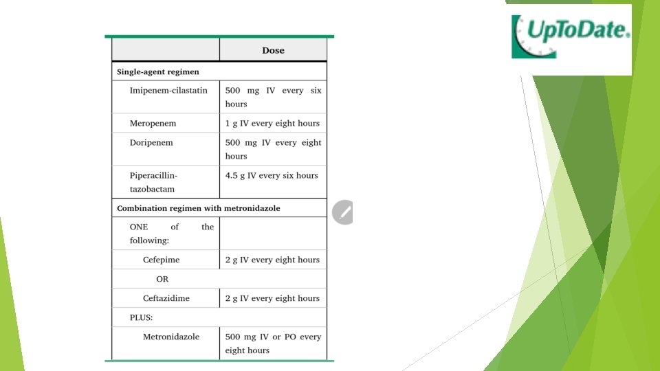

Empiric antibiotic regimens for health care associated intra -abdominal infections in adult

What treatments other than antibiotics and biliary drainage are effective for severe cholangitis? The administration of recombinant human soluble thrombomodulin may be considered for severe cholangitis complicated with disseminated intravascular coagulation. (Level D) Severe cholangitis is often complicated by DIC. Septic DIC is treated with anticoagulants such as heparin, antithrombin III, and protease inhibitors. the value of anticoagulants in severe cholangitis is unclear

Suetani et al. divided 69 patients diagnosed with acute cholangitis-induced DIC on the basis of the TG 13 diagnostic criteria into 33 who were treated with r. TM and 36 who were not, and found that the DIC resolution rate was significantly better in patients given r. TM (83. 3% vs. 52. 8%, P < 0. 01), but that there was no significant difference in mortality (13. 3% vs. 27. 8%, P = 0. 26) (CS). but as there were no serious side-effects, the use of r. TM in patients with severe cholangitis complicated by DIC may be considered.

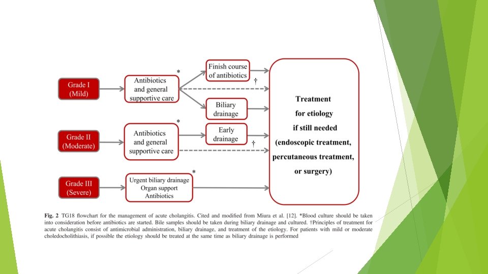
Flowchart for the management of acute cholangitis and cholecystitis Acute cholangitis should be managed in accordance with its severity. Biliary drainage and antibiotics are the two key pillars of the treatment of acute cholangitis. In some cases of acute cholangitis, acute cholecystitis may also be present; in this event the treatment strategy should be decided in consideration of the severity of both and the patient’s general status (CPG). If blood culture has not been performed as part of the initial response, it should be carried out before antibiotic administration. If biliary drainage is performed, bile samples must always be sent for culture.
 Mild acute cholangitis is defined as cholangitis that does")
Grade I (mild acute cholangitis) Mild acute cholangitis is defined as cholangitis that does not meet the TG 18 severity assessment criteria for moderate or severe cholangitis below (CPG) In most cases initial treatment including antibiotics is sufficient, and most patients do not require biliary drainage. However, biliary drainage should be considered if a patient does no respond to initial treatment. EST and subsequent choledocholithotomy may be performed at the same time as biliary drainage.
 Moderate acute cholangitis is cholangitis that is not severe")
Grade II (moderate acute cholangitis) Moderate acute cholangitis is cholangitis that is not severe but requires early biliary drainage. moderate cholangitis is assessed if at least two of the following five criteria are met: 1. WBC ≥ 12, 000 or <4, 000, 2. temperature ≥ 39°C, 3. 4. age ≥ 75 years, total bilirubin ≥ 5 mg/dl, 5. albumin <(lower limit of normal value 9 0. 73 g/dl) Early endoscopic or percutaneous transhepatic biliary drainage is indicated. If the underlying etiology requires treatment, this should be provided after the patient’s general condition has improved (CPG)
 Severe acute cholangitis is cholangitis with sepsis-induced organ damage.")
Grade III (severe acute cholangitis) Severe acute cholangitis is cholangitis with sepsis-induced organ damage. severe cholangitis is assessed if any one of the following criteria is met: 1. cardiovascular dysfunction (requiring the use of dopamine ≥ 5 μg/kg per min or noradrenaline), 2. neurological dysfunction (disturbance of consciousness), 3. respiratory dysfunction (Pa. O 2/Fi. O 2 ratio <300), 4. renal dysfunction (oliguria or serum creatinine >2. 0 mg/dl), 5. hepatic dysfunction (PT-INR >1. 5), 6. coagulation disorder (platelet count <100000 /ml)

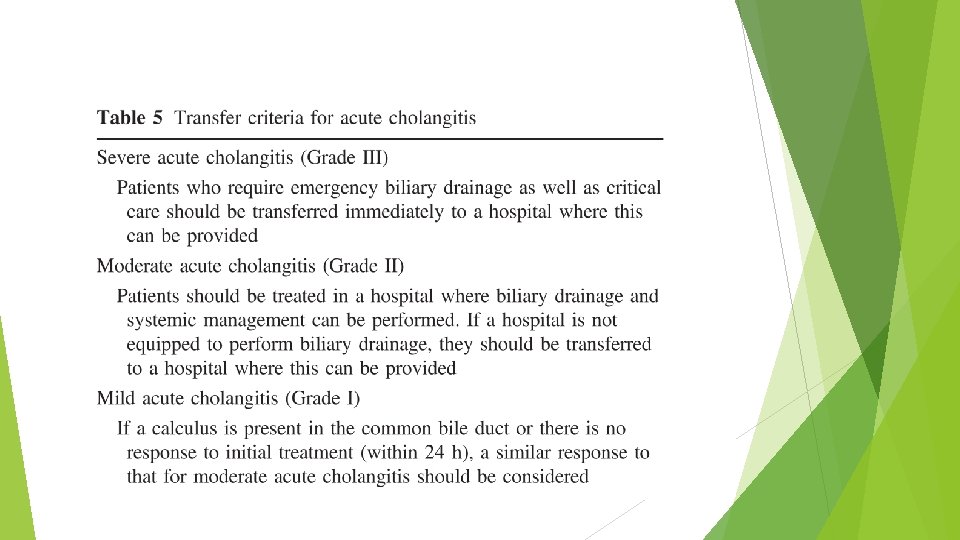
Transfer criteria If a hospital is not equipped to perform endoscopic or percutaneous transhepatic biliary drainage or provide intensive care, patients with moderate or severe cholangitis should preferably be transferred to a hospital capable of providing these treatments, irrespective of whether or not they are actually required
- Slides: 32