Today Quiz Respiratory system part 1 Organs of


 bronchus Left lung")


 • Muscular passage from nasal cavity to larynx • Three regions of")
 Regions of the pharynx")
 • Pharyngotympanic tubes open into the nasopharynx • Tonsils of the pharynx")

 • Routes air and food into proper channels • Plays a")
 • Thyroid cartilage • Largest of the hyaline cartilages • Protrudes")
 • Vocal folds (true vocal cords) • Vibrate with expelled air")

 • 4 -inch-long tube that connects larynx with bronchi • Walls are")

")
 Bronchi • Formed by division of the trachea • Each bronchus enters")


 Tree Divisions • All but the smallest of these passageways have reinforcing")

")
 Light micrograph of")


. Red blood cell Capillary")


 • Completely mechanical process that depends on volume changes")
 • Two phases • Inspiration = inhalation • Flow")
 • Inspiration • Diaphragm and external intercostal muscles contract")


 • Expiration • Largely a passive process that depends")

 • Normal pressure within the pleural space is always")


 • Amount of air that")


 5, 000 4, 000 3, 000 2, 000 1, 000")
 Movements • Can be caused by reflexes or voluntary actions •")
- Slides: 55
Today • Quiz • Respiratory system part 1
Organs of the Respiratory System • Nose • Pharynx • Larynx • Trachea • Bronchi • Lungs—alveoli
Nasal cavity Nostril Oral cavity Pharynx Larynx Trachea Right main (primary) bronchus Left lung Right lung Diaphragm
Functions of the Respiratory System • Gas exchanges between the blood and external environment • Occur in the alveoli of the lungs • Passageways to the lungs purify, humidify, and warm the incoming air
The Nose • The only externally visible part of the respiratory system • Air enters the nose through the external nostrils (nares) • Interior of the nose consists of a nasal cavity divided by a nasal septum Remember that if any area is open to the outside (even if it’s inside), it is lined with mucous membranes (wet/moist membranes)
Cribriform plate of ethmoid bone Sphenoidal sinus Posterior nasal aperture Nasopharynx • Pharyngeal tonsil • Opening of pharyngotympanic tube • Uvula Oropharynx • Palatine tonsil • Lingual tonsil Laryngopharynx Esophagus Trachea Frontal sinus Nasal cavity • Nasal conchae (superior, middle and inferior) • Nasal meatuses (superior, middle, and inferior) • Nasal vestibule • Nostril Hard palate Soft palate Tongue Hyoid bone Larynx • Epiglottis • Thyroid cartilage • Vocal fold • Cricoid cartilage (b) Detailed anatomy of the upper respiratory tract
The Nose • Olfactory receptors are located in the mucosa on the superior surface • The rest of the cavity is lined with respiratory mucosa, which: • Moistens air • Traps incoming foreign particles
The Nose • Lateral walls have projections called conchae • Increase surface area • Increase air turbulence within the nasal cavity • The nasal cavity is separated from the oral cavity by the palate • Anterior hard palate (bone) • Posterior soft palate (unsupported)
Paranasal Sinuses • Cavities within bones surrounding the nasal cavity are called sinuses • Sinuses are located in the following bones: • • Frontal Sphenoid Ethmoid Maxillary
Cribriform plate of ethmoid bone Sphenoidal sinus Posterior nasal aperture Nasopharynx • Pharyngeal tonsil • Opening of pharyngotympanic tube • Uvula Oropharynx • Palatine tonsil • Lingual tonsil Laryngopharynx Esophagus Trachea Frontal sinus Nasal cavity • Nasal conchae (superior, middle and inferior) • Nasal meatuses (superior, middle, and inferior) • Nasal vestibule • Nostril Hard palate Soft palate Tongue Hyoid bone Larynx • Epiglottis • Thyroid cartilage • Vocal fold • Cricoid cartilage
Paranasal Sinuses • Functions of the sinuses: • Lighten the skull • Act as resonance chambers for speech • Produce mucus that drains into the nasal cavity
Pharynx (Throat) • Muscular passage from nasal cavity to larynx • Three regions of the pharynx: 1. Nasopharynx—superior region behind nasal cavity 2. Oropharynx—middle region behind mouth 3. Laryngopharynx—inferior region attached to larynx • The oropharynx and laryngopharynx are common passageways for air and food
Pharynx • Nasopharynx • Oropharynx • Laryngopharynx (a) Regions of the pharynx
Pharynx (Throat) • Pharyngotympanic tubes open into the nasopharynx • Tonsils of the pharynx • Pharyngeal tonsil (adenoid) is located in the nasopharynx • Palatine tonsils are located in the oropharynx • Lingual tonsils are found at the base of the tongue
Cribriform plate of ethmoid bone Sphenoidal sinus Posterior nasal aperture Nasopharynx • Pharyngeal tonsil • Opening of pharyngotympanic tube • Uvula Oropharynx • Palatine tonsil • Lingual tonsil Laryngopharynx Esophagus Trachea Frontal sinus Nasal cavity • Nasal conchae (superior, middle and inferior) • Nasal meatuses (superior, middle, and inferior) • Nasal vestibule • Nostril Hard palate Soft palate Tongue Hyoid bone Larynx • Epiglottis • Thyroid cartilage • Vocal fold • Cricoid cartilage (b) Detailed anatomy of the upper respiratory tract
Larynx (Voice Box) • Routes air and food into proper channels • Plays a role in speech • Made of eight rigid hyaline cartilages and a spoon-shaped flap of elastic cartilage (epiglottis) https: //www. youtube. com/watch? v=Lqd. FL 0 u 2 HLY https: //www. youtube. com/watch? v=C 7 Cpk. Eszks. I https: //www. youtube. com/watch? v=Tsh-0 Vv. AGQA
Larynx (Voice Box) • Thyroid cartilage • Largest of the hyaline cartilages • Protrudes anteriorly (Adam’s apple) • Epiglottis • Protects the superior opening of the larynx • Routes food to the posteriorly situated esophagus and routes air toward the trachea • When swallowing, the epiglottis rises and forms a lid over the opening of the larynx
Larynx (Voice Box) • Vocal folds (true vocal cords) • Vibrate with expelled air • The glottis consists of the vocal cords and the slitlike pathway (opening)
Figure 13. 2 b Basic anatomy of the upper respiratory tract, sagittal section. Cribriform plate of ethmoid bone Sphenoidal sinus Posterior nasal aperture Nasopharynx • Pharyngeal tonsil • Opening of pharyngotympanic tube • Uvula Oropharynx • Palatine tonsil • Lingual tonsil Laryngopharynx Esophagus Trachea Frontal sinus Nasal cavity • Nasal conchae (superior, middle and inferior) • Nasal meatuses (superior, middle, and inferior) • Nasal vestibule • Nostril Hard palate Soft palate Tongue Hyoid bone Larynx • Epiglottis • Thyroid cartilage • Vocal fold • Cricoid cartilage (b) Detailed anatomy of the upper respiratory tract
Trachea (Windpipe) • 4 -inch-long tube that connects larynx with bronchi • Walls are reinforced with C-shaped hyaline cartilage, which keeps the trachea open (patent) • Lined with ciliated mucosa • Cilia beat continuously in the opposite direction of incoming air • Expel mucus loaded with dust and other debris away from lungs
Figure 13. 3 a Structural relationship of the trachea and esophagus. Posterior Mucosa Submucosa Esophagus Trachealis muscle Lumen of trachea Seromucous gland in submucosa Hyaline cartilage Adventitia (a) Anterior
Figure 13. 3 b Structural relationship of the trachea and esophagus. (b)
Main (Primary) Bronchi • Formed by division of the trachea • Each bronchus enters the lung at the hilum (medial depression) • Right bronchus is wider, shorter, and straighter than left • Bronchi subdivide into smaller and smaller branches
Figure 13. 1 The major respiratory organs shown in relation to surrounding structures. Nasal cavity Nostril Oral cavity Pharynx Larynx Trachea Right main (primary) bronchus Left lung Right lung Diaphragm
Lungs • Occupy most of the thoracic cavity • Heart occupies central portion called mediastinum • Apex is near the clavicle (superior portion) • Base rests on the diaphragm (inferior portion) • Each lung is divided into lobes by fissures • Left lung—two lobes • Right lung—three lobes
Coverings of the Lungs • Serosa covers the outer surface of the lungs • Pulmonary (visceral) pleura covers the lung surface • Parietal pleura lines the walls of the thoracic cavity • Pleural fluid fills the area between layers to allow gliding and decrease friction during breathing • Pleural space (between the layers) is more of a potential space
Figure 13. 4 a Anatomical relationships of organs in the thoracic cavity. Intercostal muscle Rib Trachea Parietal pleura Pleural cavity Visceral pleura Lung Thymus Apex of lung Right superior lobe Horizontal fissure Right middle lobe Oblique fissure Right inferior lobe Heart (in pericardial cavity of mediastinum) Diaphragm Base of lung Left superior lobe Oblique fissure Left inferior lobe (a) Anterior view. The lungs flank mediastinal structures laterally.
Figure 13. 4 b Anatomical relationships of organs in the thoracic cavity. Vertebra Posterior Esophagus (in posterior mediastinum) Root of lung at hilum • Left main bronchus • Left pulmonary artery • Left pulmonary vein Right lung Parietal pleura Visceral pleura Left lung Pleural cavity Thoracic wall Pulmonary trunk Pericardial membranes Sternum Heart (in mediastinum) Anterior mediastinum Anterior (b) Transverse section through the thorax, viewed from above.
Bronchial (Respiratory) Tree Divisions • All but the smallest of these passageways have reinforcing cartilage in their walls • Conduits to and from the respiratory zone • • • Primary bronchi Secondary bronchi Tertiary bronchi Bronchioles Terminal bronchioles
Respiratory Zone Structures • Respiratory bronchioles • Alveolar ducts • Alveolar sacs • Alveoli (air sacs)
Figure 13. 5 a Respiratory zone structures. Alveolar duct Respiratory bronchioles Terminal bronchiole (a) Diagrammatic view of respiratory bronchioles, alveolar ducts, and alveoli Alveolar duct Alveolar sac
Figure 13. 5 b Respiratory zone structures. Alveolar duct Alveolus (b) Light micrograph of human lung tissue, showing the final divisions of the respiratory tree (120×) Alveolar pores
The Respiratory Membrane • Thin squamous epithelial layer lines alveolar walls • Alveolar pores connect neighboring air sacs • Pulmonary capillaries cover external surfaces of alveoli • Respiratory membrane (air-blood barrier) • On one side of the membrane is air, and on the other side is blood flowing past • Formed by alveolar and capillary walls
The Respiratory Membrane • Gas crosses the respiratory membrane by diffusion • Oxygen enters the blood • Carbon dioxide enters the alveoli • Alveolar macrophages (“dust cells”) add protection by picking up bacteria, carbon particles, and other debris • Surfactant (a lipid molecule) coats gas-exposed alveolar surfaces
Figure 13. 6 Anatomy of the respiratory membrane (air-blood barrier). Red blood cell Capillary Endothelial cell nucleus Alveolar pores Capillary Macrophage Nucleus of squamous epithelial cell Respiratory membrane Alveoli (gasfilled air spaces) Squamous Red blood Surfactantcell in secreting cell epithelial cell of alveolar wall capillary O 2 CO 2 Alveolus Alveolar epithelium Fused basement membranes Capillary endothelium
Four Events of Respiration 1. Pulmonary ventilation—moving air into and out of the lungs (commonly called breathing) 2. External respiration—gas exchange between pulmonary blood and alveoli • Oxygen is loaded into the blood • Carbon dioxide is unloaded from the blood
Four Events of Respiration 3. Respiratory gas transport—transport of oxygen and carbon dioxide via the bloodstream 4. Internal respiration—gas exchange between blood and tissue cells in systemic capillaries
Mechanics of Breathing (Pulmonary Ventilation) • Completely mechanical process that depends on volume changes in the thoracic cavity • Volume changes lead to pressure changes, which lead to the flow of gases to equalize pressure
Mechanics of Breathing (Pulmonary Ventilation) • Two phases • Inspiration = inhalation • Flow of air into lungs • Expiration = exhalation • Air leaving lungs
Mechanics of Breathing (Pulmonary Ventilation) • Inspiration • Diaphragm and external intercostal muscles contract • The size of the thoracic cavity increases • External air is pulled into the lungs as a result of: • Increase in intrapulmonary volume • Decrease in gas pressure • Air is sucked into the lungs
Figure 13. 7 a Rib cage and diaphragm positions during breathing. Changes in anterior-posterior and superior-inferior dimensions Ribs elevated as external intercostals contract External intercostal muscles Changes in lateral dimensions Full inspiration (External intercostals contract) Diaphragm moves inferiorly during contraction (a) Inspiration: Air (gases) flows into the lungs
Pressure relative to atmospheric pressure Figure 13. 8 Changes in intrapulmonary pressure and air flow during inspiration and expiration. +2 +1 Inspiration Expiration Intrapulmonary pressure 0 − 1 − 2 (a) Volume of breath Volume (L) 0. 5 0 − 0. 5 (b)
Mechanics of Breathing (Pulmonary Ventilation) • Expiration • Largely a passive process that depends on natural lung elasticity • As muscles relax, air is pushed out of the lungs as a result of: • Decrease in intrapulmonary volume • Increase in gas pressure • Forced expiration can occur mostly by contraction of internal intercostal muscles to depress the rib cage
Mechanics of Breathing (Pulmonary Ventilation) • Normal pressure within the pleural space is always negative (intrapleural pressure) • Differences in lung and pleural space pressures keep lungs from collapsing • Atelectasis is collapsed lung • Pneumothorax is the presence of air in the intrapleural space
Figure 13. 7 b Rib cage and diaphragm positions during breathing. Changes in anterior-posterior and superior-inferior dimensions Changes in lateral dimensions Ribs depressed as external intercostals relax External intercostal muscles Diaphragm moves superiorly as it relaxes (b) Expiration: Air (gases) flows out of the lungs Expiration (External intercostals relax)
Pressure relative to atmospheric pressure +2 +1 Inspiration Expiration Intrapulmonary pressure 0 − 1 − 2 (a) Volume of breath Volume (L) 0. 5 0 − 0. 5 (b)
Respiratory Volumes and Capacities • Normal breathing moves about 500 ml of air with each breath • This respiratory volume is tidal volume (TV) • Many factors affect respiratory capacity • • A person’s size Sex Age Physical condition
Respiratory Volumes and Capacities • Inspiratory reserve volume (IRV) • Amount of air that can be taken in forcibly over the tidal volume • Usually around 3, 100 ml • Expiratory reserve volume (ERV) • Amount of air that can be forcibly exhaled after a tidal expiration • Approximately 1, 200 ml
Respiratory Volumes and Capacities • Residual volume • Air remaining in lung after expiration • Allows gas exchange to go on continuously, even between breaths, and helps keep alveoli open (inflated) • About 1, 200 ml
Respiratory Volumes and Capacities • Vital capacity • The total amount of exchangeable air • Vital capacity = TV + IRV + ERV • 4, 800 ml in men; 3, 100 ml in women • Dead space volume • Air that remains in conducting zone and never reaches alveoli • About 150 ml
Respiratory Volumes and Capacities • Functional volume • Air that actually reaches the respiratory zone • Usually about 350 ml • Respiratory capacities are measured with a spirometer
6, 000 Milliliters (ml) 5, 000 4, 000 3, 000 2, 000 1, 000 0 Inspiratory reserve volume 3, 100 ml Tidal volume 500 ml Expiratory reserve volume 1, 200 ml Residual volume 1, 200 ml Vital capacity 4, 800 ml Total lung capacity 6, 000 ml
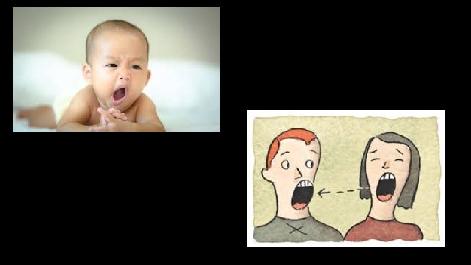
Nonrespiratory Air (Gas) Movements • Can be caused by reflexes or voluntary actions • Examples: • • • Cough and sneeze—clears lungs of debris Crying—emotionally induced mechanism Laughing—similar to crying Hiccup—sudden inspirations Yawn—very deep inspiration https: //www. youtube. com/watch? v=h 53 Q 0 tsl. R 9 Y