Today is a great day to learn about

Today is a great day to learn about your BRAIN!!!

Human Brain

How do we know about the brain? • EEG- electrical activity of brain • CAT- 3 D images • MRI- image of radio waves/ Hydrogen atom energy • PET- visual image of trace chemical activity after injection of radioactive substance
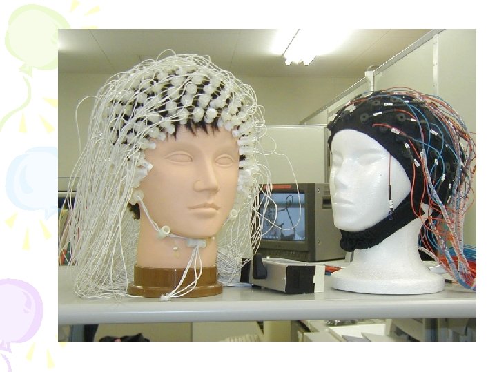
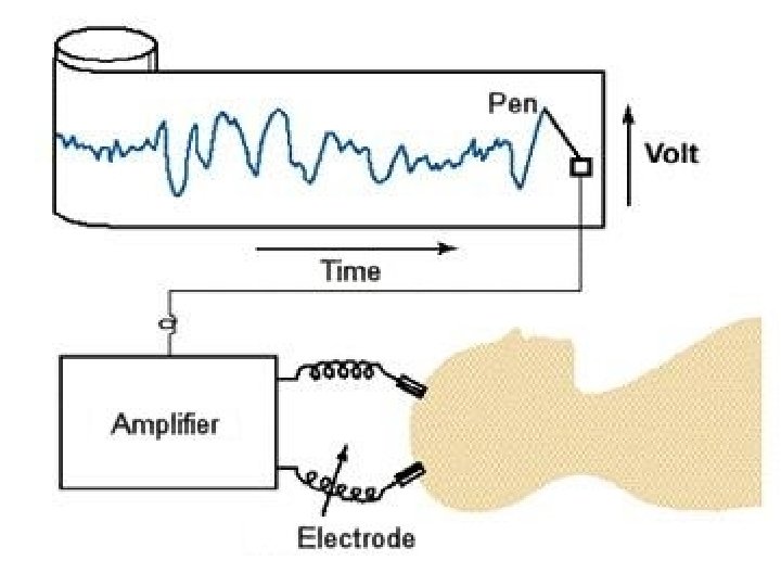
 • Electrodes are placed on the scalp that amplify recordings")
• Electroencephalogram (EEG) • Electrodes are placed on the scalp that amplify recordings of the waves of electrical activity across the brain’s surface
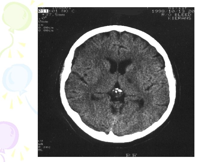
 • A series of X-ray photographs")
• Computed Tomography (CT or CAT Scan) • A series of X-ray photographs taken from different angles and combined by computer into a composite representation of the brain
 • A visual display of brain activity")
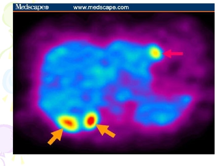
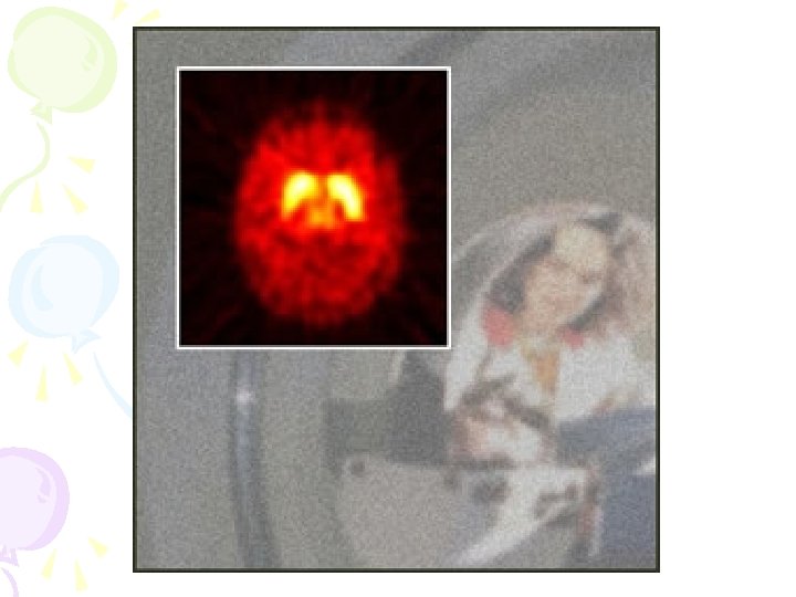
• Position Emission Tomography (PET Scan) • A visual display of brain activity that detects where a radioactive form of glucose goes while the brain performs a given task
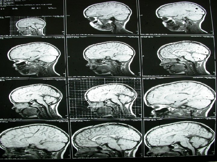
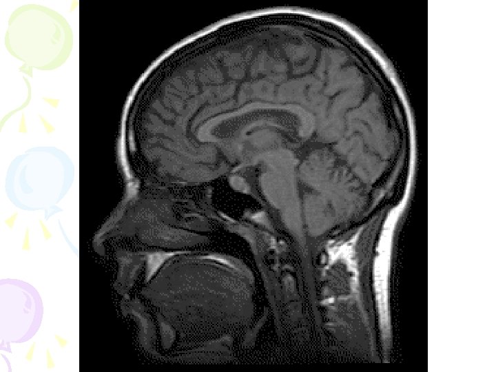
 • A technique that uses magnetic fields and")
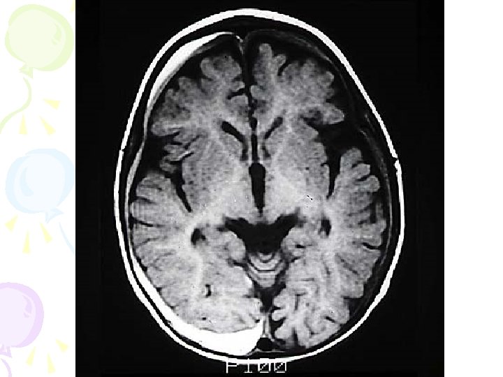
• Magnetic Resonance Imaging (MRI) • A technique that uses magnetic fields and radio waves to produce computer-generated images that allow us to see structures within the brain

• Accidents • Case study analysis of victims of suffer from a brain injury, resulting in variations in normal behavior • IE. Phineas Gage

• Lesions • Lesioning is the removal or destruction of part of the brain. • IE. Lobotomy

Let’s Review • Get the most important idea worksheet • At your tables come up with what you think is the most important idea and then 5 supporting ideas

THE BRAIN AND ITS FUNCTIONS

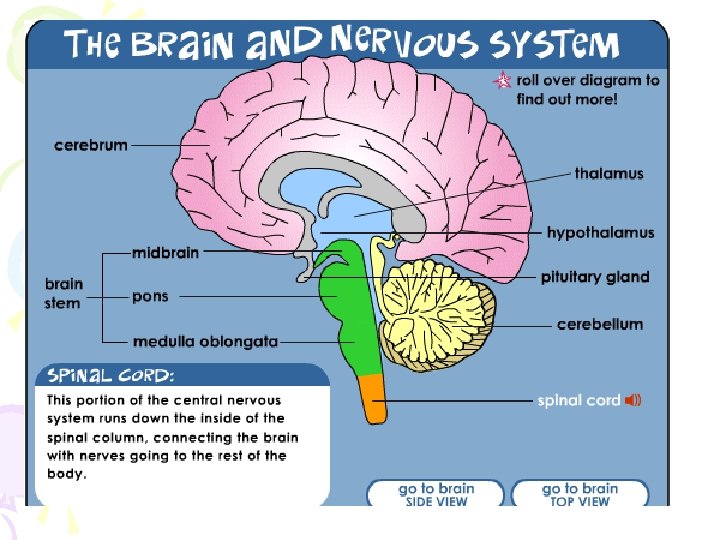
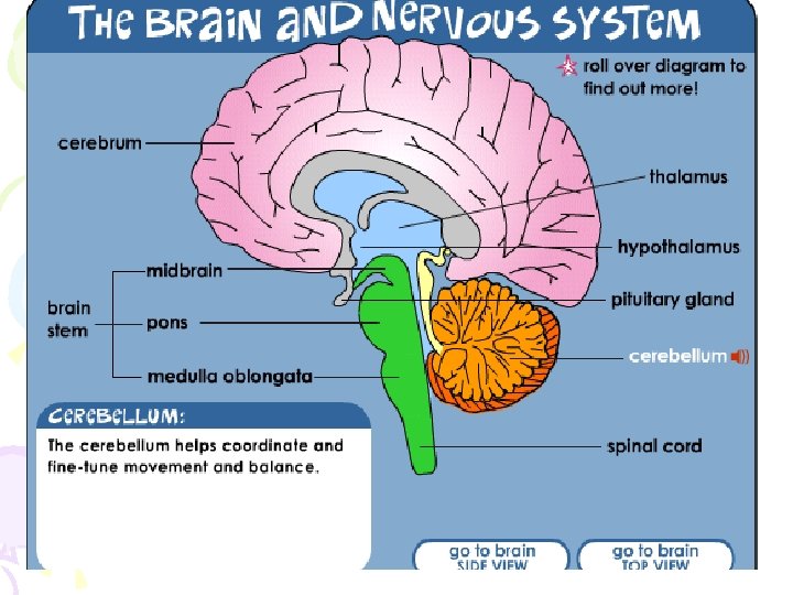
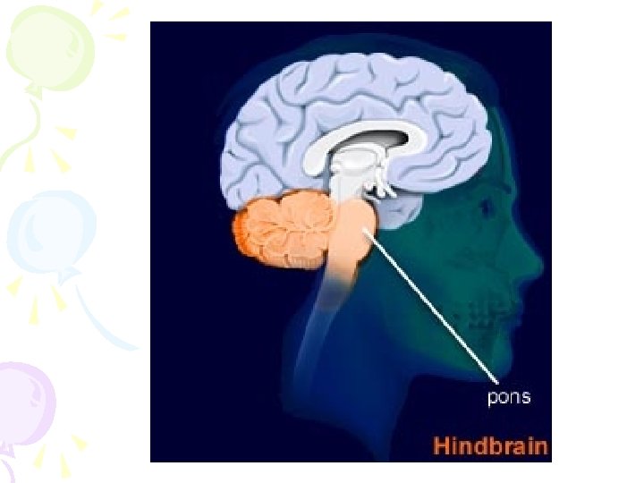
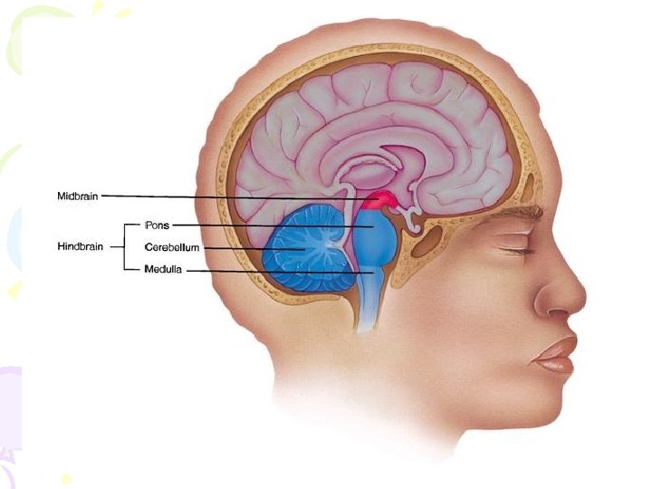
• Brain Stem • Medulla • Pons • cerebellum • Reticular Formation • Spinal cord • Midbrain

Spinal cord • Connects the brain to the body • Spinal reflexes occur here

Brainstem • The oldest part of the brain • Is responsible for automatic survival functions • Located where the spinal cord swells and the brain just begins


Midbrain • The MIDBRAIN is also responsible for behaviors associated with hearing and sight • Pupil dilation and eyeball movement
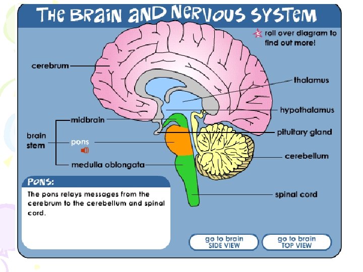

PONS • The PONS is responsible for helping to regulate breathing, to help with sleep and wake cycles, and controls facial expressions
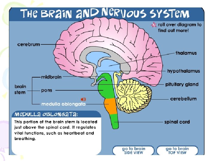

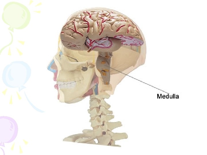
Medulla • The base of the brainstem • Controls life-supporting functions like heartbeat and breathing • Damage to this area can lead to death.
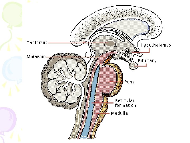

Medulla • The point at which the spinal cord enters the skull is called the MEDULLA • The MEDULLA controls heartbeat and breathing, blood pressure, and attention

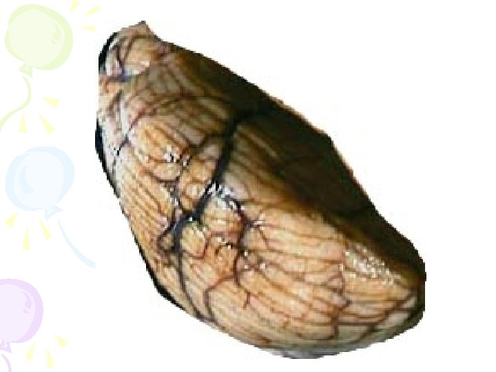
Cerebellum • Latin for the “little brain” • Located in the rear of the brain • Helps coordinate voluntary movements and balance • If damaged, the person could perform basic movements but would lose fine coordination skills.

Reticular formation • The major area of the Midbrain is the RETICULAR FORMATION • The RETICULAR FORMATION extends from the spine to the thalamus, and is responsible for arousal/wakefulness and attentiveness
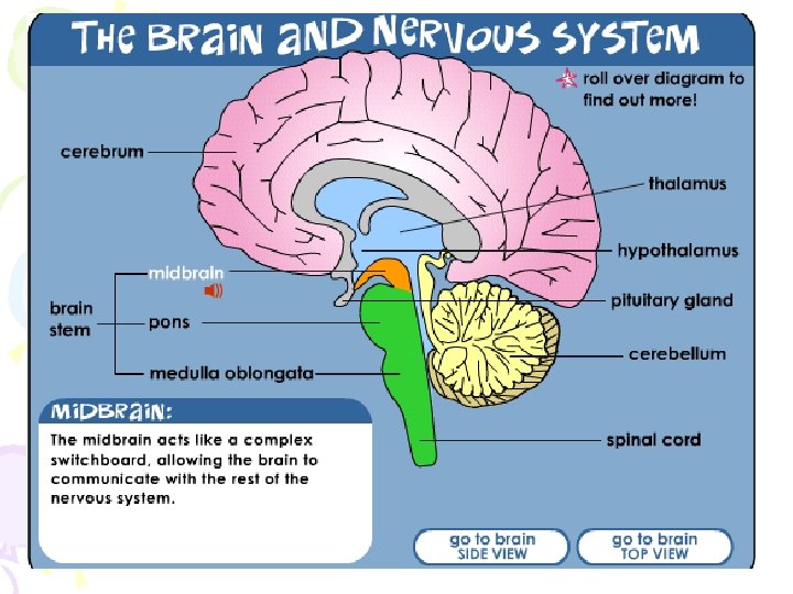

• The MIDBRAIN is also responsible for behaviors associated with hearing and sight • Pupil dilation and eyeball movement

Okay let’s teach • Time to move • Shake hands with 4 different people. • Next touch 6 different walls • Stop await instructions

Partner Up • Get a simile summary for the group of two • You may pick any part of the hindbrain and make up a simile • Have fun with this!

• Welcome to the Limbic System

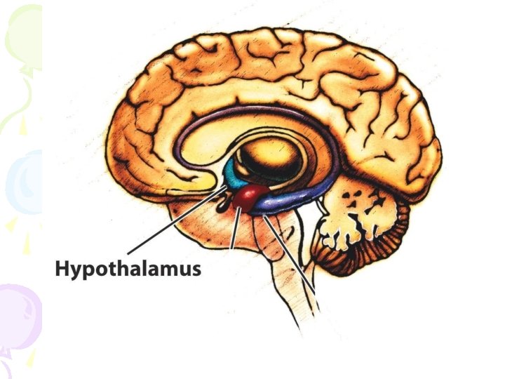
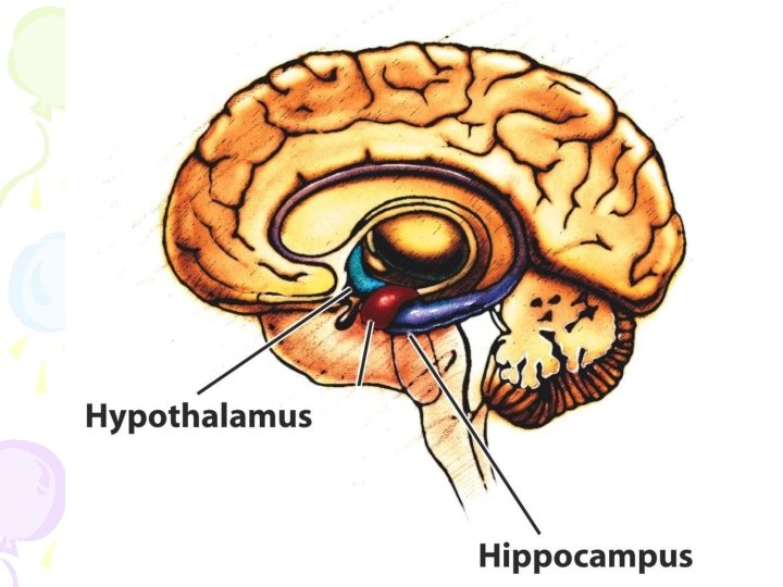
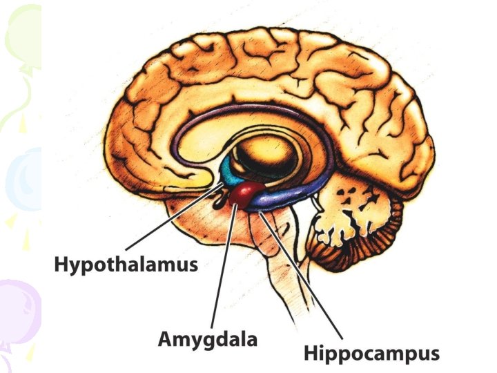
Limbic System • A ring of structures around the thalamus; at the border of the brainstem and cerebral cortex • Helps regulate memory, aggression, fear, hunger, and thirst • Includes the hypothalamus, hippocampus, and amygdala
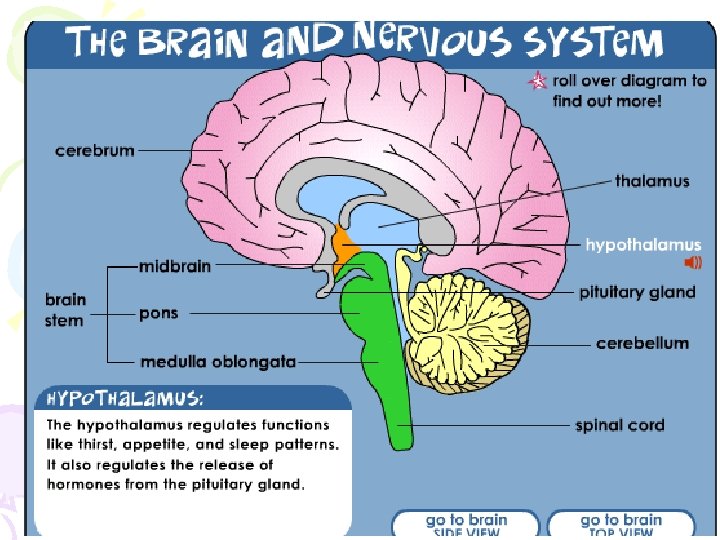

Hypothalamus • Located directly under the front of the thalamus • Regulates eating, drinking, body temperature, and the fight or flight reactions to stress • Plays a role in emotions, pleasure, and sexual function
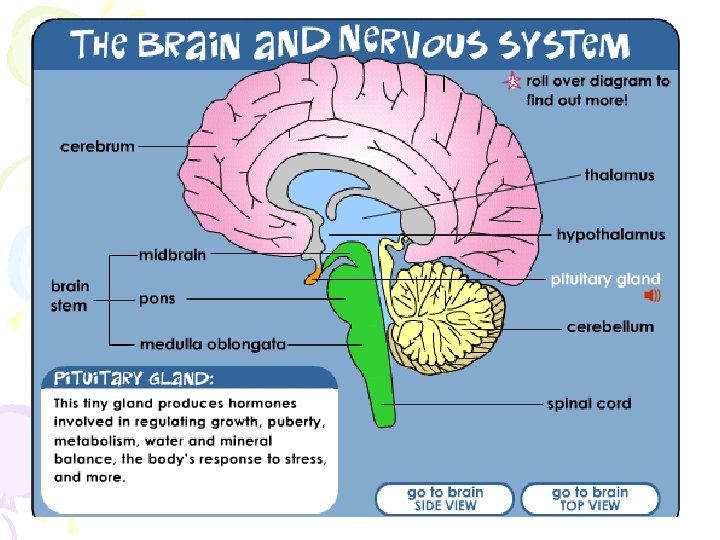

Pituitary Gland • Master gland • Part of the endocrine system • Regulates all glands within the body

Hippocampus • Wraps around the back of the thalamus • Plays a role in processing new memories for permanent storage • Looks something like a seahorse – Hippo is Greek for “horse. ”

Amygdala • Two almond shaped structures • Controls emotional responses such as fear and anger
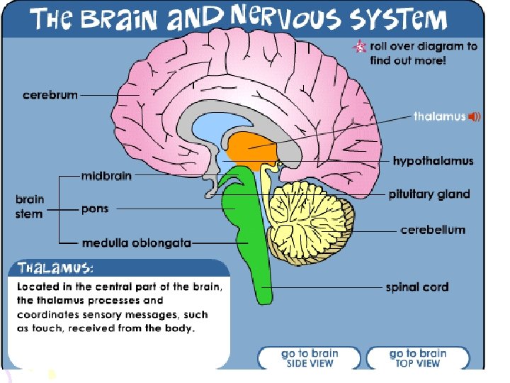

Thalamus • Sits atop the brainstem • The brain’s sensory switchboard -directs messages to the sensory receiving areas in the cortex • Excluding smell • Thalamus is Greek for “inner chamber. ”

Time to process

Cerebrum / Cerebral Cortex


Cerebrum • Longest part of brain • Two hemispheres • Responsible for voluntary movement, speech, emotion, memory intelligence and memory processing

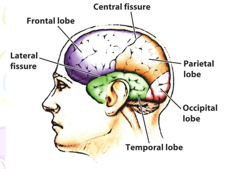
Cerebral Cortex • The body’s ultimate control and information processing center • Covers the brain’s lower level structures • Contains an estimated 30 billion nerve cells • Divided into four lobes
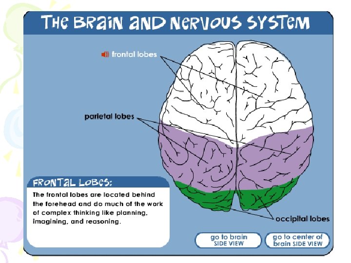
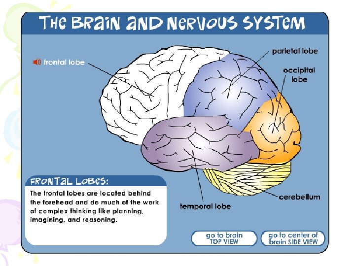

Frontal Lobes • The portion of the cerebral cortex lying just behind the forehead • Is involved in making plans and judgments
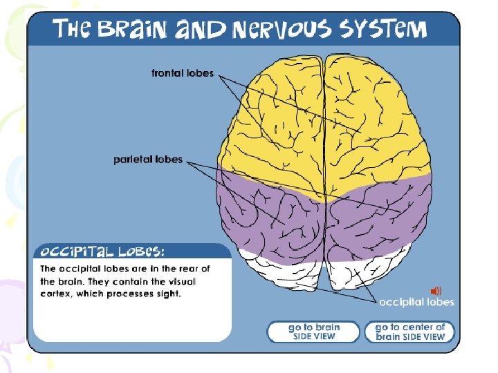

Occipital Lobe • The primary visual processing area • Located in the back of the head
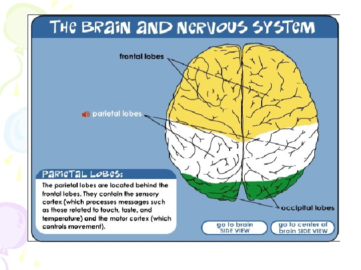
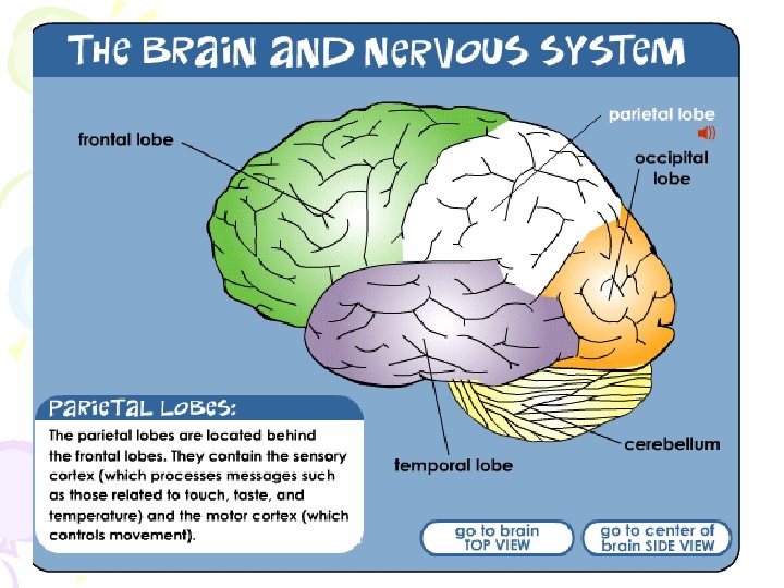

Parietal Lobes • Regions available for general processing, including mathematical reasoning • Designated as the association lobes • Behind the frontal lobes • Processes taste, smell, and feeling • Motion – motor cortex
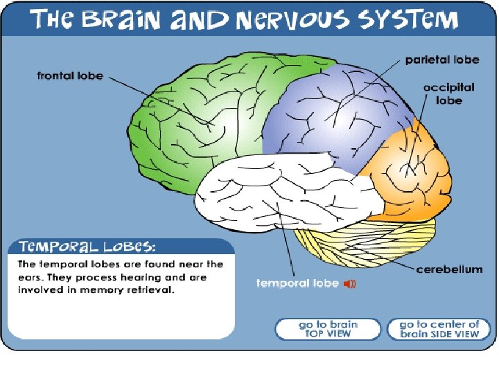

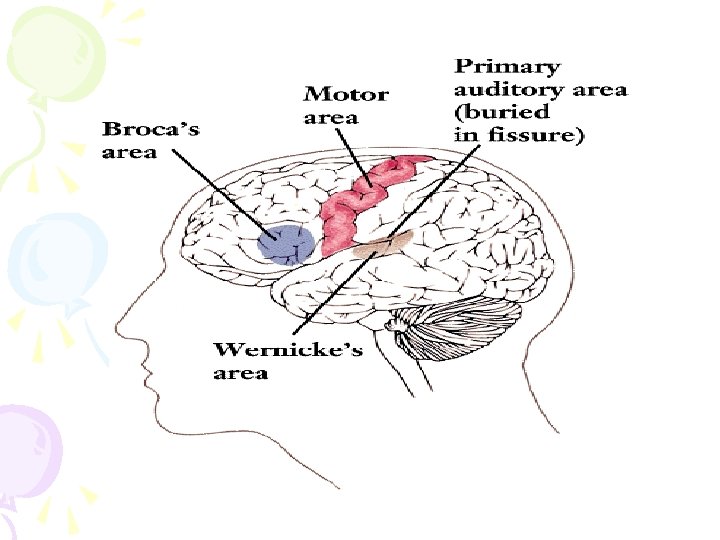
Temporal Lobes • Includes the auditory cortex where sound information is processed • Memories are processed and stored here • Located roughly above the ears

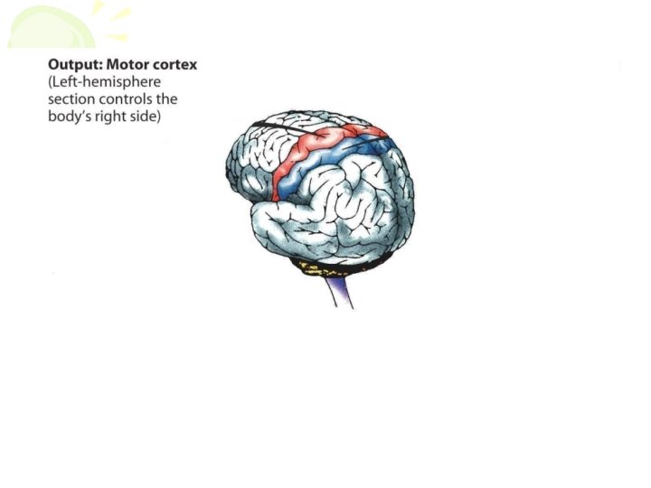
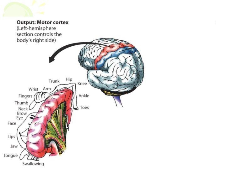
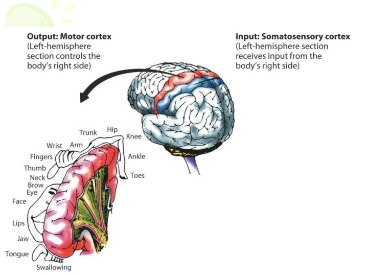
Motor Cortex • Area at the rear of the frontal lobes • Controls voluntary movement • Different parts of the cortex control different parts of the body. • The motor cortex in the left hemisphere controls the right side of the body and visa versa.

Somatosensory Cortex • Located in the front of the parietal lobes • Registers and processes body senses • Soma is Greek for “body. ”

Module 8: The Brain Hemispheric Differences

Hemispheric Differences • “Left-brained” and “right-brained” debunked • Brain is divided into two hemispheres but works as a single entity. • Both sides continually communicate via the corpus callous, except in those with split brains.

Module 8: The Brain Hemispheric Differences: Language and Spatial Abilities

The Brain’s Left Hemisphere • For most people, language functions are in the left hemisphere. • For a small percentage of people, language functions are in the right hemisphere.

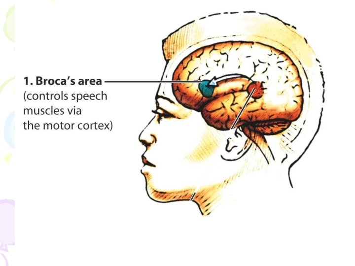
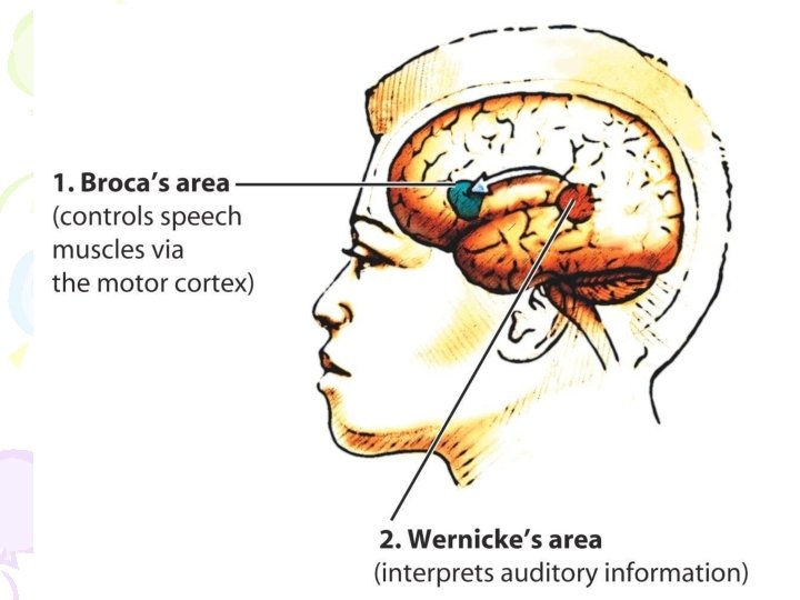
Broca’s Area • Located in the frontal lobe and usually in the left hemisphere • Responsible for the muscle movements of speech • If damaged the person can form the ideas but cannot express them as speech

PET Scan of Broca’s Area

Wernicke’s Area • Located in the temporal lobe • Involved in language comprehension and expression; our ability to understand what is said to us • Usually in the left temporal lobe

PET Scan of Wernicke’s Area

The Brain’s Right Hemisphere • Houses the brain’s spatial abilities • Our spatial ability allows us to perceive or organize things in a given space, judge distance, etc. • Helps in making connections between words

Module 8: The Brain Plasticity

Plasticity • The ability of the brain tissue to take on new functions • Greatest in childhood • Important if parts of the brain are damaged or destroyed

The End
- Slides: 92