TIU Faculty of Science Medical Analysis Department Microbial

TIU - Faculty of Science Medical Analysis Department Microbial Physiology Heshu J. Ahmed/Assist. Lecturer _______________________ Fungal Culture – Stage /1 st Semester Heshu. jalal@tiu. edu. iq https: //tiu. edu. iq/ 2020 - 2021

Introduction • To confirm clinical suspicions to establish fungal cause of disease. To help in: • Choosing a therapeutic agent • Monitoring the course of disease • Confirming mycological cure

Types and collection of specimens • Specimen collection depends on the corresponding disease. • Very important to proceed for a final diagnosis.
 Superficial Mycosis • Dermatophytic lesion- Spreads outward in a concentric fashion – scrape")
A) Superficial Mycosis • Dermatophytic lesion- Spreads outward in a concentric fashion – scrape outwards from the edge of the lesion with a scalpel blade at 90 degree angle or use Cellophane tape (when scaling is less). • Scalp lesion – Scraping with a blunt scalpel, including hair stubs, scales & contents of plugged follicles. Cutting hair is seldom useful.
 Superficial Mycosis • Onychomycosis- Stop antifungal one week prior to collection. Sample should")
A) Superficial Mycosis • Onychomycosis- Stop antifungal one week prior to collection. Sample should be taken near the base of the nail as fungus in distal ends is not viable: include full thickness of the nail. • Mucosal infections – mucosal scrapings are preferred over swabs.
 Subcutaneous mycosis • Pus aspirates and biopsy are valuable. Biopsy should be avoided")
B) Subcutaneous mycosis • Pus aspirates and biopsy are valuable. Biopsy should be avoided in sporotrichosis as it leads to spread of infection and hinder healing.
 Systemic Mycosis • • Pus Biopsy Feces Urine Sputum CSF Blood Scrapings or")
C) Systemic Mycosis • • Pus Biopsy Feces Urine Sputum CSF Blood Scrapings or swabs from the edge of lesions.

Diagnosis • • • Direct examination Fungal culture Serological tests Skin tests PCR & other molecular methods

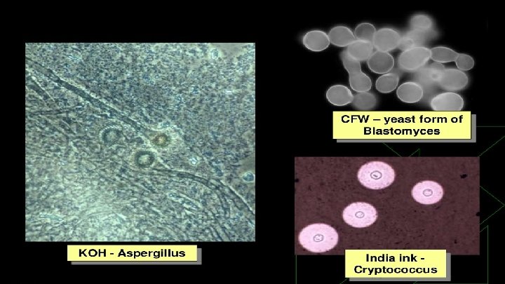
Direct Examination • Very decisive in the diagnosis of fungal infections. • Wet Mounts • Slide and tube KOH mounts- 10 or 20 % of KOH for 5 -20 minutes helps in the digestion of proteins debris and dissolving keratin. • Calcofluor white- fluorescent stain- stain binds to glucan and chitins which are abundant in fungal cell well. • India Ink – capsulated fungi.

Direct Examination • Gram stain- Fungi are gram positive. • Histopathology • Superficial infection, subcutaneous & systemic infections • Routine Stain- Hematoxylin & Eosin (HE) • Special Stains- PAS ( periodic acid), GMS , Mayer’s mucicarmine, Gridley’s stain. • Fluorescent – antibody staining- to detect fungal Ag in clinical specimen such as pus, blood, CSF, tissue sections.
 Corn Meal Agar (CMA) Bird seed")
Fungal Culture • • Sabouraud Dextrose Agar (SDA) Corn Meal Agar (CMA) Bird seed agar Brain Heart Infusion (BHI) agar

Fungal Culture • Temperature requirement • Majority of fungi – 37 C • Superficial mycosis – 30 C • Dimorphic mycosis – 25 & 37 C • Incubation time • At least 4 weeks • Usually positive cultures are obtained in 7 -10 days • Candida & Aspergillus – 24 to 72 hrs

Fungal Culture • Specimens should be cultured on agar slants: • • Safe Require less space More resistant to drying during prolonged incubation Blood cultures should be incubated in to biphasic blood culture bottles
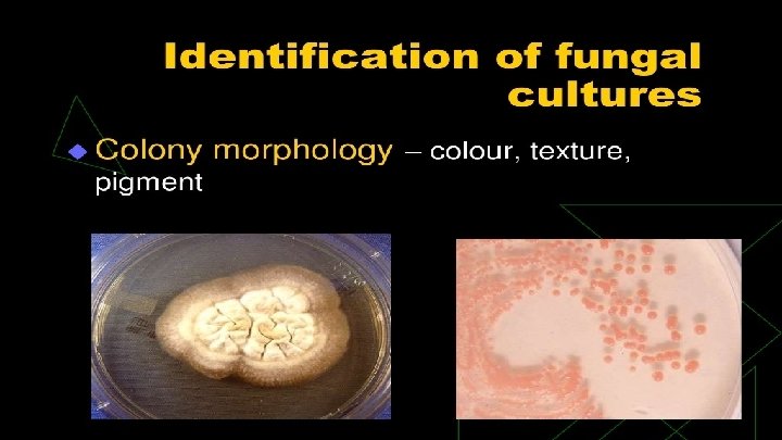

Identification of fungal cultures • Colony morphology – color, texture, pigment • Fungal Morphology- under microscope- using Lactophenol Cotton Blue (LPCB) stain • Composition of LPCB • Lactic acid – preserves fungal structure • Phenol- kills any living organism • Glycerol- prevents drying • Cotton blue- imparts blue color to structure

Identification of fungal cultures • Special culture techniques- slide culture to see sporing structures & spore arrangement, CHROM agar for candida specific. • Biochemicals – ability to assimilate carbon & nitrogen, sugar fermentation.

Serology • Detection of Ab or Ag in serum or body fluids § § Agglutinations Immunodiffusion Indirect fluorescent Ab detection ELISA, RIA

Skin tests Detects delayed hypersensitivity. Shown by occurrence of induration and erythema withing 24 to 72 hours following intradermal inoculation of antigen. • • Histoplasmosis Candidiasis Blastomycosis Sporotrichosis Histoplasmin Candidin Blastomycin Sporotrichin

Other Methods • • • PCR- Polymerase Chain Reaction RFLP- Restriction fragment length Protein electrophoresis Nucleic acid probes Serotyping Karyotyping

Thank you
- Slides: 21