Tissues Groups of cells with similar structure and


 Diagram: Transitional Photomicrograph: Transitional epithelium")


- Slides: 17
• Tissues – Groups of cells with similar structure and function – Four primary types • Epithelial tissue (epithelium) • Connective tissue • Muscle tissue • Nervous tissue Body Tissues
Epithelial Tissues • Locations – Body coverings – Body linings – Glandular tissue • Functions – Protection – Absorption – Filtration / Excretion – Secretion
Epithelium Characteristics • Cells fit closely together and often form sheets – Tight junctions • The apical surface is the free surface of the tissue • The lower surface of the epithelium rests on a basement membrane (acellular) • Avascular (no blood supply) • Regenerate easily if well nourished – Through what process?
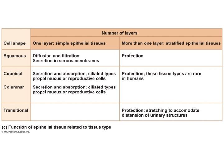
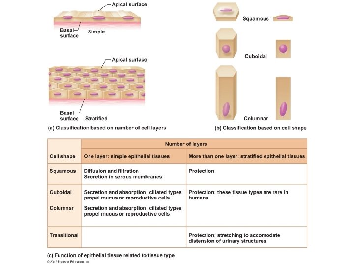
Classification of Epithelia • Number of cell layers – Simple—one layer – Stratified—more than one layer Basal surface Apical surface Simple Apical surface Basal surface Stratified (a) Classification based on number of cell layers Figure 3. 17 a
Classification of Epithelia • Shape of cells – Squamous • flattened – Cuboidal • cube-shaped – Columnar • column-like
Simple Epithelia • Simple squamous – Single layer of flat cells – Location - usually forms membranes • Lines body cavities • Lines lungs and capillaries – Functions in diffusion, filtration, or secretion in membranes – g http: //ww w. youtube. com/watch ? v=df 3 RL 0 Ki. Ug
Simple Epithelia • Simple cuboidal – Single layer of cube-like cells – Locations • Common in glands and their ducts (ex: salavary glands) • Forms walls of kidney tubules • Covers the ovaries - Functions – secretion & absorption Simple cuboidal epithelial cells Basement membrane Nucleus of simple cuboidal epithelial cell Basement membrane (b) Diagram: Simple cuboidal Connective tissue Photomicrograph: Simple cuboidal epithelium in kidney tubules (250×).
Simple Epithelia http: //www. youtube. c om/watch? v=u 7 y. Gj 6 i 5 l. BA • Simple columnar Video shows brush – Single layer of tall cells border of small intestine – Often includes mucus-producing goblet cells – Location - lines digestive tract – Functions in secretion and absorption; ciliated types propel mucus or reproductive cells; those located in intestines contain microvilli to increase the surface area for more absorption. Simple columnar epithelial cell Goblet cell Nucleus of simple columnar epithelial cell Basement membrane (c) Diagram: Simple columnar Basement membrane Connective tissue Photomicrograph: Simple columnar epithelium of the small intestine (430×).
Simple Epithelia http: //www. youtube. com/ watch? v=mi. EElu. Vlem. Q • Pseudostratified columnar Video shows epithelial tissue lining trachea – Single layer, but some cells are shorter than others – Often looks like a double layer of cells but all cells rest on the basement membrane – Location - respiratory tract, where it is ciliated – Functions in absorption or secretion; contain goblet cells for secretion of mucus http: //www. youtube. com/watch? v=FQwqhblxz 3 I Cilia Pseudostratified epithelial layer Basement membrane (d) Diagram: Pseudostratified (ciliated) columnar Basement membrane Connective tissue Photomicrograph: Pseudostratified ciliated columnar epithelium lining the human trachea (430×).
Stratified Epithelia • Stratified squamous – Cells at the apical surface are flattened – Functions as a protective covering where friction is common – Locations - lining of the: • Skin • Mouth • Esophagus Nuclei Stratified squamous epithelium Basement membrane (e) Diagram: Stratified squamous Photomicrograph: Stratified squamous epithelium lining of the esophagus (140×). Basement membrane Connective tissue
Stratified Epithelia • Stratified cuboidal—two layers of cuboidal cells; functions in protection • Stratified columnar —surface cells are columnar, cells underneath vary in size and shape; functions in protection Both rare in humans
Stratified Epithelia • Transitional epithelium – Composed of modified stratified squamous epithelium – Shape of cells depends upon the amount of stretching – Functions in stretching and the ability to return to normal shape – Location - lines organs of the urinary system
Transitional epithelium Basement membrane Transitional epithelium Connective tissue (f) Diagram: Transitional Photomicrograph: Transitional epithelium lining of the bladder, relaxed state (215×); surface rounded cells flatten and elongate when the bladder fills with urine. Figure 3. 18 f
Glandular Epithelium • Gland – One or more cells responsible for secreting a particular product – Secretions contain protein molecules in an aqueous (water-based) fluid
Glandular Epithelium • Two major gland types – Endocrine gland • Ductless since secretions diffuse into blood vessels • All secretions are hormones – Exocrine gland • Secretions empty through ducts to the epithelial surface • Include sweat and oil glands