Tissues Chapter 5 http asweknowit netimagesedudwa 520 tissues


 • Shape of")


")
 Adipose Reticular dense 2.")
 tissue (fig 5 -13) – Stretchable –")
 – Contains mainly fat cells")
 – 3 D web of")
")


- Slides: 36
Tissues Chapter 5 http: //asweknowit. net/images_edu/dwa 5%20 tissues. jpg
4 Types of Tissues All tissues can be classified into four major categories based on structure and function: 1. Epithelial: Covers and protect body surfaces, lines body cavities, moves substances in and out of blood (secretion, excretion & absorption), form glands 2. Connective: support, connection, transport, protection 3. Muscle: moves the body & its parts; specialized for contractility 4. Nervous: provides communication between body parts and coordinates body functions
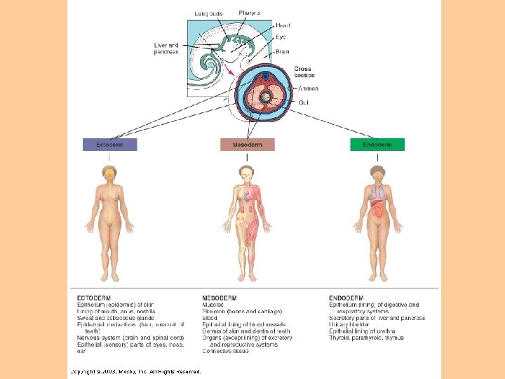
Embryonic Development • Zygote becomes a blastocyst through mitotic division • Cells of the blastocyst regroup into primary germ layers – Endoderm, mesoderm, ectoderm – Gastrulation – Histogenesis
Epithelial Tissue Subdivided into 2 types: 1. Membranous – Covers the body & some of its parts – Lines body cavities (pleural, pericardial, peritoneal), blood vessels, respiratory, digestive and genitourinary tracts 2. Glandular – Form the secretion units of the endocrine & exocrine glands
Epithelial Tissue Functions of epithelial tissues: 1. Protection – Ex: skin protects body from injury & disease-causing micro-organisms 2. Sensory – Epithelial structures that specialize in sensory functions found in skin, nose, eye, ear 3. Secretion – Glandular epithelium secrete hormones, digestive juices & sweat 4. Absorption – Ex: gut absorbs nutrients; exchange of respiratory gases 5. Excretion – Ex: kidney tubules concentrate & excrete urine and other waste products
Epithelial Tissue • Basement membrane – Thin, noncellular layer of adhesive – Connects epithelial tissue and underlying connective tissue • Avascular – “without” vascular – Epithelial cells do not have blood vessels – Oxygen & nutrients diffuse from capillaries through connective tissue & basement membrane to epithelial cells
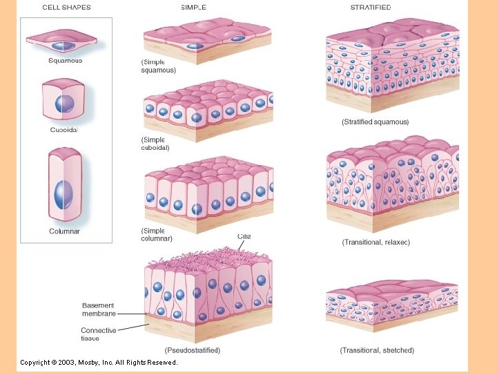
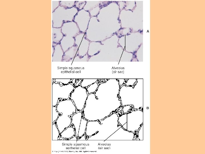
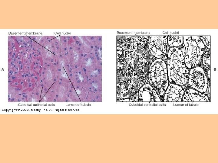
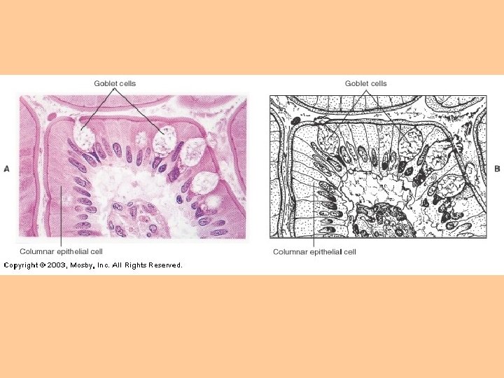
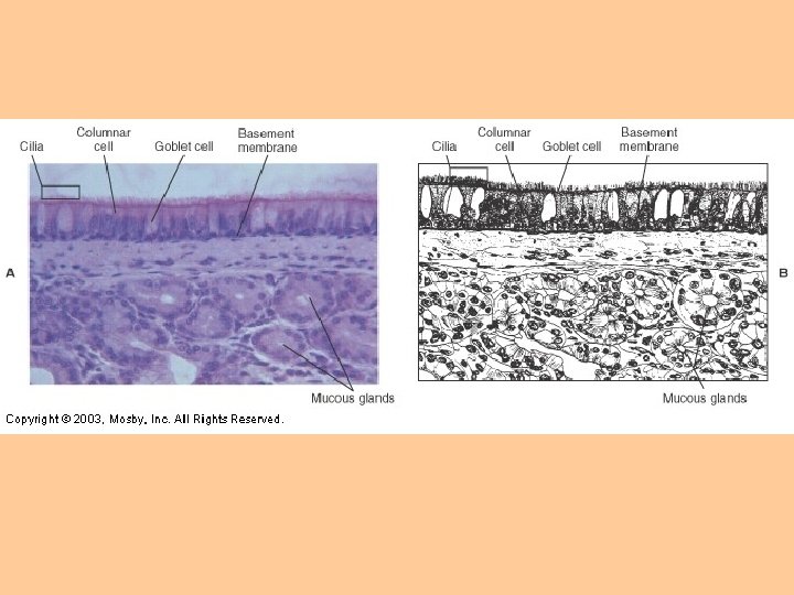
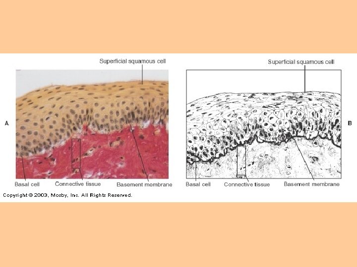
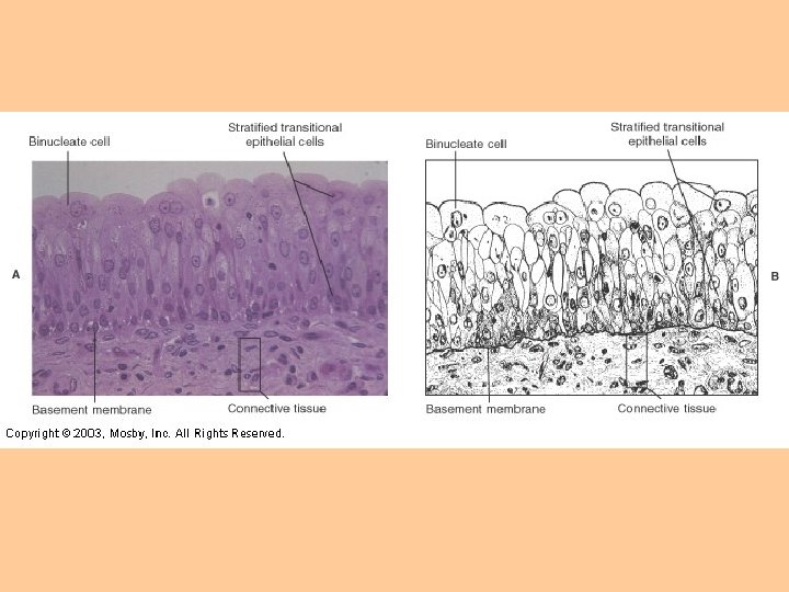
Classification of Membranous Epithelial Tissue • Cell Shape – Squamous: flat, plate-like – Cuboidal: cube-shaped; larger cytoplasm – Columnar: narrow and cylinder-shaped – Pseudostratified: single-layered; all cells touch the basement membrane but may not extend to the top of the membrane • Layers of Cells – Simple: single layer – Stratified: cells are layered on top of one another – Transitional: cell shape & layers differ
Glandular Epithelium • Specialized for secretory activity • Unicellular glands – Single celled – Ex: goblet cells • Multicellular glands – Function in clusters, solid cords or specialized follicles
Endocrine vs Exocrine • All glands are classified as endocrine or exocrine • Exocrine glands – Discharge/secrete into ducts – Ex: salivary glands • Endocrine glands – “ductless glands” – Secrete hormones directly into blood or interstitial fluid – Ex: pituitary and thyroid glands
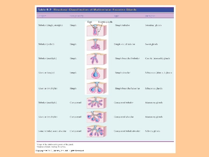
Structural Classification of Exocrine Glands • (Table 5 -2, p. 133) • Shape of gland: – Tubular – Alveolar (sac-like) • Complexity of gland: – Simple (one duct) – Compound – > 2 ducts (branched)
Functional Classification of Exocrine Glands 1. Apocrine – – Collect secretory products at apex (tip) Apex of cell pinches off Cell repairs itself & repeats process Ex: milk-producing mammary glands 2. Holocrine – – – Collect secretory product inside the cell Rupture to release (self-destructs) Ex: sebaceous glands (oil glands) 3. Merocrine – – – Discharge through plasma membrane This type applies to most exocrine glands Ex: salivary glands
Figure 5 -12, p. 132
Connective Tissue • Most widespread tissue in the body • Functions: – Connection – Support – Transport – Protection – Insulation
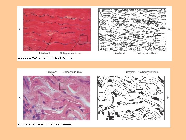
Characteristics of Connective Tissue • • Common origin – mesoderm Matrix – Intercellular material – Few cells, fibers, fluid, ground substance (material between cells) – Fibers: 1. Collagenous fibers 2. Reticular fibers 3. Elastic fibers
Fibers 1. Collagenous fibers – – – “white fibers” Made of collagen (fibrous protein) Tough, strong 2. Reticular fibers – – – Delicate Reticulin – protein Support small structures (ex: capillaries) 3. Elastic fibers – – – Extensible & elastic Elastin – protein Found in “stretchy” tissue (ex: cartilage of the external ear)
Classification of Connective Tissue 1. Fibrous – – Loose (areolar) Adipose Reticular dense 2. Bone 3. Cartilage – – – Hyaline Fibrocartilage elastic 4. Blood **Reference Table 5 -3, pp. 134 -135**
Fibrous Connective Tissue 1. Loose connective (areolar) tissue (fig 5 -13) – Stretchable – most abundant connective tissue in the body – Connects adjacent structures • • Ex: btwn other tissues and organs Ex: superficial fascia
Fibrous Connective Tissue 2. Adipose tissue (fig 5 -14) – Contains mainly fat cells – Supportive/protection pads around kidneys & other body structures – Storage deposit for excess food – Insulating material, conserves body heat
Fibrous Connective Tissue 3. Reticular Tissue (Fig 5 -16) – 3 D web of reticular fibers – Forms the framework of the spleen, lymph nodes & bone marrow – Meshwork filters harmful substances out of the blood
Fibrous Connective Tissue 4. Dense Fibrous Tissue (fig 5 -17, 5 -18, 5 -19) • Densely packed fibers • Regular Dense CT – – Fibers arranged in regular, parallel rows Collagen fibers Flexible, strong Tendons (muscle to bone) & ligaments (bone to bone) • Irregular Dense CT – Fibers intertwine – Withstand stress from any direction – Ex: dermis (inner layer of skin); outer capsule of kidney & spleen
Bone Tissue • We will cover this when we cover the skeletal system • Just know that bone is a type of connective tissue
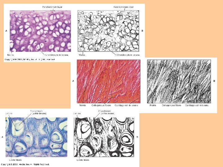
Cartilage • Only 1 cell type – chondrocyte – Located in lacuna • Avascular – receive nutrients via diffusion • Injuries to cartilage heal slowly due to poor nutrient delivery
Cartilage - Types 1. Hyaline cartilage – – – Most common Covers ends of long bones (where joints articulate) Found in supporting rings of respiratory tubes 2. Fibrocartilage – – – Strongest, most durable Intervertebral disks Menisci in knee joint 3. Elastic cartilage – – – Fine elastic fibers High degree of flexibility External ear
Blood • • Unusual type of connective tissue No ground substance Matrix = plasma (55%) Formed elements = blood cells (45%) – Erythrocytes – RBCs – Leukocytes – WBCs – Thrombocytes – platelets • Transport function – Respiratory gases, nutrients, waste products
Anthony’s Textbook of Anatomy and Physiology 17 th Edition. Thibodeau, Gary A. Ph. D and Patton, Kevin T. Ph. D. Mosby, Inc.