Tissue The Living Fabric Tissue The Living Fabric

Tissue: The Living Fabric

Tissue: The Living Fabric • Individual body cells are specialized, with each type performing specific functions that help maintain homeostasis and benefit the body as a whole. • Groups of cells that are similar in structure and perform a common or related function are called tissues (tissu = woven). • Four primary tissue types interweave to form the “fabric” of the body. These basic tissues are epithelial (ep″i-the′le-ul), connective, muscle, and nervous tissue, and each has numerous subclasses or varieties. • If we had to assign a single term to each primary tissue type that would best describe its general role, the terms would most likely be covering (epithelial), support (connective), movement (muscle), and control (nervous).
, is a sheet of")
Epithelial Tissue • Epithelial tissue, or an epithelium (plural: epithelia), is a sheet of cells that covers a body surface or lines a body cavity (epithe = laid on, covering). • It occurs in the body as (1) covering and lining epithelium and (2) glandular epithelium. • Covering and lining epithelium forms the outer layer of the skin, dips into and lines the open cavities of the cardiovascular, digestive, and respiratory systems, and covers the walls and organs of the closed ventral body cavity. • Glandular epithelium fashions the glands of the body.

• Epithelia form boundaries between different environments. For example, the epidermis of the skin lies between the inside and the outside of the body, and epithelium lining the urinary bladder separates underlying cells of the bladder wall from urine. • In its role as an interface tissue, epithelium accomplishes many functions, including (1) protection, (2) absorption, (3) filtration, (4) excretion, (5) secretion, and (6) sensory reception. • The epithelium of the skin protects underlying tissues from mechanical and chemical injury and bacterial invasion and contains nerve endings that respond to various stimuli acting at the skin surface (pressure, heat, etc. ). The epithelium lining the digestive tract is specialized to absorb substances; and that found in the kidneys performs nearly the whole functional “menu”—excretion, absorption, secretion, and filtration. Secretion is the specialty of glands.

Special Characteristics of Epithelium 1. Polarity • All epithelia have an apical surface, an upper free surface exposed to the body exterior or the cavity of an internal organ, and a lower attached basal surface. • Although some apical surfaces are smooth and slick, most have microvilli, fingerlike extensions of the plasma membrane. Microvilli tremendously increase the exposed surface area, and in epithelia that absorb or secrete substances (those lining the intestine or kidney tubules, for instance. Some epithelia, such as that lining the trachea, have motile cilia that propel substances along their free surface. • Lying adjacent to the basal surface of an epithelium is a thin supporting sheet called the basal lamina (lam′ĭ-nah; “sheet”). The basal lamina acts as a selective filter that determines which molecules diffusing from the underlying connective tissue are allowed to enter the epithelium. The basal lamina also acts as a scaffolding along which epithelial cells can migrate to repair a wound.

Special Characteristics of Epithelium 2. Specialized contacts • Except for glandular epithelia, epithelial cells fit close together to form continuous sheets. Adjacent cells are bound together at many points by lateral contacts, including tight junctions and desmosomes. The tight junctions help keep proteins in the apical region of the plasma membrane from diffusing into the basal region, and thus help to maintain epithelial polarity. 3. Supported by connective tissue • All epithelial sheets rest upon and are supported by connective tissue. Just deep to the basal lamina is the reticular lamina, a layer of extracellular material containing a fine network of collagen protein fibers that “belongs to” the underlying connective tissue. Together the two laminae form the basement membrane. The basement membrane reinforces the epithelial sheet, helping it to resist stretching and tearing forces, and defines the epithelial boundary.

Special Characteristics of Epithelium 4. Avascular but innervated • Although epithelium is innervated (supplied by nerve fibers), it is avascular (contains no blood vessels). • Epithelial cells are nourished by substances diffusing from blood vessels in the underlying connective tissue. 5. Regeneration • Epithelium has a high regenerative capacity. Some epithelia are exposed to friction and their surface cells rub off. • Others are damaged by hostile substances in the external environment (bacteria, acids, smoke). • If and when their apical-basal polarity and lateral contacts are destroyed, epithelial cells begin to reproduce themselves rapidly. As long as epithelial cells receive adequate nutrition, they can replace lost cells by cell division.

Classification of Epithelia • Each epithelium is given two names. The first name indicates the number of cell layers present; the second describes the shape of its cells. • Based on the number of cell layers, there are simple and stratified epithelia. • Simple epithelia are composed of a single cell layer. They are typically found where absorption and filtration occur and a thin epithelial barrier is desirable. • Stratified epithelia, consisting of two or more cell layers stacked one on top of the other, are common in high-abrasion areas where protection is important, such as the skin surface and the lining of the mouth.
 Classification on the basis of number of layers. (b) Classification")
Classification of Epithelia (a) Classification on the basis of number of layers. (b) Classification on the basis of cell shape: • Squamous cells (skwa′mus) are flattened and scalelike (squam = scale); • cuboidal cells (ku-boi′dahl) are boxlike, approximately astall as they are wide; and • columnar cells (kŏ-lum′nar) are tall and column shaped.

Classification of Epithelia • The nucleus of a squamous cell is a flattened disc; • that of a cuboidal cell is spherical; and • a columnar cell nucleus is elongated from top to bottom and usually located close to the cell base.

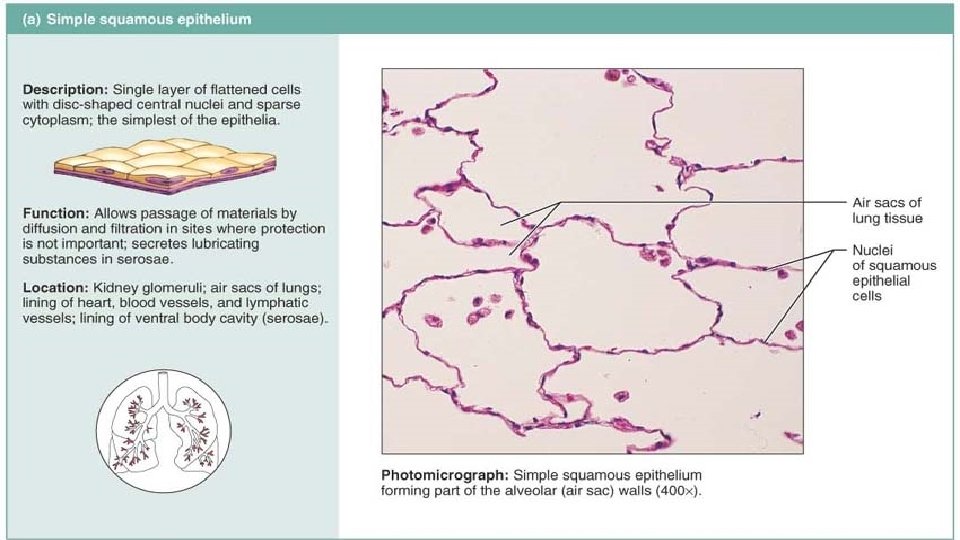
Simple Epithelia Simple Squamous Epithelium • The cells of a simple squamous epithelium are flattened laterally, and their cytoplasm is sparse. When the cells are cut perpendicular to their free surface, they resemble fried eggs seen from the side, • In the kidneys, simple squamous epithelium forms part of the filtration membrane; in the lungs, it forms the walls of the air sacs across which gas exchange occurs. • Two simple squamous epithelia in the body have special names that reflect their location. Endothelium (en″do-the′le-um; “inner covering”) provides a slick, friction-reducing lining in lymphatic vessels and in all hollow organs of the cardiovascular system—blood vessels and the heart. Capillaries consist exclusively of endothelium, and its exceptional thinness encourages the efficient exchange of nutrients and wastes between the bloodstream and surrounding tissue cells. Mesothelium (mez″o-the′le-um; “middlecovering”) is the epithelium found in serous membranes lining the ventral body cavity and covering its organs.

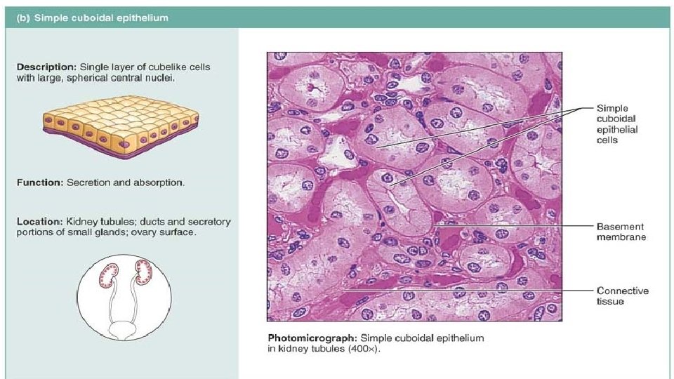
Simple Epithelia Simple Cuboidal Epithelium • Simple cuboidal epithelium consists of a single layer of cells as tall as they are wide. • The spherical nuclei stain darkly, causing the cell layer to look like a string of beads when viewed microscopically. • functions of simple cuboidal epithelium are secretion and absorption. This epithelium forms the walls of the smallest ducts of glands and of many kidney tubules.

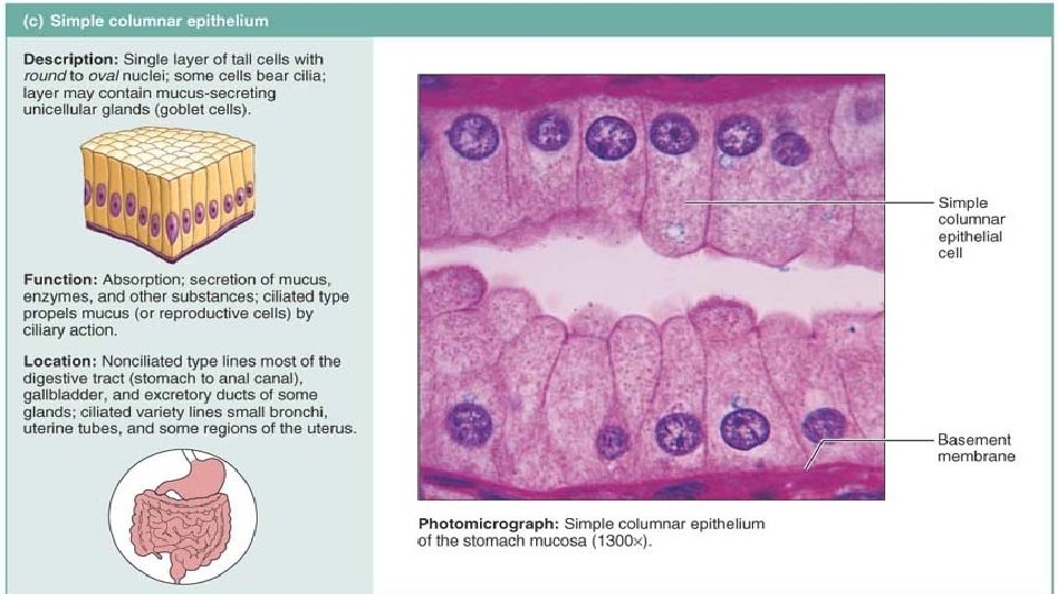
Simple Epithelia Simple Columnar Epithelium • Simple columnar epithelium is seen as a single layer of tall, closely packed cells, aligned like soldiers in a row. • It lines the digestive tract from the stomach through the rectum. Columnar cells are mostly associated with absorption and secretion, and the digestive tract lining has two distinct modifications that make it ideal for that dual function: (1) dense microvilli on the apical surface of absorptive cells and (2) goblet cells that secrete a protective lubricating mucus. • Some simple columnar epithelia display cilia on their free surfaces, which help to move substances or cells through an internal passageway.
. (a) Photomicrograph of a mucussecreting goblet")
Simple Columnar Epithelium Goblet cells (unicellular exocrine glands). (a) Photomicrograph of a mucussecreting goblet cell in the simple columnar epithelium lining the small intestine (approx. 2000×). (b) Diagram of a goblet cell. Notice the secretory vesicles and well-developed rough ER and Golgi apparatus.
")
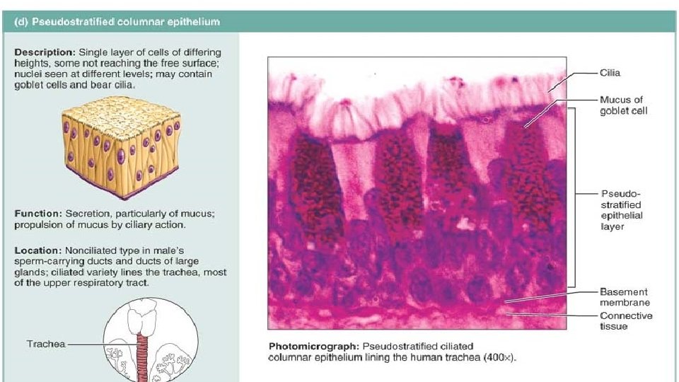
Simple Epithelia Pseudostratified Columnar Epithelium • The cells of pseudostratified columnar epithelium (soo″do-stră ′tĭ-fīd) vary in height. All of its cells rest on the basement membrane, but only the tallest reach the free surface of the epithelium. Because the cell nuclei lie at different levels above the basement membrane, the tissue gives the false (pseudo) impression that several cell layers are present; hence “pseudostratified. ” The short cells are relatively unspecialized and give rise to the taller cells. • This epithelium, like the simple columnar variety, secretes or absorbs substances. • A ciliated version containing goblet cells lines most of the respiratory tract. Here the motile cilia propel sheets of dust-trapping mucus superiorly away from the lungs.

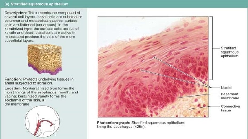
Stratified Epithelia Stratified Squamous Epithelium • Stratified squamous epithelium is the most widespread of the stratified epithelia. • Composed of several layers, it is thick and well suited for its protective role in the body. Its free surface cells are squamous. This epithelium is found in areas subjected to wear and tear, and its surface cells are constantly being rubbed away and replaced by division of its basal cells. • To avoid memorizing all its locations simply remember that this epithelium forms the external part of the skin and extends a short distance into every body opening that is directly continuous with the skin. The outer layer, or epidermis, of the skin is keratinized (ker′ah-tin″īzd), meaning its surface cells contain keratin, a tough protective protein. (The epidermis is discussed in Chapter 5. ) The other stratified squamous epithelia of the body are nonkeratinized.

Stratified Epithelia Stratified Cuboidal and Columnar Epithelia • Stratified cuboidal epithelium is quite rare in the body, mostly found in the ducts of some of the larger glands (sweat glands, mammary glands). It typically has two layers of cuboidal cells. • Stratified columnar epithelium also has a limited distribution in the body. Small amounts are found in the pharynx, the male urethra, and lining some glandular ducts. This epithelium also occurs at transition areas or junctions between two other types of epithelia. Only its apical layer of cells is columnar.

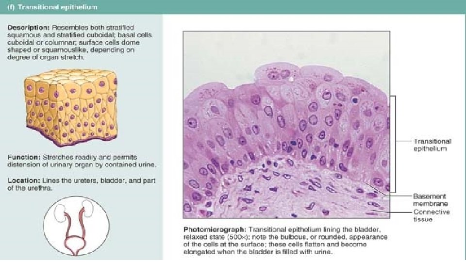
Stratified Epithelia Transitional Epithelium • Transitional epithelium forms the lining of hollow urinary organs, which stretch as they fill with urine. • Cells of its basal layer are cuboidal or columnar. • The apical cells vary in appearance, depending on the degree of distension of the organ. When the organ is distended with urine, the transitional epithelium thins from about six cell layers to three, and its domelike apical cells flatten and become squamouslike. The ability of transitional cells to change their shape (undergo “transitions”) allows a greater volume of urine to flow through a tubelike organ. In the bladder, it allows more urine to be stored.

Glandular Epithelia • A gland consists of one or more cells that make and secrete (export) a particular product. • This product, called a secretion, is an aqueous (water-based) fluid that usually contains proteins, but there is variation—for example, some glands release a lipid- or steroid-rich secretion. • Secretion is an active process; glandular cells obtain needed substances from the blood and transform them chemically into a product that is then discharged from the cell. Notice that the term secretion can refer to both the gland’s product and the process of making and releasing that product. • Glands are classified as endocrine (“internally secreting”) or exocrine (“externally secreting”) depending on where they release their product, and as unicellular (“one-celled”) or multicellular (“many-celled”) based on the relative cell number making up the gland. Unicellular glands are scattered within epithelial sheets.

Endocrine Glands • Because endocrine glands eventually lose their ducts, they are often called ductless glands. • They produce hormones, regulatory chemicals that they secrete by exocytosis directly into the extracellular space. From there the hormones enter the blood or lymphatic fluid and travel to specific target organs. • Each hormone prompts its target organ(s) to respond in some characteristic way. For example, hormones produced by certain intestinal cells cause the pancreas to release enzymes that help digest food in the digestive tract. • Their secretions are also varied, ranging from modified amino acids to peptides, glycoproteins, and steroids. Since not all endocrine glands are epithelial derivatives, we defer consideration of their structure and function

Exocrine Glands • Exocrine glands are numerous, and many of their products are familiar. All exocrine glands secrete their products onto body surfaces (skin) or into body cavities • the unicellular glands directly (by exocytosis) and the multicellular glands via an epithelium-walled duct that transports the secretion to the epithelial surface. • Exocrine glands are a diverse lot. They include mucous, sweat, oil, and salivary glands, the liver (which secretes bile), the pancreas (which synthesizes digestive enzymes), and many others.
 gland")
Unicellular Exocrine Glands • The only important example of a unicellular (or one-celled) gland is the goblet cell. True to its name, a goblet cell is shaped like a goblet (a drinking glass with a stem). • Goblet cells are sprinkled in the epithelial linings of the intestinal and respiratory tracts amid columnar cells with other functions. In humans, all such glands produce mucin (mu′sin), a complex glycoprotein that dissolves in water when secreted. Once dissolved, mucin forms mucus, a slimy coating that both protects and lubricates surfaces

Multicellular Exocrine Glands • Compared to the unicellular glands, multicellular exocrine glands are structurally more complex. • They have two basic parts: an epithelium-derived duct and a secretory unit (acinus) consisting of secretory cells. In all but the simplest glands, supportive connective tissue surrounds the secretory unit and supplies it with blood vessels and nerve fibers, and forms a fibrous capsule that extends into the gland proper and divides the gland into lobes.

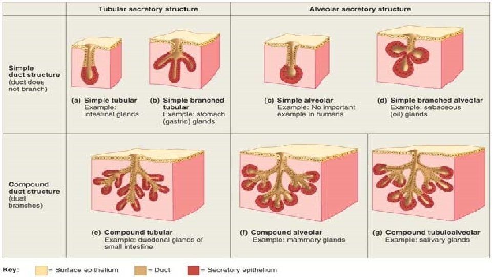
Multicellular Exocrine Glands Structural classification • On the basis of their duct structures, multicellular exocrine glands are either simple or compound. • Simple glands have an unbranched duct, whereas compound glands have a branched duct. • The glands are further categorized by their secretory units as 1. tubular if the secretory cells form tubes; 2. alveolar (al-ve′o-lar) if the secretory cells form small, flasklike sacs (alveolus = “small hollow cavity”); and 3. tubuloalveolar if they have both types of secretory units.

Multicellular Exocrine Glands Modes of secretion • Multicellular exocrine glands secrete their products in different ways, so they can also be described functionally. • Most are merocrine glands (mer′o-krin), which secrete their products by exocytosis as they are produced. The secretory cells are not altered in any way. The pancreas, most sweat glands, and salivary glands belong to this class. • Secretory cells of holocrine glands (hol′o-krin) accumulate their products within them until they rupture. (They are replaced by the division of underlying cells. ) Because holocrine gland secretions include the synthesized product plus dead cell fragments (holos = all), you could say that their cells “die for their cause. ” Sebaceous (oil) glands of the skin are the only true example of holocrine glands.

Sub-Chapter Test
- Slides: 33