Tissue Specific Exercises Tendon Tendinopathy Strong evidence exists


 in people")


 • Rapid onset generally related to load")


 Long history of minimal symptoms")
 Past history")


 showed its involvement in")


 used a high")


- Slides: 64
Tissue Specific Exercises Tendon
Tendinopathy • Strong evidence exists that the inflammation paradigm is inaccurate. • The term “tendinitis” is therefore inaccurate. • The term “tendinopathy” as a generic descriptor of tendon problems is more accurate • Evidence is mounting for a biochemical cause of pain as well as mechanical discontinuity of collagen fibers
Tendinopathy Histological examination of tendinopathy: • Absence of inflammatory cells • Poor healing response • Non-inflammatory intratendinous collagen degeneration • Fiber disorientation and thinning • Scattered vascular ingrowth Frank inflammatory lesions and granulation tissue are infrequent and are mostly associated with tendon ruptures. Tendinopathy can be viewed as a failure of the cell matrix to adapt to the stresses being put on the tissue.
Palpation tenderness • Tendons may be painful to palpation (especially in athletes) in people who do not have tendinopathy • Palpation pain does not predict diagnosis • Like other chronic conditions, there can be a disconnect between clinical presentation and abnormalities on imaging • Pain may be present without imaging change and vice versa.
UE/LE tendinopathy discrepancy • There may be important differences between UE and LE tendinopathies in terms of the contribution from peripheral tissue or central drivers • The role of central sensitization has been highlighted for rotator cuff and lateral elbow tendinopathy • Preliminary evidence that central sensitization does not occur in Achilles tendinopathy
Why the difference? • The importance of UE function for self-care and occupational activities, might contribute to these differences in central sensitization between UE and LE tendinopathies • All nociceptive input is evaluated in terms of threat • Increased threat associated with UE tendinopathy (e. g. negative thoughts and feelings arising due to fear of pain with daily activities, fear of tendon rupture, fear of unemployment) likely amplifies the pain experience
Three stages of tendinopathy • Model describes 3 distinct cases, but in reality it is a continuum • Reactive tendinopathy • Tendon dysrepair or failed healing • Degenerative tendinopathy
Evidence of Transition along the Continuum
Reactive tendinopathy • Proliferative response in the cells and ground substance following acute overloading of the tendon • The cells are activated and proliferated. They produce more proteoglycans which makes the tendon thicker • Purpose: temporarily decrease the force per unit area.
• The activated cells produce nociceptive substances, which trigger the pain response and possibly neurogenic mediated inflammation. • The integrity of the collagen is mostly maintained at this stage • If the process is not reversed, it will progress to the tendon disrepair stage
Clinical Presentation • Younger (15 -25 years) • Rapid onset generally related to load • Load substantially exceeds tendon’s previous exposure • Swelling and soft tissue proliferation • Easily aggravated by exercise, slow to settle • Painful • Uncommon
Summary • The reactive phase is a short term adaptation to tendon overload that thickens the tendon, reduces the stresses and increases stiffness • Potential to revert back to normal exists if overloading is decreased or there is sufficient time between loading sessions to allow for tendon adaptation
Tendon disrepair • Greater breakdown of the matrix • Marked increase in proteoglycans and collagen. • The increase in proteoglycans causes separation of the collagen and disorganization of the matrix • Tissue changes are often localized
Degenerative tendinopathy • Progression of both matrix and cell changes, including areas of cell death (tendinosis) • Large areas of disorganization in the matrix • Localized areas of degeneration surrounded by healthy tissue and areas of tissue in other stages of pathology • The areas of degeneration have very little capacity to recover. • Degenerative tendons can show improved function despite not returning to normal size or structure
Degenerative Pathology
Clinical Presentation Degenerative • • Older (30 -60 years) Long history of minimal symptoms Variable swelling and lumps/bumps Often exhibit unloading strategies or atrophy Not painful Common Not seen clinically, because they don’t hurt!
Clinical Presentation Reactive on Degenerative • • • Older (40 -60 yrs) Past history with load related exacerbations Onset after overload Variable swelling Painful Very common
Medication Risk Factors Certain medications play a role in causing or exacerbating tendinopathies • Statins Statin-induced tendinopathy was first reported in the 2000 s. • The predominant musculoskeletal complaint is myalgia, but tendinopathy is also a side effect • The incidence of statin-induced tendinopathy is low: 2% of all complications
Medication Risk Factors • Fluoroquinolones are a class of synthetic antibiotics, including commonly used medications such as Cipro and Levaquin. • Tendinopathy side effect not widely recognized until the early 2000’s • Estimated rate of tendinopathy as a side effect is 2% • Achilles tendon being most commonly affected • Onset is usually acute (8 days after beginning treatment)
Medication Risk Factors • Corticosteroids can impair local collagen synthesis, resulting in tendon atrophy, reduction of tensile strength, and increased chance of failure • Local steroid injections in the vicinity of a tendon are discouraged due to the concerns with respect to rupture
Imaging Best done when: • The case is complicated and chronic • PT has failed • Examination has identified differential diagnosis in need of exclusion
Diagnostic ultrasound • To establish a bottom line • To look for involvement of paratenon, bursa and plantaris muscle • Not good for imaging the fat pad. If you suspect fat pad involvement, you need to do an MRI
Imaging • No evidence to support the notion that the tendon does improve structurally on imaging as a result of good PT • The relation between objective changes on imaging, and complaints of pain, is tenuous at best • Functionally they will improve, but minimal changes will be noted on imaging
Why is that? • Changes in the pain system take place faster than the restoration of the tendon structure. • The healing process for the tendon structure is simply different from that of pain, but no less important • This puts even more importance on adherence to a tendon training home program long after they have been discharged from PT
What causes tendon pain? • The clinical presentation of tendinopathy is not just defined by local tendon pathology (structure) but also by the integration of the pain system and motor impairments. Coombes BK, Bisset L, Vicenzino B. A new integrative model of lateral epicondylalgia. Br J Sports Med 2009: 43: 252 -258
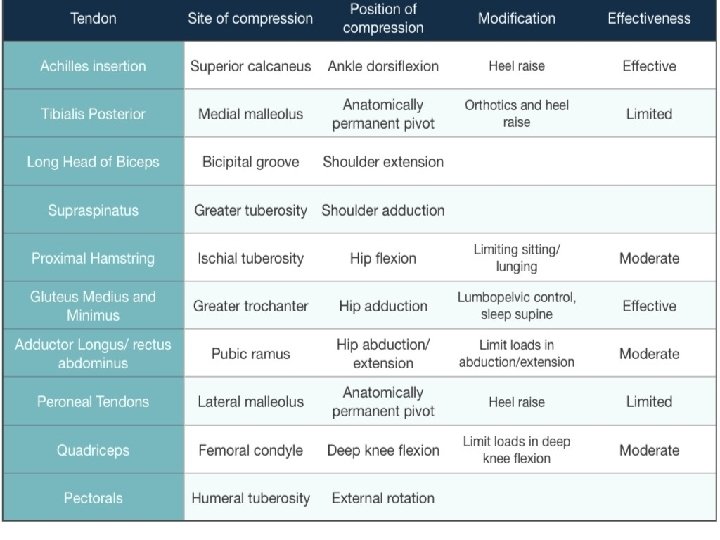
Differential Dx Achilles tendinopathy • When patient fails to improve with traditional approaches to treatment and has symptoms of pain on the medial aspect of the Achilles tendon, consider involvement of the plantaris tendon
Plantaris • Functions as a weak plantar flexor at the ankle and knee flexor • Originates from the lateral aspect of the femur and inserts onto the medial aspect of the calcaneus • The insertion point of the plantaris muscle can vary with up to 9 different insertion points • It is also possible to not have a plantaris muscle at all • Therefore, it has long been considered a non-important muscle and has been given little attention other than as a good tendon to use for reconstruction-type surgeries.
Plantaris involvement • A study from Alfredson et al (2011) showed its involvement in “Achilles pain • Due to the proximity with which the plantaris tendon and the Achilles tendon can be situated, peritendinous tissue can become irritated or compressed against the Achilles tendon, creating pain along the medial aspect of the Achilles.
Plantaris Involvement • Can be connected to the Achilles tendon • Plantaris is stiffer than Achilles, and can become tendinopathic first • Usually well localized medial Achilles pain on load • Chronic ones can present like Achilles • Can be symptomatic at any stage, usually with overload
Plantaris involvement • If patient is not improving with 6 weeks of a solid tendon loading program, and the complaints of pain are mainly over the medial portion of the Achilles tendon, consider involvement of the plantaris tendon • Ultrasound imaging can be used to confirm the painful structure. Treatment: ü surgical intervention to scrape the tendons ü removal of the plantaris tendon ü injection of a substance to help separate the tissues.
Treatment - Medication • Use of NSAIDs has been controversial • Reduces pain, but has been reported to slow tissue healing • However: in reactive tendinopathy slowing the healing process might actually be desirable, to slow cellular upregulation and excessive ground substance manufacture • In the reactive phase, it is recommended to take NSAIDs for 2 -4 weeks, 600 mg TID
Treatment - Medication • Corticosteroids may be appropriate in the reactive phase • Peritendinous injections have been clinically accepted, but it is not known currently whether peritendinous injection actually induces cell and matrix changes within the tendon. • NSAIDs and corticosteroid injections provide short-term pain relief, but long-term effectiveness has not been established (Andres and Murrell 2008) • PRP can be beneficial for chronic degenerative tendons, where you are stuck in progressing
Adjunct treatments • Electrotherapy, iontophoresis, cold laser and massage have limited evidence for their effectiveness • May be used as needed but should never form the basis of a tendon treatment
Treatment - Therapy • First and foremost, address the underlying biomechanical fault that causes the tendon to be a stress riser in the first place. • If the biomechanical fault is not addressed, it is nearly impossible to create an ideal environment for tendon healing to take place. • For example, in Achilles tendon problems, this is usually a combination of talocrural and subtalar hypomobilities in combination with soft tissue restrictions and motorcontrol problems in the mid/hindfoot.
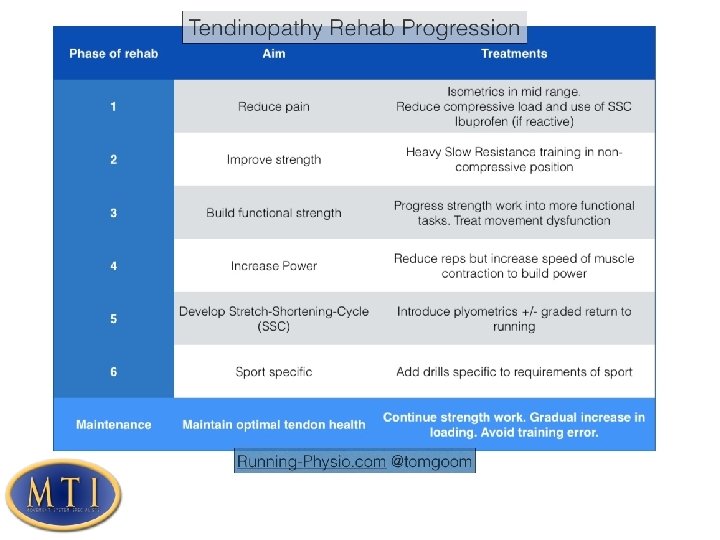
Exercise • Best intervention for tendinopathy • Only stimulus with the capacity to positively affect tendon matrix • Application should vary according the presentation • Reactive tendinopathy requires relative rest from high tendon load activities and prescription of isometric exercise for pain • In contrast, disrepair and degenerative pathology require progressive loading to moderate cell response and address functional deficits • Exercise brings strain on the tendon, which fuels the healing response
What is proper loading for injured tendons? • Little evidence comparing different loading programs and establishment of treatment guidelines • Need to reduce the abusive load if the capacity of the musculotendinous unit has decreased because of injury. • It is vital to find the baseline training level that does not provoke the tendon and to take this as the point to start the rehab process • Tendon becomes stiffer and better able to absorb tensile loads as a result of tendon rehab
Tendon response to proper loading At the microscopic level, as a tendon heals: • vessels and nerves regress • collagen fibers become stronger • Tendon becomes less thickened, more resistant to load, and less prone to re-injury (i. e. , recovering a more normal stress-strain curve)
Measuring tendon capacity • No easy measures of tendon capacity • Even extensively pathological tendons appear to have the capacity to tolerate very high sporting loads • The mechanisms by which the tendon increases capacity in response to training loads are unclear, as the tissue pathology may not change • Perhaps we are merely increasing the capacity of the normal part of the tendon
Pain response • Don’t get too preoccupied with the pain intensity the patient presents with. • More important to pay attention to treatment response. • No increased pain for longer than 30 -45 minutes after treatment (or workout) is finished • Any worsening of edema, morning stiffness, or delayed-onset pain should be closely monitored and controlled
Aggressive forms of therapy • Some authors have advocated reinjuring the tendon through treatments such as intratendinous needling, injections, aggressive soft tissue therapy, etc. • These approaches may improve the patient’s symptoms in the short term, but could result in long-term damage to the tendon. • No evidence that backs up this type of approach • Inn addition, this rationale is not well supported by current knowledge of tendon tissue pathology
Eccentric training programs • Common intervention when rehabilitating Achilles tendinopathy • Systematic reviews have identified low methodological quality among studies on this intervention • Additionally, there is substantial variability in the protocol used for eccentric training
Eccentric Training Programs Reported benefits of eccentric training programs: • • • Modulation of neurological stretch response Perturbations of tendon force Increased shear forces between the tendon and paratenon structures Pain modulation Adaption of mechanoreceptor activity Despite these data, it is unclear whether it is important to isolate the eccentric component of the exercise rather than performing a concentric-eccentric exercise.
Eccentric Training Programs • Not the be all end all • Not essential for tendon rehab • Very useful as a part of the rehab process to maximize loading on the tendon, and improve the ability of the tendon to handle plyometric activities • Cannot be all you do!
Proper exercise build up • Isometrics • Concentric/eccentric • Heavy load eccentric (where they cannot lift it concentrically) • Plyometrics (Stretch-Shortening-Cycle)
Exercise – Reactive Phase • Emphasize unloading type activities • Includes activities like Total Gym, biking, using an unloading device to walk/run, but also simple measures like using a heel lift to take the strain of a reactive Achilles tendon • In this phase you want to reduce the strain, as well as the compression on the tendon.
Exercise – Reactive Phase • Low load isometric exercises work well in the reactive phase • Research shows the following effects with isometric exercise: ü reduces tendon pain immediately for 45 minutes post intervention ü increases maximum voluntary muscle contraction
Exercise – Reactive Phase • Much smaller decrease in pain and no change in cortical inhibition were found after isotonic exercises • However, an RCT by van Ark et al (2015) showed no difference between isometric and isotonic exercises after four weeks. • Besides decrease in tendon pain they also found a decrease in cortical inhibition, which is present at elevated levels in patellar tendinopathy.
Exercise – Reactive Phase • Pain reduction observed following isometric exercise may be due to: ü Cortical changes observed and motor neuron pool recruitment ü Changes at a tissue level
Clinical Implications • Isometric exercise may be used to reduce pain and motor inhibition in the early stages of rehabilitation • Provides an important option for painful tendons that are difficult to load without aggravating symptoms, or potentially pre-strength training sessions in later stages. • There also may be a role for using isometric exercise to reduce motor inhibition and improve responses to strength training, as it is currently known that deficits persist despite rehabilitation.
Optimum Prescription • Not known yet • Current consensus for reactive tendinopathy: patient should be able to hold the isometric contraction for 30 -60 seconds with 30%-40% bodyweight at midrange • Progress to maintaining a 60 second contraction with full bodyweight.
Optimum Prescription • An RCT by van Ark et al (2015) used a high percentage of repetition maximum (80% 1 RM) for isometric as well as isotonic exercises in later stages • Beneficial effects from rehabilitation require high load per repetition • This improves muscle strength and neural activation as well
Optimum Prescription • Essential patient masters this before progressing to any type of eccentric activity • If they cannot, they are not able to go through the full range with proper muscle control, and they have no business progressing to eccentric activity.
Low load vs high load isometrics • Low load isometrics are ideally suited to control pain, but do not affect the tendon tissue directly • For that you need high load isometrics, as only they are able to introduce sufficient strain to stimulate tendon healing.
Progressing Once the patient is out of the reactive phase, the training schedule will look as follows: • 3 x/week heavy • 2 x/week endurance • Progress to incorporating plyometrics 2 x/week. Once that is tolerated well, you can truly do separate days of plyometric workouts alone • For endurance training, try to do calf raises until complete fatigue, up to 70 reps. For starters, 4050 reps is good
Later stages • The tendon needs to be heavily loaded to get the appropriate strain to facilitate healing • Get them in the Gym • Exercises like squats in Smith machine are good (regular squats and jump squats) • Heavy loading isometrics • Most will do a combo of endurance training and heavy loading
Patellar tendon • Be careful not to go over 70 -80 degrees of knee flexion with the heavy loading exercises • This includes squatting and leg press • The tensile forces on the patellar tendon become extremely high at that point, and the risk of exacerbation is very real • Still need to work out the patellar tendon at that endrange of course, but you do that with functional activities, or with low loading
Patellar tendon • Leg extensions are a very good exercise for patellar tendon rehab • Not a functional activity, but it is great to develop significant strain on the tendon tissue, necessary to provide stimulus for healing
Length of training program • Effective treatment for tendinopathy will result in significant (up to 80%) reduction in pain within 12 weeks • Confirms the relative plasticity of the pain system • However, that does not mean that from a structural standpoint the tendon is better as well • This can take significantly more time
Length of training program • Rehabilitation should be continued until the tendon regains full capacity to store and release energy without immediate or latent pain • Follow by a maintenance program if returning to competitive sport
Maintenance Program • Once the patient is done with the supervised portion of the rehab, they need to be put on a maintenance program • This needs to consist of daily low load isometrics for pain control, combined with endurance work • Heavy load isometrics are done 1 x/week
Maintenance Program • Remember it is all about increasing tissue capacity • Taking time off from tendon training means that the tissue capacity is going to decrease • This means tendon training needs to continue in the off season, as well as after a big race
Schematic of tendon rehabilitation Improving tendon capacity with progressive loads Introducing compressive loads are critical within each stage The start and end points of rehabilitation will vary between each patient