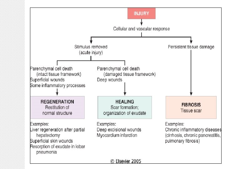
Tissue Repair Regeneration of injured cells by cells

Tissue Repair

Regeneration of injured cells by cells of same type, for example regeneration of skin/oral mucosa Replacement by fibrous tissue (fibroplasia, scar formation) Both require cell growth, differentiation, and cell-matrix interaction
 cells: ◦ Normally little proliferation but remain capable")
Varieties of Proliferative Potential �Stable (quiescent) cells: ◦ Normally little proliferation but remain capable of more rapid cell division following injury. ◦ Liver, kidney, pancreas, endothelium, fibroblasts ◦ Chances of regeneration are GOOD
 cells: ◦ Replace dying cells ◦ Epithelial")
Varieties of Proliferative Potential �Labile (always dividing) cells: ◦ Replace dying cells ◦ Epithelial cells of the skin, oral cavity, exocrine ducts, and GI tract; endometrial and bone marrow cells. ◦ Chances of regeneration are EXCELLENT
 cells: ◦ Not capable of proliferation. ◦")
Varieties of Proliferative Potential �Permanent (non-dividing ) cells: ◦ Not capable of proliferation. ◦ Irreversible injury leads only to scar ◦ Nerve cells, myocardium, skeletal muscle,

Cell Cycle

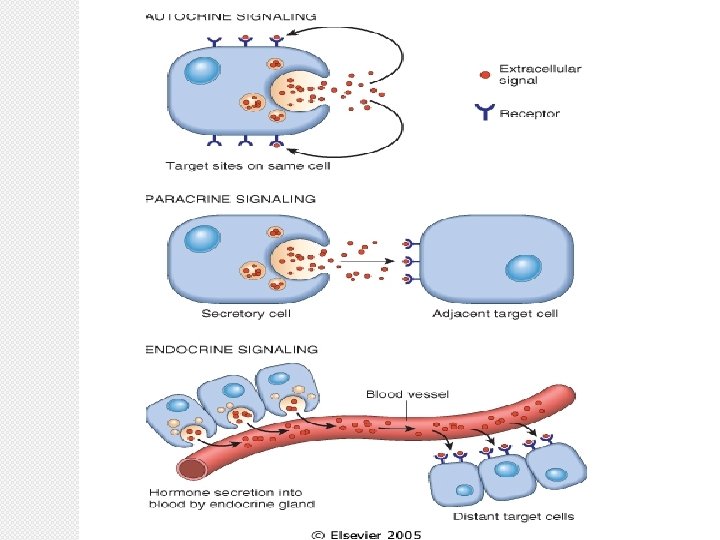
Cell Signaling Patterns �Autocrine = ligand is secreted and detected by same cell �Paracrine = ligand is secreted and separately detected by neighboring cells �Endocrine = ligands (usually hormones) are secreted into the vasculature to affect distant target cells

Wound healing
 �This occurs in clean, incised wound with good apposition")
Healing by first intension (primary) �This occurs in clean, incised wound with good apposition of the edges.

24 hours

3 to 7 days

WEEKs
 �This occurs in open wound, particularly when there has been")
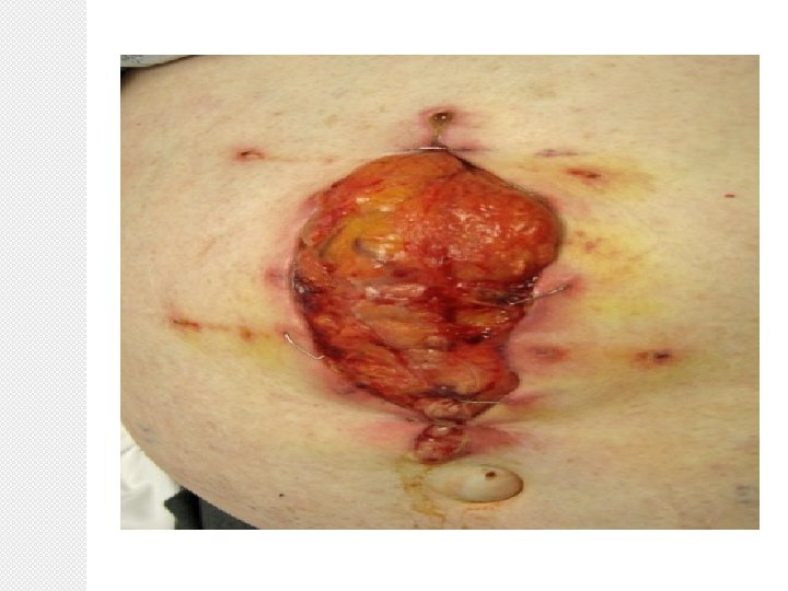
Healing by second intension(secondary) �This occurs in open wound, particularly when there has been significant loss of tissue , necrosis or infection

24 hours

3 to 7 days

WEEKs

FIBROSIS �Fibrosis, in general, refers to any fibroblast proliferation with deposition of excess extracellular matrix which is mostly collagen. �Leads to functional loss. �It is the end result of wound healing

This is a healing biopsy site on the skin seen a week following the excision, The skin surface has reepithelialized, and below this is granulation tissue with small capillaries and fibroblasts forming

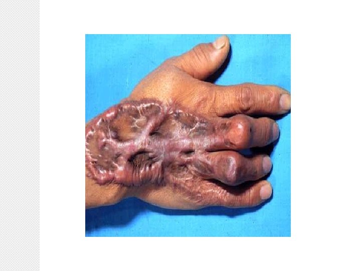
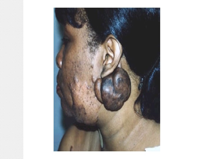
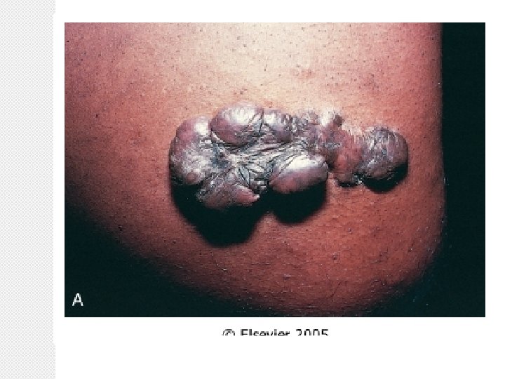
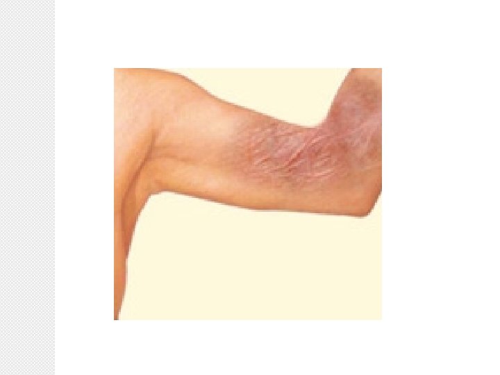
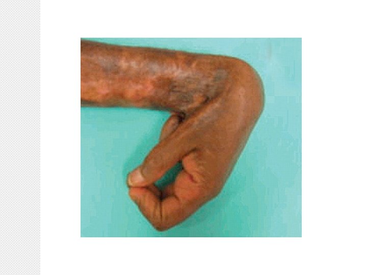
Complications of Wounds • Deficient scar formation – wound dehiscence/ ulceration • Excess repair – keloid formation • Excess contraction – joint contractures/ intra-abdominal adhesions
- Slides: 27