Tissue Adhesive A New Tool for the Vascular


 ➢Think about VAD insertion and management • Application of skin")


* ➢ 2 -octyl-cyanoacrylate (OCA)* • Quick drying • Longer drying")


 ISD n=31")


- Slides: 45
Tissue Adhesive: A New Tool for the Vascular Access Toolbox CORA VIZCARRA RN, BSN, MBA, CRNI, VA-BC What’s in your toolbox?
Disclosures/Disclaimers • Consultant/speaker for Adhezion Biomedical • Brand names to provide examples of products used in the evidence presented.
Learning Objectives ➢Review of vascular access device complications. ➢Define the components and characteristics of tissue adhesives. ➢Describe the purposes of tissue adhesives when used with VADs. ➢Analyze the clinical outcomes with tissue adhesives used with VADs.
Vascular Access ➢ 1 – 2 Billion PIVs annually world wide • >300 million in the US ➢Over 30 million CVC’s, PICCs, Midlines Protect the Patient! Educate the Clinician! Save the Line!
Go a ls & O bje ➢Protect the integrity of skin c tive ➢Protect puncture site from skin organisms s ➢Reduce VAD movement and dislodgment Vascular Access Devices (VAD) ➢Reduce unplanned dressing changes
Vascular Access Devices (VAD) ➢Think about VAD insertion and management • Application of skin antiseptics o Agents o Application methods and techniques • Puncture of skin and vein wall – create a surgical wound • Securing and stabilizing the VAD o Securing from accidental dislodgement o Stabilizing to prevent side-to-side or to-and-fro motion • Application of medical adhesives o Tape/Dressings
Vascular Access Complications • Infection – Systemic and Local • Vessel Trauma/Thrombosis • Skin Damage • Catheter Migration • Catheter Failure Requiring Early Replacement
Peripheral Catheter Failure ➢Up to 63% failure across 8 RCTs 1 ➢All study types, all causes – minimum failure 30%, maximum 95% • Included infiltration/extravasation, occlusion, accidental removal, phlebitis, and infection Helm, et. al. 2015
Peripheral IV Complications • Phlebitis -- Incidence reports of 14. 7% to 16. 1% • Precipitated by mechanical, chemical and infectious causes • Movement of the body relative to the secured catheter - Direct trauma to the intima • Infiltration – Most common form of failure; Incidence 15. 7% to 33. 8% • Results from erosion or penetration of the catheter through the vessel wall • Even in non-joint regions, inadequate device securement can lead to catheter tip motion and consequent injury to vessel wall Helm, et. al. 2015
Peripheral IV Complications • Occlusion -- Incidence of 2. 5% to 32. 7% • Device kinking • Catheter migration into a dead-end position within the vessel wall without frank infiltration • Dislodgement -- Incidence of 3. 7% to 50% • Study by Jackson; 3296 PIV restarts over 6 months • Catheter dislodgement 50% of the failures • Inadequate securement; tubing catching on clothing, etc. • Current securement devices add bulk to the catheter-dressing complex and extend adhesive surface area, Helm, et. al. 2015
Peripheral Catheter Infection ➢New Systematic Review • PIVC-BSI = 0. 18% in 85063 peripheral catheters • Mean of 22% (range 7%-60%) of 7860 nosocomial CRBSI • Mean of 38% (range 12% to 64%) S. aureus CRBSI from infected peripheral catheters 2 ➢Arterial Catheters – 1. 7/1000 catheter days 3 Mermel, et al. 2017, Maki, et al. 2006
Midlines and CVAD Infections ➢CRBSI • Midlines – 0. 2/1000 catheter days 3 • PICCs – 0. 12 – 2. 3/1000 catheter days 4, 5 • 3. 1/1000 catheter days (Ullman, Pediatrics; 2015) 6 • • CVCs – 0. 1 – 4. 8/1000 catheter days 3 Maki, et al. 2006, Raiy et al. 2010, Kang et al. 2017, Ulllman et al. 2015
Central Line Complications ➢Oozing/blood leakage at insertion site • Non-routine dressing changes are common: Average 22. 8%, 43% of respondents have > 25% Early Dressing Changes (AVA 2017 survey) • 7 – 24. 7% Oozing and 3. 8% leaking 6 • 0. 6% major bleeding; 2. 8 – 5. 4% minor bleeding 7 Lueng et al. 2011, Vinson et al. 2014
Central Line Complications ➢Accidental Withdrawal • 4. 2% 8 Qui et al. 2014
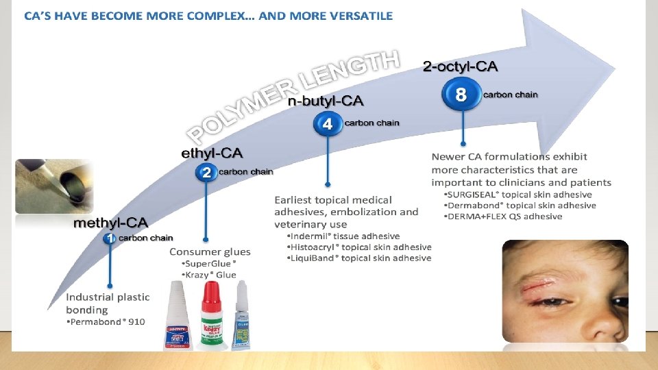
Tissue Adhesive ➢Can adding tissue adhesive to our toolbox make a difference in these outcomes? ➢What is tissue adhesive? • Glue - cyanoacrylate, (CA) a liquid monomer 9 o Polymerizes when exposed to moisture present in air, liquid, or tissue o Exothermic process – releases energy when the molecules come together ❖May release a small amount of heat Januchowski et al. 2014
Tissue Adhesive ➢N-butyl-cyanoacrylate (BCA)* ➢ 2 -octyl-cyanoacrylate (OCA)* • Quick drying • Longer drying time • Rigid/Brittle • More cytotoxic • Higher tensile strength & more flexible • Stronger thermal reaction • Less cytotoxic • Requires minimum 24 hours before fully water resistant • Reduced thermal reaction *Adhezion Biomedical, Internal Testing • Immediately water-resistant
Tissue Adhesive ➢Antimicrobial activity of different cyanoacrylate formulations • First generation products • Most were effective against gram positive bacteria • Second generation products (2 -octyl and octyl blends) • Most are effective against gram positive • Two of newer formulations; published data demonstrating broad-spectrum activity against Gram Positive, Gram Negative, Yeast, and Fungi 10, 11 Prince et al. 2017
Tissue Adhesive – Uses with VADs ➢Early in vitro testing demonstrated suitability of tissue adhesive for VADs 12 ➢ 4 purposes identified • Enhanced securement of VADs • Wound closure by a protective barrier • Minimizes oozing at puncture site • Infection prevention by immobilizing and killing bacteria Simonova et al. 2012
Tissue Adhesive – Current Evidence ➢Peripheral IV catheters ➢Peripheral arterial catheters ➢Central venous access devices ➢Epidural catheters
Tissue Adhesive – Peripheral IV Catheters ➢ 4 arm pilot randomized trial in adults on medical-surgical units 13 • Catheter failure, premature removal due to complication Standard polyurethane (SPU) control group Bordered polyurethane Sutureless securement (SSD) + SPU TA + SPU Number 21 20 23 21 # failed 8 5 5 3 Failure rate 6. 92 3. 82 3. 14 2. 40 Adverse Events 0 0 0 4 Marsh, N. , et al. 2015.
Tissue Adhesive – Peripheral IV Catheters ➢ 2 arm randomized trial in adult emergency patients 14 • Assessed failure at 48 hours, modes of failure BPU + tape TA + BPU + tape Number 190 179 Failure No (%) 52 (27%) 31 (17%) Dislodgement 26 (14%) 13 (7%) Phlebitis 9 (5%) 6 (3%) Occlusion 20 (11%) 15 (8%) Bugden, S. , et al. , 2016
Pilot Trial with Tissue Adhesive on PIVs Range of Dwell Time Average Dwell Time Number of Patients Age Range Complications Leaking Phlebitis Occlusion Infiltration Pulled out by patient Control Group 4 h 9 min – 164 h 1 min Study Group 5 h 32 min – 329 h 26 min 35 hours 73. 2 hours N=35 N=25 24 d – 24. 4 years 2. 5 months – 20. 2 years 16 (46%) 5 0 3 4 4 8 (32%) 4 1 2 1 0 Presented with permission from D. Doellman, Cincinnati Children’s Hospital
Tissue Adhesive - PICCs in Pediatrics ➢ 3 arm, single centre Pilot RCT 15 ➢Primary outcome: PICC failure (cessation of function prior to completion of therapy) ➢Compared: 1. Bordered polyurethane (BPU) dressing + sutureless securement device (SSD) 2. Integrated securement dressing (ISD) 3. Tissue adhesive (TA) + bordered polyurethane (BPU) dressing Kleidon, et. al. 2017
Tissue Adhesive - PICCs in Pediatrics BPU + SSD n=32 2 (6%) ISD n=31 2 (6%) TA + BPU n=32 1 (3%) Results PICC failure ➢Overall failure rate (5%) Incident rate/1000 catheter days 8 8 3 Dwell time 8 7 7 Adverse skin events* 5 (16%) 3 (10%) 10 (31%) Parental satisfaction (0 -10) 7. 6 9. 7 8. 5 ➢Lower incident rates in TA group ➢Skin issues higher in TA group ➢N - Butyl *Itchiness, rash, skin tear, blister or bruising at any time during study
Tissue Adhesive - PICCs in Pediatrics • Dressing Changes Days to First Change BPU + SSD n=32 3. 5 ISD n=31 2. 5 TA + BPU n=32 5. 5 Bleeding 19 14 5 Lifting 7 0 3 Leakage 1 1 0
Tissue Adhesive - Tunnelled CVADs in Pediatrics ➢ 4 arm, 2 centre pilot RCT 16 ➢Primary outcome: CVAD failure ➢Compared: 1. Bordered polyurethane (BPU) dressing + suture 2. Sutureless securement device (SSD) + suture + BPU 3. Tissue adhesive (TA - at exit wound and under catheter bifurcation) + BPU 4. Integrated securement-dressings (ISD) + suture Ullman, AJ. , et al. 2017
Tissue Adhesive - Tunnelled CVADs in Pediatrics ISD+ suture n=12 SSD+suture +BPU n=13 BPU+suture (control) n=11 TA+ BPU n=12 CVAD failure 2 (17%) 1 (8%) 0 0 Complications 1 (8%) 2 (15%) 0 0 2 (17%) 1 (8%) 2 (18%) 0 10 25 17 4 Results ➢ Lower non-routine dressing changes ➢ High staff approval on application ➢ High parental satisfaction on Adverse skin event* removal but not staff satisfaction Non-routine dressing *rash, blister, itchiness
Tissue Adhesive - Acute Care IJ CVADs ➢ 4 -arm randomized trial elective cardiac surgical patients, 5 th arm added 17 • 8. 5 Fr, 20 cm quad lumen or 7 Fr, 16 cm triple lumen, all chlorhexidine impregnated, all inserted by IJ vein. Suture + BPU Suture + lattice pad dressing SSD + SPU TA + Suture + SPU 55 56 55 23 30 Catheter failure 2 (4) (%) 1 (2) 4 (7) 4 (17) 0 (0) Median dwell time in hours 68. 2 67. 8 69 72. 2 Number Rickard CM, 2016 69
Tissue Adhesive – All CVADs ➢Pittiruti, M. , et al. Cyanoacrylate Glue and Central Venous Access Device Insertion 18 ➢Poster – AVA 2016 Scientific Meeting • 513 non-tunneled PICCs and CICCs • 114 tunneled PICCs, CICCs, and FICCs • 802 implanted ports ➢ 100% effective in prevention of post-insertion bleeding ➢ 10 fold reduction in CRBSI Pittiruit, et al. AVA Scientific Meeting
Tissue Adhesive in CR-BSI Prevention Bundle ➢Added elements to their existing CR-BSI Prevention Bundle 19 ➢US pre-puncture evaluation (Rc. Ce. VA) ➢Tunneled the exit site ➢Sealed exit site with tissue adhesive at the time of insertion ➢No CHG sponge dressing at time of insertion; added at 1 st dressing change ➢Consistent use of transparent dressings ➢Simulation based training program for all inserters Biacucci, et al. 2017
Tissue Adhesive in CR-BSI Prevention Bundle • Conducted in a PICU from June 2009 – June 2014 • 1150 catheter days; 648 in the study group and 503 in the control • CR-BSI rate dropped from 15/1000 catheter days to 1. 5/1000 catheter days • 2. 2 day longer dwell • Comments in conclusion about tissue adhesive; • “… sealing the exit site. . reduces risk of extraluminal contamination … and reduces bleeding at puncture site and prevents the “in and out” motion may reduce local damage to the endothelium and reduce risk of thrombosis. ”
Application of Tissue Adhesive for PIVs ➢All prep solutions on skin must be thoroughly dry ➢Apply to catheter-skin junction ➢ Apply under catheter hub/extension set
Application of Tissue Adhesive for PIVs ➢ Gently press hub for a few seconds ➢ Add additional adhesive to catheter-skin junction to flow around hub and create a seal
Application to Central Lines ➢Apply adhesive directly to the insertion site to • reduce micromovement • create a protective barrier/seal around the insertion site • Reduce leaking/oozing ➢Apply in conjunction with other modalities to enhance securement • Apply after placement of sutures or subcutaneous securement devices • Could be applied over the skin puncture sites from sutures or devices • No consensus on timing of application in conjunction with adhesive backed mechanical securement devices
Videos courtesy of Matt Ostroff/St. Joseph’s Medical Center
Videos courtesy of Matt Ostroff/St. Joseph’s Medical Center
Recent Testimonials Before Pictures used with permission from patient After
Reapplication for long-term use ➢Many questions still unanswered
Adhesive removal ➢Commercially available adhesive removers are capable of loosening cyanoacrylate quickly ➢PDI ➢Uni-solve ➢Remove ➢Detachol ➢Active ingredients: • Paraffin • Petrolatum • D-Limonene • Propanol • Esters of IPA
Take Home Message ➢Tissue adhesive benefits • Enhanced catheter securement • Seal around puncture site o Decrease contamination of site o Reduced oozing/leaking from puncture site • Studies demonstrate feasibility of the concept and suggests reduction of complications ➢Large studies are in progress ➢Promoting skin integrity and reducing VAD complications is critical aspect of patient care with any type of VAD
Thank You for Your Attention
References 1. Helm, R. E. , et al. , Accepted but Unacceptable: Peripheral IV Catheter Failure. Journal of Infusion Nursing, 2015. 38(3): p. 189203. 2. Mermel, L. A. , Short-term Peripheral Venous Catheter–Related Bloodstream Infections: A Systematic Review. Clinical Infectious Diseases, 2017: p. cix 562. 3. Maki, D. , et al. , The risk of bloodstream infection in Adults with Different Intravascular Devices: A Systematic Review of 200 Published Prospective Studies. Mayo Clin Proc, 2006. 81(9): p. 1159 -1171. 4. Raiy, A. , et al. , Peripherally inserted central venous catheters in an acute care short-term intravenous catheters. Am J Infect Control. 2010. 38(2): p. 149 -53. setting: A safe alternative to high-risk 5. Kang, J. , et al. Peripherally inserted central catheter-related complications in cancer patients: a prospective catheter days. J Vas Acces. 2017. 18(2): p. 153 -157. study of over 50, 000 6. Ullman, A. , et al. Complications of central venous access devices; a systematic review. Pediatrics. 2015. Nov 136(5): e. 1331 – 44. 7. Lueng, TK. , et al. Cancer Nrsg. 2011. A retrospective study on the long term placement of peripherally inserted central catheters and the importance of nursing care and education. 34(1); p. E 25 -30.
References 8. Vinson, DR. , et al. Bleeding complications of central venous catheterization with abnormal hemostasis. Am J of Emerg Med. 2014. 32(7): p. 737 -42. 9. Qui, XX. , et al. , Incidence, risk factors, and clinical outcomes of peripherally inserted central catheter spontaneous dislodgement in oncology patients: A prospective cohort study. Int J Nrs Stud. 2014. 51(7). P. 955 -63. 10. Januchowski, R. and O. W Jordan Ferguson III, The clinical use of tissue adhesives: a review of the literature. Osteopathic Family Physician, 2014. 6(2). 11. Prince, D. et al. , Antibacterial effect and proposed mechanism of action of a topical surgical adhesive. AJIC. 2017. 12. Prince, D. et al. , Immobilization and Death of Bacteria by Flor Seal® Microbial Sealant. International Journal of Pharmaceutical Science Invention. 2017. 6(6). P 45 -49. 13. Simonova, G. , et al. , Cyanoacrylate tissue adhesives – effective securement technique for intravascular catheters: in vitro testing of safety and feasibility. Anaesth Intensive Care, 2012. 40(3): p. 460 -6. 14. Marsh, N. , et al. , Securement methods for peripheral venous catheters to prevent failure: a randomised controlled pilot trial. The journal of vascular access, 2015. 16(3): p. 237 -244.
References 15. Bugden, S. , et al. , Skin Glue Reduces the Failure Rate of Emergency Department-Inserted Peripheral Intravenous Catheters: Randomized Controlled Trial. Ann Emerg Med, 2016. 68(2): p. 196 -201. 16. Kleidon T. , et al. A pilot randomized controlled trial of novel dressing and securement techniques in 101 paediatric patients. Journal of Vascular and Interventional Radiology 2017. Ullman, AJ. , et al. Innovative dressing and securement of tunneled central venous access devices inpediatrics: A pilot randomized control trial. BMC Cancer. 2017. 17: 595. 18. C. M. Rickard, R. , BN, Grad Dip Crit Care Nurs, Ph. D, FACN, FAAHMS a, �, , et al. , A four-arm randomised controlled pilot trial of innovative solutions for jugular central venous access device securement in 221 cardiac surgical patients. Journal of Critical Care, 2016. 19. Pittiruti, M. , et al. Cyanoacrylate Glue and Central Venous Catheter Insertion. AVA 2016 Scientific Meeting. Poster Abstract. 20. Biasucci, D. G. , et al. Targeting zero catheter-related bloodstream infections in pediatric intensive care unit: a retrospective matched case-control study, J Vas Access, 2017.