Tips for doing well in neonatology section of


 expected normal is 1.")


-Endocardial cushion defect, VSD – Extremities-single palmer")
 – Holoprosencephaly, cleft lip/palate, coloboma, sloping")
 – Cardiac (common, VSD, PDA, PS), clenched hand, overlap")


")
 and central thru")

")


 – p. H<7, AS<3 at 5 min, neurological sequelae")


 maternal causes (DM, hyperparathyroidism), perinatal causes")


 • Who – All <32 w – All with")


- Slides: 128
Tips for doing well in neonatology section of Pediatric Boards Shantanu Rastogi MD, MMM Neonatologist, Maimonides Medical Center Associate Professor of Clinical Pediatrics SUNY-HSC at Brooklyn
Some general points • There are no negative marks and hence no questions SHOULD be left unanswered • When the answers are not clear in the first reading of the question then and only then try the method of exclusion to get to the best possible answer
Rastogi’s Rule • Common presentations of common diseases • Rare presentations of common diseases • Common presentations of rare diseases • Rare presentations of rare diseases
FETAL WELL BEING
Biophysical profile • NST, fetal body movements, breathing, fetal tone, amniotic fluid volume • Scores of 10 is well fetus, 2 is certain fetal asphyxia, 4 or 6 needs frequent reevaluation for delivery
Electronic FHR monitoring • Normal – FHRv of 6 -15 bpm, basis of nonstress test-reactive/positive test is normal • Abnormal patterns – Tachycardia, >160, infection – Bradycardia, <110, head compression – Loss of FHRv, hypoxia
Decelerations Early, mirror image of uterine contractions, head compression Variable, irregular, umbilical cord compression Late , occurs 10 s after uterine contraction and last longer, uterine placental insufficiency
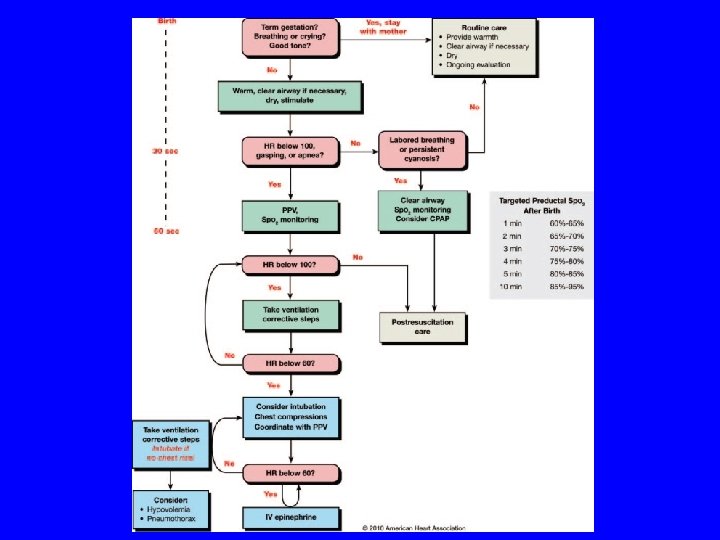
RESUSCITATION
Case • FT baby is delivered vaginally has HR of 90/m and no respiratory efforts, central cyanosis, flaccid and has no reflexes. • What is the Apgar score? • What is the first step during resuscitation? • What is the subsequent step?
Case continued • After 1 min of IPPV the HR is 50/min • What is the next step?
Case • You are preparing to attend the vaginal delivery of an infant at 40 weeks gestation. Artificial ROM was 8 hours ago, which revealed meconium-stained amniotic fluid. What is the next step. . .
Question 1: Time for the saturation to reach (85 -95%) expected normal is 1. 2. 3. 4. 1 minute 5 minutes 10 minutes 60 minutes
Recent changes for the NRP • Temp. control: normothermia, prevention of hyperthermia • Suctioning – Clear AF: only if obstruction, IPPV needed – MSAF: intubate only in non vigorous babies • Use of Pulse oximetry: normal sats. by 10 minutes of life • Use of RA: To use blended O 2, can use RA in term, 100% O 2 only if >90 sec. of ineffective resuscitation
• Use of CO 2 detectors: obstructions, confirm ET placement • Epinephrine administration: IV, 0. 010. 03 mg/kg, no high dose • Induced therapeutic hypothermia • Stopping resuscitation: 10 min. without HR
INFECTIONS
GBS • The incidence of sepsis has ↓ due to the ↓ in GBS sepsis, but that from gram negative remain the same • CDC guidelines-2010: – Screen all women at 35 -37 wks of gestation, specific method to obtain and culture specimen – Consider mother to be GBS positive if previously positive and h/o invasive neonatal infection in prior preg. – No prophylaxis required for elective c-section even if GBS positive and not in labor – Clarification of penicillin sensitivity
GBS • Risk Factors: – Intrapartum Prophylaxis to mother -if GBS positive OR unknown with <37 weeks GA, ROM ≥ 18 h, maternal fever ≥ 100. 4 F, NAAT +ve OR GBS bacteriuria OR H/O previous invasive neonatal GBS infection – Mn of the baby-Symptomatic OR if mother had adequate IP and baby is <37 wks /ROM >18 h do CBC/BCx (* If IP <4 h and if baby >37 wks /ROM <18 h-no CBC/BCx). • Clinical presention – Early-<7 DOL, pneumonia>sepsis>CNS, ascending infection – Late onset->7 DOL, less mortality but more CNS involvement and sequelae, deep infection as cellulitis, arthritis, osteomyelitis
Other bacterial infections • E. coli/Klebsiella sp. ↑ in % in neonatal sepsis, not in absolute numbers. Clinically present with progression through 3 classical stages of shock • Listeria- Gram + rod, in unpasteurized milk, cheese, raw vegetables and uncooked meat – Early onset: <7 d, transplacental, chocolate colored amniotic fluid, preterm deliveries, sepsis/pneumonia – Late onset: >7 d, nosocomial, meningitis with mononuclear cells
Initial drug of choice • Ampicillin and Aminoglycoside
Other congenital infections Clinical presentation CMV Rubella Toxoplasmosis LBW +++ ++ + Liver/Spleen ++++ ++ + Jaundice +++ + ++ Petechiae +++ ++ + CHD + +++ Cataract +++ + Retinopathy + +++ Cerebral calcification Periventricular+ cortical++ Microcephaly ++ +
Management of Syphilis VDRL FTA Inference Mother Baby - - + +/- + + - - + + No or prozone False+ Mother/B disease Treated disease
Conjunctivitis • Time of onset will give the clue to the cause: – Hours-chemical-silver nitrate – DOL 2 -5 -Nisseria gonorrheae-purulent, emergency, needs IV antibiotics – DOL 5 -14 -Chlamydia-bilateral, cough
CHROMOSOMAL DISEASES
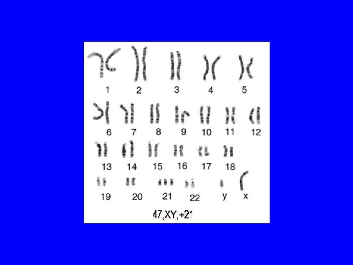
Trisomy 21 • Types– 94% non-disjunction – 3 -5% translocation – 2% mosaic. Commonest cause in both old and young mothers is non-disjunction • Recurrence risk– If no translocation- 1% risk till mat. age is 37 y – if mat. translocation-10 -15% – if pat. translocation-5%
Trisomy 21 • Defects – Cardiac (40 -50%)-Endocardial cushion defect, VSD – Extremities-single palmer crease, 5 th finger has hypoplastic middle phalange and clinodactyly – Face-slanting palpebral fissure, Brushfield spots, epicanthic folds, short neck, flat occiput – GI- duedenal atresia, Hirschsprung Disease – Neurology- hypotonia, MR, – Other –hypothyroidism, leukemia, hip dysplasia
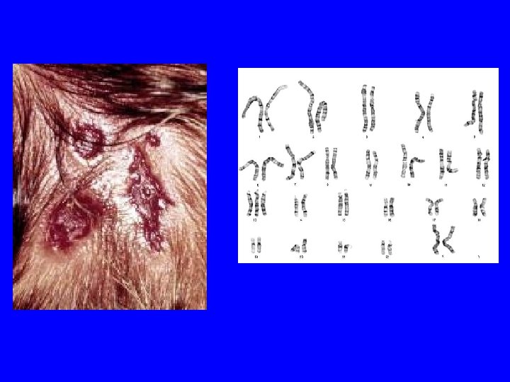
Other chromosomal anomalies • Trisomy 13 (Patau-MIDLINE deformities) – Holoprosencephaly, cleft lip/palate, coloboma, sloping forehead, cutis aplasia, VSD, polydactyly, hyperconvex nails, persistence of fetal Hb
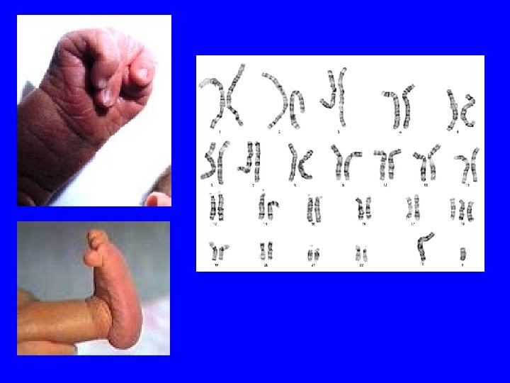
• Trisomy 18 (Edward) – Cardiac (common, VSD, PDA, PS), clenched hand, overlap of 2 nd over 3 rd and 5 th over 4 th finger, rocker bottom feet, small mouth/eyes/palpebral fissures, short sternum, hernia, cryptorchidism
PULMONARY
Question 2 • A 28 w GA male infant weighing 1500 g experiences respiratory distress requiring ventilation soon after birth. Ventilator parameters are: SIMV of 40 /min, 22/5 pressures and Fi. O 2 of 0. 8 to maintain a Pa. O 2 of 60 mm Hg. CXR shows. . .
Surfactant is administered. What is the most probable observation over next few hours? 1. Reduction in spontaneous respiratory rate 2. Decreased PIP requirement 3. Pulmonary hemorrhage 4. Pneumothorax 6
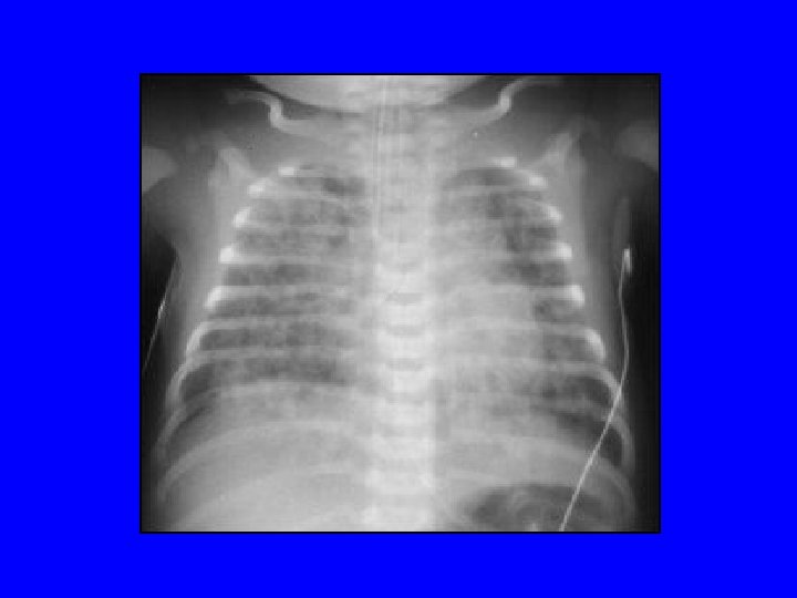
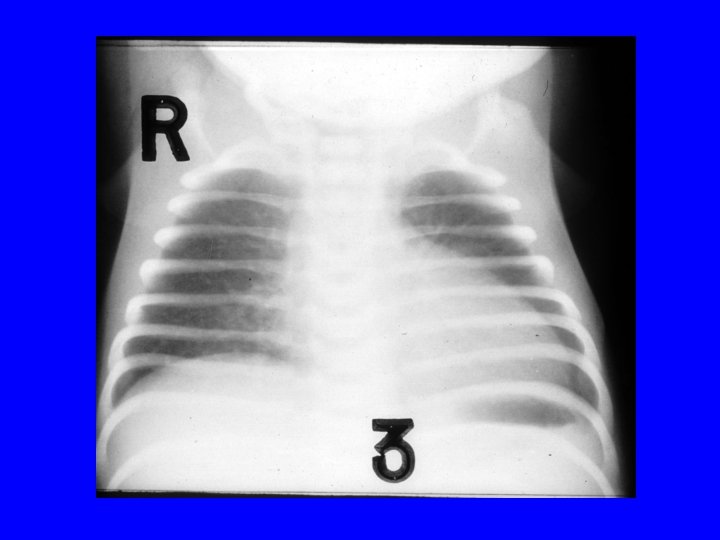
RDS-Surfactant Deficiency • Clinical course: Peak-1 to 3 d and recovery starts with onset of diuresis • Risk Factors: Low GA, male gender , Mat. DM, perinatal depression • RDS in term: SPB def, IDM, Beckwith Weideman syndrome, congenital syphilis • Pathology: Hyaline membrane (cellular debris in fibrinous matrix) • Treatment-surfactant replacement, supportive • Complications: pneumothorax • DD for reticulogranular CXR- GBS pneumonia, PAPVR
Case • An infant is delivered by c/s at 34 wk GA. There is no h/o ROM, maternal fever, or abn. FHR. The infant requires IPPV with a bag-mask and 100% O 2 in the delivery room. AS are 5 and 7. UA p. H was 7. 23. He is admitted to the WBN, but develops respiratory distress within an hour of life. RA ABG shows 7. 10/ 80/50/-10 with CXR
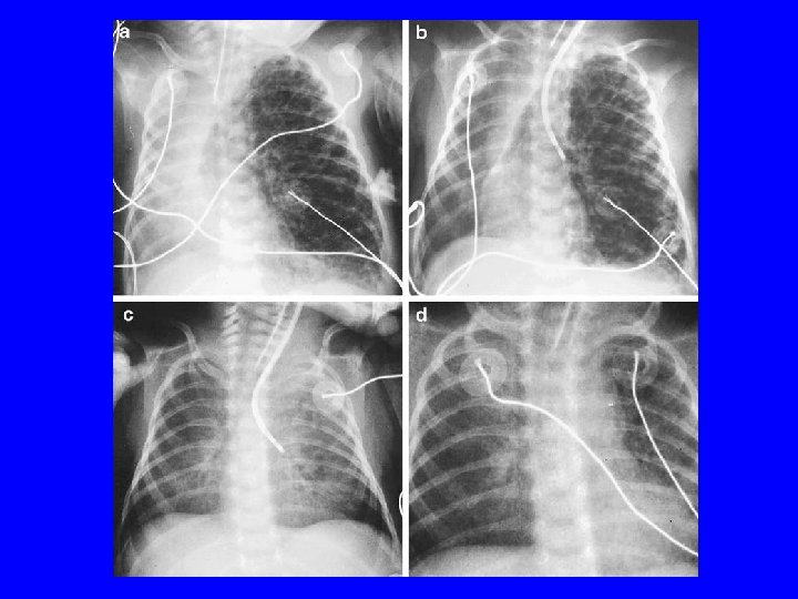
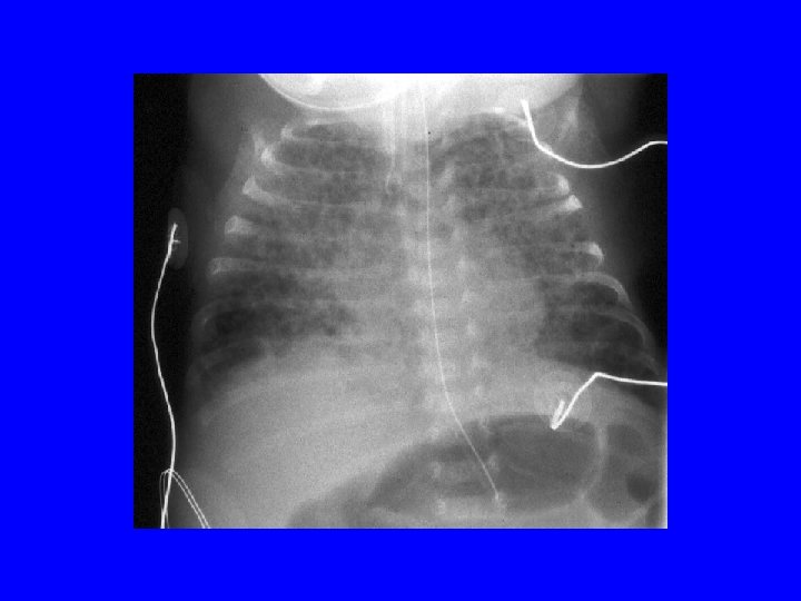
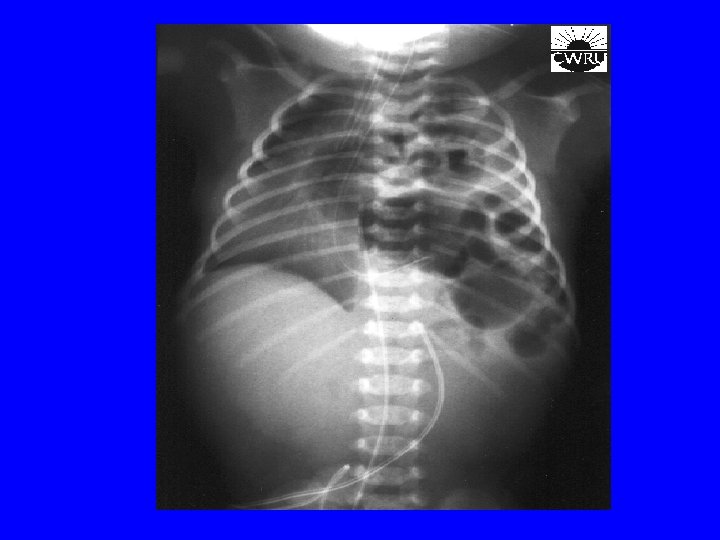
Air leak syndromes • Pneumothorax – Air between parietal and visceral pleura – Risk Factors-Aspiration (MAS), parenchymal diseases (RDS), PPV (high PIP) – Presentation can vary from • Tension: severe RD, bradycardia, apnea, hypotension with mediastinal shift • large leak: respiratory distress • small leak: usually accidental finding – Complication: IVH by ↓ venous return , SIADH – Diag. : asymmetrical air entry, transillumination – Rx: EMERGENCY if tension-needle aspiration followed by chest tube, supportive
Airleak Syndromes • Pneumopericardium- air in pericardial sac – If large- muffled HS, venous congestion, decreased CO – Rx if symptomatic-pericardial aspiration, high mortality
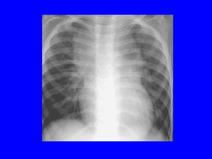
• Pneumomediastinum- air in mediastinum – Usually after IPPV or difficult intubation, high PIP – Muffled heart sounds, CXR-sail sign – Usually needs supportive treatment
• Pulmonary Interstitial Emphysema-air in interstitial space in the lung tissue – Usually preterm with RDS and on ventilation – Rx: decrease MAP, if unilateral-selective intubation/blocking of bronchus
Broncho-Pulmonary Dysplasia • BPD- 36 wk of corrected GA with oxygen requirement • Mechanical trauma (Baro- and Volu-trauma) to susceptible lungs leading to inflammation • Injury is increased in PT due to low levels of antiproteases and antioxidants • Poor compliance, increased WOB, pulmonary hypertension, RVH • Radiographic: honeycomb appearance • Rx- nonspecific as supportive care, inc. calories (120 -150 cal/kg/day), diuretics, bronchodilators, steroids
Apnea of Prematurity • Cessation of air flow for >20 s usually with cyanosis/bradycardia • Types: Central (no effort, no air flow), Obstructive (no airflow despite effort), Mixed • Cause: prematurity (usually after 12 h of life), infection, maternal med. (narcotics, magnesium), infant med. (prostaglandin), CNS disorders (IVH) • Treatment: Treat underlying disease, methylxanthines (caffeine) , CPAP, mechanical ventilation
Transient Tachypnea of Newborn • It is a diagnosis of exclusion • Cause: delayed absorption of lung fluid • Risk factors: elective c/s, IDM, perinatal depression, precipitous delivery • Radiograph: Fluid in the minor fissure • Rx: supportive, resolves 2 -3 days
Meconium aspiration syndrome • Definition: MSAF+RD+CXR changes • Clinical: usually post-term, severe respiratory distress • Complications: pulmonary hypertension, airleak syndromes • CXR: snow storm appearance • Prevention: suctioning of meconium as per NRP protocol • Rx: respiratory support, correcting acidosis, antibiotics, surfactant
Case • A FT infant is delivered vaginally to mother with DM. He develops cyanosis and respiratory distress immediately following birth that requiring IPPV with 100% oxygen. There is no improvement and is admitted to the NICU. His BW is 4, 500 g, has saturation of 70 in the right hand 45 in the left leg, with precordial lift and a loud S 2. CXR reveals decreased pulmonary blood flow. The MOST likely cause of respiratory distress in this infant…
Pulmonary Hypertension • Cause – Maladaptation: normal vasculature but vasoconstriction (hypoxia, hypothermia, polycythemia, pneumonia) – Maldevelopment: abnormal structure of pulmonary vascular bed (chronic intrauterine hypoxia, pulmonary hypoplasia) • Rx- respiratory support (no hyperventilation), correcting acidosis, i. NO, sildanefil, ECMO
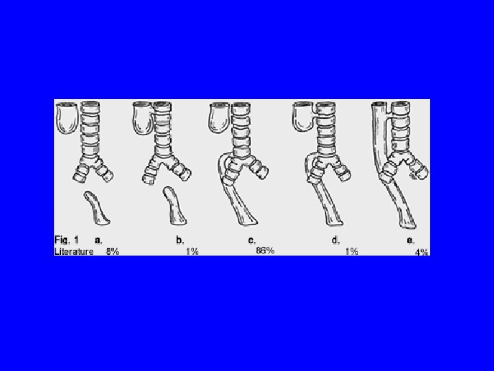
Congenital diaphragmatic hernia • Types: Posterolateral thru Foramen of Bochdalek (L>>R) and central thru Foramen of Morgagni • Clinically can present in-utero as hydrops, after birth as RD due to lung hypoplasia, scaphoid abdomen • Complications: related to lung hypoplasia and pulmonary hypertension (PH) • Treatment: IMMEDIATE intubation, correcting p. H and delayed surgical repair. May require ECMO
CARDIOLOGY
Congenital Heart Disease-Some Facts • Incidence 8/1000 live births (excluding PDA in PT newborns) with 25% have other associated abnormalities • Suspect: cyanosis with minimal respiratory distress
Congenital Heart disease -some facts • VSD- commonest CHD • TOF- commonest cyanotic HD beyond neonatal period • TGA-commonest cyanotic HD in first week of life • HLHS- 2 nd commonest cyanotic HD in first week of life and commonest cause of cardiac mortality during that period
Presentation of CCHD • 5 T’s, DO, ESP- TGA, TOF, TAPVR, Tricuspid atresia, Truncus arteriosus, DORV, Ebstein’s Anomaly, Single ventricle and Pulmonary atresia • HLHS Vs. sepsis: usually HLHS presents after the duct is closed by 48 -72 h and neonate presents with cardiac failure to the ER with no murmur. If no high risk factors for infection always consider the diagnosis of HLHS
Maternal conditions and CHD • Maternal drugs – Aspirin/Indomethacin-PH/PDA closure, Lithium-Ebstein’s anomaly, Ethanol-VSD • Maternal diseases – Lupus-Cong. Heart block (anti Ro, anti La Ab), Diabetes (VSD-commonest, TGA, ventricular hypertrophy-most specific)
NEUROLOGY
SCa. LP Injuries • In Sub. Cutaneous tissue- caput succedaneum, soft, crossed midline/sutures, usually with molding, resolves over several days • Beneath Galea Aponeurotica in Loose areolar tissuesubgaleal, can move to neck and behind ear, can cause anemia, hypotension, jaundice, resolves in 2 -4 wk • Sub. Periosteal- cephalhematoma, confined to suture lines, firm, 10% have skull fracture, jaundice, resolve in weeks to months
A 2 day old preterm infant who was born at GA of 26 wk and BW of 650 g on RA CPAP. Her blood pressure has dropped acutely, and developed seizures. There B/L equal breath sounds, no murmur, hypotonia, a bulging anterior fontanelle, and lethargy. Lab. shows severe anemia, metabolic acidemia, and hyperglycemia. This is most probably related to 1. 2. 3. 4. Severe hemolysis Adrenal hemorrhage Intraventricular hemorrhage Sepsis/Meningitis 6
Intraventricular Hemorrhage • From: germinal matrix • Incidence: ↑ with ↓GA e. g. <1 kg 30%, 1 -1. 25 kg 15%, 1. 25 -1. 5 kg 8% • Timing- 50% in 24 h and 90% in 72 h present with S/S of anemia + CNS involvement. • Prognosis: Poor with increasing grades • Complication: hydrocephalous/PVL
Birth Asphyxia • Definition (ACOG) – p. H<7, AS<3 at 5 min, neurological sequelae (HIE), multiple organ dysfunction • HIE staging ( Sarnat Stages) – Stage 1: hyperactive CNS with sympathomimetic activity, 100% normal – Stage 2: decreased CNS activity, parasympathomimetic activity, seizures, 80% normal – Stage 3: variable presentation, seizures rare, burst suppression EEG, 100% severe sequelae
Cerebral palsy • Non progressive neurological deficit • Incidence: 2 -5/1000 • Clinicopathological patterns: – Selective neuronal necrosis- HIE, diffuse damage, Quadriplegia, MR, seizures – Parasagittal cerebral injury- ↓ perfusion, necrosis in watershed areas of carotids, weakness of proximal muscles U>L – Focal or multifocal ischemia- meningitis, trauma, thrombotic syndromes, as hemiplegia, seizures, cognitive defects – Status Marmoratus- kernicterus, basal ganglia, choreoform movements
Brachial plexus injury Roots Incidence Typical S/S Differentiate Associated Erb-Duchenne C 5 -7 Common Waiters tip Palmer grasp + C 4/5 (phrenic nerve), C 7 (scapular winging) Klumpke C 8 -T 1 Rare Ape hand T 1( Horner’s syndrome)
Hydrocephalous • 2 types: – Obstructive- common, commonest cause is post hemorrhagic HC, others are aqueductal stenosis, Dandy-Walker Syndrome (cystic dilatation of 4 th ventricle with hypoplasia of vermis) – Communicating-usually after bleeds, infections, NTD, Arnold Chiari malformation • Treatment: LP, shunts
Case • Mother is rushed in for stat c/s for abruption. Apgars are 2, 3, 8 - improving after IPPV, chest compression and fluid resuscitation. Admitted to NICU. Mother had uneventful antenatal course. Baby has not passed urine and serum sodium is 125. He develops generalized tonic clonic seizures and bradycardia after 12 h requiring phenobarb. • What is the most likely cause for these seizures
Neonatal Seizures • Types – Subtle-most frequent, oral, facial ocular activity, may be associated with changes in HR, resp. BP and sats. – Multifocal clonic-one limb migrating to another – Focal clonic- may represent focal disease – Tonic-change in posture, more in preterm – Myoclonic • Many causes- asphyxia, metabolic, infection, trauma, malformation • Initial drug of choice is phenobarb
Hearing Screening • Discussing neonatal hearing screening with medical students on rounds in WBN. The statement that you are MOST likely to include in your discussion is that – A. an infant should be tested while asleep – B. intervention in children who have hearing impairment should begin at 12 months of age – C. normal neonatal hearing screening results should be confirmed by repeat testing at 6 months of age – D. otoacoustic emission is the definitive procedure for testing hearing in newborn – E. visual reinforcement audiometry currently is used as a screening test in newborns
METABOLIC/ENDOCRINE
Inborn errors of metabolism • When to suspect: just about any S/S specially if the initial usual diagnosis e. g. sepsis is not responding to the usual forms of treatment e. g. antibiotics. • Specific smells – – Sweaty feet-Isovaleric acidemia/Glutaric aciduria Male cat urine-Glycinuria Maple syrup odor-Branched chain aa Musty odor-PKU
Hyperammonemia-confirm A. Metabolic acidosis Hypoglycemia No ketosis Ketonuria 1. Fatty acid oxidation defects 2. Organic acidurias Normal lactate 1. Butyric acidosis 2. Glutaric aciduria High lactate 1. 2. 3. Congenital lactic acidosis MMA, PA, Iso. VA Multiple carboxylase def.
Hyperammonemia-confirm B. Normal p. H and glucose → Plasma and urine aa Citrulline Abnormal Normal-rare 1. Congenital lysine intolerence 2. Rett syndrome Trace Test Orotic acid Low: CPS def High: OTC def. Mildly High Very High 1. Arginosuccinic acidemia 2. Citrullinemia Argininosuccinate synthetase def.
Temperature Regulation • Neonates more prone to heat loss as – ↓skin thickness- radiant+ conductive loss – ↓subcut. fat – ↓peripheral vasoconstriction leading to ↓heat conservation – Immature autonomic nervous system – ↑BSA to wt. -radiant heat loss • Convective Incubators-large radiant loss( ↓by double wall) + small evaporative loss( ↓by inc. humidity) and small conductive heat loss (↓by rubber mattress) • Radiant Warmer-large convective and evaporative loss, ↓by covering by Saran wrap
Hypothyroidism • Commonest cause-thyroid dysgenesis • Early presentations-prolonged jaundice, large post. fontanelle • Others-umbilical hernia, macroglossia, hypotonia, goiter • Diagnosis- by newborn screening-low T 4 and high TSH • Rx-levothyroxine
Case • Baby delivered after difficult vaginal delivery to a mother with gestational diabetes poorly controlled by insulin. Baby was 4300 g and was send to WBN where he developed tachypnea and jitteriness. • What is the Differential Diagnosis for tachypnea and jitteriness for this baby?
Hypoglycemia- IDM • Commonest presentation of IDM and can primarily present as tachypnea (Other IDM related causes are RDS, TTN, CHD (VSD) , birth asphyxia birth trauma and hypocalcemia) • Other common presentations of IDM are polycythemia and jaundice • Specific malformations-Hypertrophic Obstructive Cardiomyopathy d/t asymmetrical ventricular septal hypertrophy and caudal agenesis syndrome
Hypocalcemia • Types – Early (till 72 h) maternal causes (DM, hyperparathyroidism), perinatal causes (prematurity, asphyxia, infections) – Late (after 72 h) hypoparathyroidism, hypomagnesemia, vitamin D def. • S/S: If symptomatic as jitteriness, high pitched cry, Chvostek/Trousseau sign, siezures, prolonged QTc • Rx: Underlying cause, Ca, Vit. D, low phosphate formula
Congenital Adrenal Hyperplasia • Commonest cause: 21 hydroxylase def. (followed by 11 beta hydroxylase def. ) • S/S with 21 OH-salt wasting in 2 nd week of life with ↑K, ↓Na and hypotension with pseudohermaphroditism in females and males may have precocious puberty. • Diagnosis: ↑ 17 OHP in amniotic fluid or serum • Rx: Antenatal-maternal glucocorticoid, Postnatal-replacement of GC/MC
HEMATOLOGY
Isoimmunization-Rhesus factor • Mother is Rh- i. e. dd (Rhesus An has 3 components C, D, E with D as the major component). Baby is Rh+ i. e. DD or Dd. • Initial pregnancy usually induces Ig. M which does not cross placenta, but next pregnancy induces Ig. G which crosses placenta easily causing hemolysis of fetal RBC. • Prevented by giving Rhogam to Rh- mother at 28 wk GA and at birth of Rh+ baby.
Isoimmunization-ABO • Incidence not influenced the number of pregnancies • Mothers with group A or B produce Ig. M antibodies and that of O produce Ig. G which easily crosses placenta • Usually milder than Rh as the antigen is on all the tissues and they capture the antibodies transferred from the mother • Has spherocytosis with B-O incomp.
Phototherapy • Mechanism of action – Configurational Photo-isomerization: 4 Z-15 Z to 4 Z 5 E – Structural Photo-isomerization: lumibilirubin – Photo-oxidation • Blue light- effective wavelength (710 -780 nm) and penetrates skin well. • If phototherapy given to baby with high direct bilirubin -bronze baby syndrome
Jaundice related to breast feeding Vs Breast feeding jaundice • Jaundice related to BF – Usually exaggerated physiological jaundice due to decreased intake • BF jaundice – Prolonged with peak of 20 -30 mg/dl by 2 wk and than normalize over 4 -12 wk – Rapid decrease after cessation of breast feeding for 24 h and rises 2 -4 mg/dl after resuming BF – Can cause kernicterus
Thrombocytopenia • Sick Vs Well baby • Commonest for well babies is Alloimmune and Autoimmune, and for sick babies is sepsis/DIC • Autoimmune – Transference of antiplatelet antibodies as that of lupus, ITP – Maternal and newborns platelets are low • Alloimmune – Transplacental transference of maternal antibodies ( like Rh disease), with normal maternal platelets – Severe, can have IC bleed, death in 20%
BB delivered at home with precipitous delivery was admitted to WBN and discharged with mother. Was exclusively breast fed. Develops fresh bleeding per rectum on DOL 4. There is no other site of bleeding or pertinent history. Clinical examination is normal. What is the first next step… 1. To order GI series 2. Start antibiotics after CBC/BCx 3. Administer a dose of phytadione 4. Emergency laparotomy 6
Hemorrhagic Disease of Newborn • Vitamin K needed for carboxylation reaction which activates the clotting factors • Newborns have ↓ Vit. K: ↓bacterial flora in intestine to produce Vit. K, immaturity of hepatic synthesis • ↓placental transference if anticonvulsants, warfarin or ATT used antenatally or when exclusively breast fed. • Types: – Early: <24 h, maternal drugs – Classic: 2 -7 d, exclusive breast feeding – Late: 2 w-6 m, hepatobiliary disease, IC Bleed
GASTROENTEROLOGY
Esophageal atresia • 30 -40% with associated abnormalities as in VATER and VACTERL associations • 4 varieties with type 3 being the commonest upper end of esophagus is atretic and lower end has fistula with trachea • Type 4 or H type is rare but commonest one from the examinations perspective: S/S as cough during feeding and recurrent aspirations • Rx-surgical-primary or delayed, can be done in stages.
Case • Baby is delivered to mother with history of polyhydramnios. Mother had irregular antenatal care. Baby did not tolerate feeds and started to have non bilious vomiting. OG tube could be passed to the stomach. AXR was ordered and shows…
Double bubble sign • Associated with duodenal atresia • High rate of association with trisomy 21, malrotation and CHD.
Case • 24 week PT AGA BG has had relatively uneventful course in NICU. At 8 weeks of life when tolerating full feeds developed abdominal distention. Feeds were stopped and AXR shows. Was treated medically. What is the commonest sequalae?
Necrotizing Enterocolitis • 10% of those born <1500 g • Predisposing factors-prematurity, feeds, infection, poor perfusion • AXR- pneumotosis intestinalis • Outcome-high mortality and morbidity such as small gut syndrome (if surgery resection is done) or strictures (if treated medically)
Congenital Hyperplastic Pyloric Stenosis • 3/1000 births, male x 5 • Related to decreased NO production • Hypochloremic, hypokalemic, metabolic alkalosis • Barium-string sign, US-bull’s eye sign • Rx-Pyloromyotomy
Case • 2 day old FT baby in WBN develops distention and has not passed meconium since birth. PMD orders an AXR which shows large dilated stacked loops with absence of air in the recto-sigmoid region. • What is the next step…
Case continued • Barium Enema which reveals gradual narrowing of the sigmoid • The likely diagnosis is… • And is confirmed by…
Hirschsprung’s Disease • 1: 5000, usually male, 80% rectosig. only • Can be associated with trisomy 21 • Failure of cranial to caudal migration of neural crest cell-↓parasymp. innervation • Diagnosis: AXR, biopsy (absent ganglion cells) • Complication: Acute bacterial colitis • Rx: single stage pull through or initial colostomy followed by correction
Meconium plug vs. ileus vs. peritonitis • Plug – benign variation of Hirschsprung’s disease – Delayed passage of meconium – Usually has small colon, IDM • Ileus – 90% have CF – bilious vomiting, obstruction, AXR-bubbles in the intestinal lumen – enema successful in 60% • Peritonitis – In utero perforation – Secondary to ileus, atresia, volvulus, gastroschisis – Usually seal spontaneously or can require surgery
Omphalocele vs. Gastroschises Incidence Chrom. Abn. Midline Covering Umb. cord Assoc. Abn. M/M Omphalocele Common Yes Involved More Gastroschises. Rare No (80% on R) No Normal Less
DERMATOLOGY
ERYTHEMA TOXICUM • Most common 30 -70% • Onset DOL 2 -3 • 1 -3 mm erythematous macule/papule-pustule • Fades in 5 -7 days • May reoccur • Benign, has eosinophils
PUSTULAR MELANOSIS • More in dark skinned • 3 stages-noninflammatory pustule, ruptured pustule with scale, hyperpigmented macule (up to 3 mm) • Benign, has neutrophils • No Rx
Question 5 • Baby born with oligohydramnios, Potters Sequence, and hypoplastic lungs requiring ECMO. Had B/L hydronephrosis diagnosed by antenatal US and the voiding cystourethrogram at second day of life shows…
The likely diagnosis is 1. 2. 3. 4. Oligohydroamnios Atonic bladder Posterior Urethral Valves Congenital Marion’s disease 6
ADDITIONAL MATERIAL
Case • A FT boy is born to a woman who has known multiple drug abuse problems. Her urine drug screen was positive for barbiturates, benzodiazepines, and opioids. The infant is delivered NSVD with AS of 8 and 9. You are asked to evaluate the infant for early discharge at 23 hours of age. Findings on physical examination are normal, with the exception of jitteriness, and the infant is not breastfeeding well. Should you clear for discharge?
Timing of Drug Withdrawal • Depends on the half life of the drug, the time before delivery drug was abused and the severity often depends on the duration of abuse during pregnancy • Usually with most of the drugs withdrawal is within 1 -2 days but with methadone can be delayed till 5 -7 days after birth due to long half life
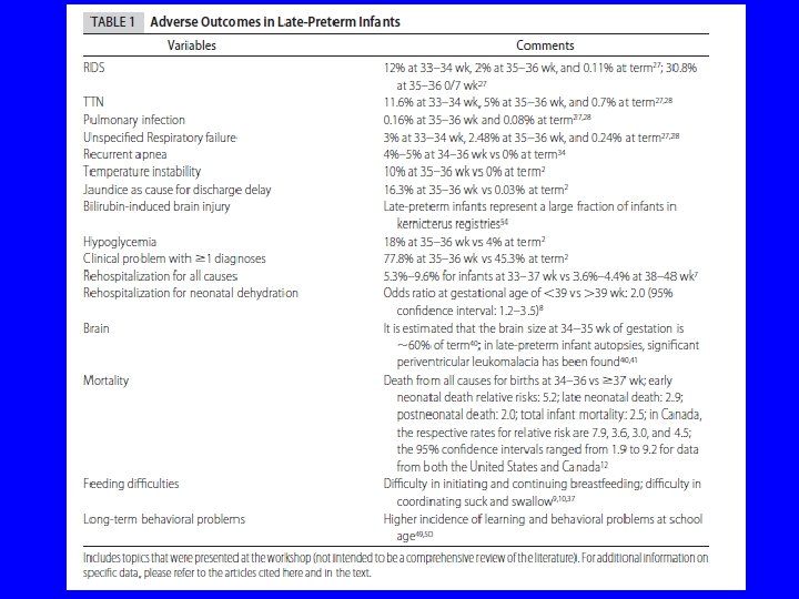
Late Preterm Infants
RSV prophylaxis guidelines (AAP 2009) • Who – All <32 w – All with CHD/CLD on medications – All ≥ 32 w to <35 w + 1 of 2 risk factors • When: Nov. 1 st to March 1 st except in FL • Number of doses – 5 for < 32 w – 3 or till 90 days old for ≥ 32 w to <35 w • Risk Factors: – Attends childcare – Siblings who are <5 y and stay in same household
Management of Hepatitis B Mat. status + unknown _ Newborn >2 kg Newborn <2 kg HBV 3 doses, 1 st<12 h HBIG 1 dose, <12 h 1 dose, <7 d HBV 4 doses, 1 st<12 h HBIG 1 dose, <12 h 3 doses, 1 st at 1 -2 M None
PDA • Normal course – physiological closure 12 -15 h , anatomic closure several months, – about 4% of term, 10% of 30 -37 wk and 50% of <30 wk do not close by 72 h and considered PDA • S/S– Term-asymptomatic, systolic or machinery murmur, bounding pulse, CHF – Preterm can also have ↓bf to the gut can cause NEC, pulmonary h’mge. and BPD • Treatment-Fluid restriction, maintaining hematocrit, ibuprofen (indomethacin), surgical correction
Case • You receive a telephone call from the mother of one of your patients, who tells you that she is 27 weeks pregnant and that her obstetrician has diagnosed a fetal arrhythmia. In discussion with the obstetrician, you learn that the fetal heart rate is 240 beats/min and that there is a 1: 1 relationship between the atrial and ventricular contraction. What advice would you give to the mother regarding the management of the baby after delivery?
Fetal arrhythmias • Bradyarrhythmias: Heart blocksfollowing clinically delivering if there is development of hydrops • Tachyarrythmias: Supra-ventricular Tachycardia-treated with giving digoxin to the mother
Some surgical procedures • • • Rashkind balloon septostomy: TGA Blalock Taussig Shunt: TOF Glenn procedure: TA/SV with PS Jantene: TGA Fontan: TA/SV Norwood: HLHS-Stage 1 -3
Norwood for HLHS • Stage 1 – Atrial septectomy – PA to Asc. aorta – BT shunt • Stage 2 – Glenn – Remove BT • Stage 3 – Fontan
Glycogen storage disease • 8 types, types 1, 2 3 are commonest • Type 1 –von Gierke (L, K, GI), Glucose 6 PO 4 ase def. , lactic acidosis, hepatomegaly, diarrhea, bleeding disorder, poor prognosis • Type 2 -Pompe (muscle, nerves), lysosomal glucosidase def. , muscle weakness, cardiomegaly, CHF, poor prognosis • Type 3 - Forbes (liver, muscle), low glucose, hepatomegaly, muscle fatigue, onset after neonatal period, good prognosis
MPS and Lipidoses • MPS- dysostosis multiplex, Alder Rielly bodies in WBC and urine MPS – Hurler-iduronidase def. , onset-1 y, cloudy cornea, HSM, coarse features, short stature, kyphosis – Hunters-iduronidase sulfatase def. , onset 1 -2 y, Xlinked, only MPS with retinal abn.
• Lipidoses – Gauchers-glucocerebrosidase def. , Gaucher cell in bone marrow, normal retina • type I-normal CNS, onset any age • type II-profound CNS involvement, onset 1 y – Niemann Pick-sphignomyelinase def. , foam cells in bone marrow • type A cherry red spot, profound CNS involvement, onset 1 m • type B normal retina, normal CNS, onset any age
Galactosemia • AR, galactokinase or Galactose-1 -PO 4 ase uridyltransferase def. • Presents when feeds are introduced as lethargy, hepatomegaly, liver failure, renal tubular acidosis • Can have cataract at birth • ↑ rsk of infection specially E. coli • Lab- ↑LFT’s, galactose in urine (reducing substance positive with negative glucose oxidase test ) • Rx- elimination of all galactose and lactose in diet
PKU and Homocystinuria • Classic PKU-AR, def. of phenylalanine hydroxylase, mousy/musty urine odor, severe MR and seizures if untreated, diagnosed by NBS, Rx by low phenylalanine diet • Homocystinuria-AR, def. of cystathianine synthetase, usually asymptomatic in neonatal period, has downward dislocated lens (DD Marfan’s), myopia, osteoporosis, scoliosis, arachnodactyly, ↓ joint mobility (DD Marfan’s), MR, seizures, thrombotic episodes, Rx ↓methionine, supplement cysteine, folate, pyridoxine
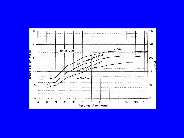
AAP guidelines for bili management • To measure bilirubin in hours of life • Aggressive phototherapy and specific follow up depending on the zone in the hourly bilirubin charts
Acknowledgement for the images • • Neonatology on web Google images Case Western Reserve University Trialsight medical media sample image from Google images • Maimonides Medical Center