Thyroid parathyroid Embryology Day 24 th gestation ass

Thyroid & parathyroid Embryology Day ~24 th gestation ass. prof dr. Asaad Mohammed kadhim Consultant General &laparoscopic Surgery
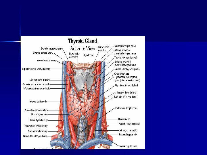

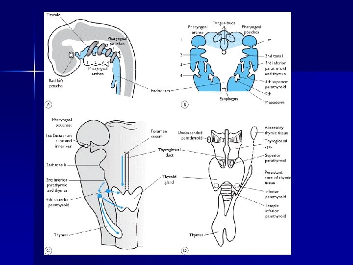
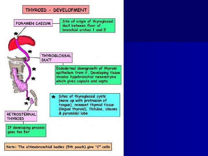
1 st endocrine gland n thyroid gland originates from between 1 st and 2 nd pharyngeal pouches n Proliferation of endodermal cells on midline floor of developing pharynx Between 2 key structures: n Initially develops 1. caudal to the “tuberculum impar” (median tongue bud) 2. ant to the copula n …-------------- called “foramen cecum” n Foramen cecum = opening of the thyroglossal duct into the tongue

initial thyroid precursor ----------- called “thyroid primordium” Starts as a simple midline thickening n forms thyroid diverticulum n initially hollow n later solidifies and becomes bilobed

Initial descent starts ant. to pharynx n Thyroid still connected to the tongue via the thyroglossal duct ( during gestational weeks 7 -10 tubular thyroglossal duct solidifies then obliterates entirely ) n Further descent – ant (ventral) to the hyoid bone and laryngeal cartilage n During descent – thyroid forms its mature shape: median isthmus connecting 2 lateral lobes n Descent COMPLETE at 7 th gestational week n

Pyramidal lobe: n in up to 50% people n persistence of the inferior end of the thyroglossal duct
 n From ultimobranchial body (last")
Parafollicular cells C cells n Secrete calcitonin (Ca regulation) n From ultimobranchial body (last structure derived from the branchial pouches) n Ultimobranchial body – from 5 th pharyngeal pouch n

Parafollicular cells n n Migrating cells from the neural crest region infiltrate ultimobranchial body (UBB) This structure (neural crest + UBB) = incorporated thyroid gland UBB fuses with the thyroid gland scattered through posterior&superior lobes in Summary: the C cells of the thyroid are of neural crest origin.

Thyroid Anomalies n Thyroglossal Sinuses Duct Cysts and – May form anywhere along the course followed by the thyroglossal duct – Most seen by 5 yr – Asymptomatic unless infected – Midline, painless, moveable neck mass

Ectopic Thyroid Gland – Lingual thyroid n Result of failure to descend n Often only thyroid tissue present – Accessory thyroid tissue n Tongue n Neck, superior or lateral to thyroid

PARATHYROID GLANDS

The upper parathyroids arise from the dorsal endoderm of the fourth branchial pouch, the ventral part of which is fused laterally to the developing thyroid gland on the floor of the primitive pharynx.

Thus some 92 per cent of the upper parathyroids remain in close contact with the posterolateral aspect of the thyroid lobes, just above and behind the level at which the recurrent laryngeal nerve crosses the inferior thyroid artery

When the upper parathyroid glands are ectopic they become progressively more dorsally displaced. As a result, the upper parathyroids may, in a small number of individuals, come to lie between the thyroid and oesophagus or behind the oesophagus in the upper posterior mediastinum

It has been suggested that parathyroid enlargements favour dorsal displacement due to the forces of deglutition and negative intrathoracic pressure. Aberrant superior glands may also be found rarely within the carotid sheath.

The lower parathyroid glands develop from the dorsal endoderm of the third branchial pouch, which also gives rise to the thymus. In contrast to the fourth-pouch derivatives, which remain fairly static during embryological development,

the structures developing in association with the third pouch undergo caudal migration, which is often excessive. The third pouch leapfrogs over the fourth and divides, leaving a discrete mass of parathyroid tissue on each side, usually within 2 cm of the lower pole of the thyroid

and the thymus as a bilobed structure in the anterior mediastinum overlying the great vessels. Excessive fragmentation produces thymic rests and accessory parathyroid glands. Failure to fragment results in intrathymic parathyroid tissue, which accounts for the location of 20 per cent of parathyroids, half of which are bilateral.

THANKS
- Slides: 23