Thymus gland Thymus gland In the medulla epithelioreticular


 Paracortical area is Formed of • T lymphocytes")


")

- Slides: 19
Thymus gland
Thymus gland
In the medulla, epithelioreticular cells form onionized structures called Hassall’s corpuscles –quite prevalent in older thymus function not very well known, but produce interleukins and so likely influence T-cell differentiation LM view
Thymus gland of adult The young thymus
Yellow bone marrow: inactive Red bone marrow: active.
Lymph Node Structure - Capsule & subcapsular sinus - Trabeculae & trabecular sinuses contain lymph, macrophages, and reticular cells - Cortex: • superficial cortex (B-cells) -primary follicles/nodules -secondary follicles/nodules (i. e. with germinal centers) • “deep” cortex (T-cells, dendritic cells) - Medulla: • medullary cords (B-cells, plasma cells) • medullary sinuses (lymph, more macrophages, plasma cells, and reticular cells)
Low power view of LN The outer part of the LN is highly cellular cortex, superficial (outer) cortex and paracortex (inner cortex) The inner part of the LN is less cellular medulla The cellular component of the LN which are T & B lymphocytes plasma cells and APCs are arranged into dense and loose arrangement. Dense cortical nodules and medullary cords Loose loosely scattered B lymphocytes, plasma cells, macrophages and lymph sinuses
Cortical nodules rounded aggregation of B lymphocytes Primary LN Small naïve B lymphocytes +macrophages Secondary LN Small naïve B lymphocytes Lymphoblast (Germinal center) + macrophage s
Secondary Lymphatic nodules When the lymph node is activated by antigen Small B lymphocytes Lymphoblast
Inner cortex ( thymus dependant area) Paracortical area is Formed of • T lymphocytes • Macrophages
Medulla formed of : 1. Medullary cord rich cells separated by medullary sinuses 2. medullary sinuses, large BV & supporting trabeculae 3. all are present in a framwork of reticular fiber Cells of medullary cord 1. plasma cell most common 2. Macrophages 3. Some B lymphocytes 1. Numerous Plasma cells 1. Marophages 2. B lymphocytes
Organization of the spleen: white pulp and red pulp White pulp: lymphatic aggregations around “central” arteries: periarterial lymphatic sheath (PALS): T-cells lymph nodules: B-cells Red pulp: cords and sinuses
As the body is exposed to antigens and the immune system mounts an immune response in the form of antibody production, lymph nodules (w/ germinal centers) appear in the white pulp of the spleen. U-M Histology Collection
Lymphoid Nodule of the spleen 1. Germinal Center+ mantle zone 2. Central artery (high endothelium)
PALS w/ secondary follicle Shown here with “central” artery cut in cross section –note that the CA has been pushed off to the side by the rapid expansion of cells in the germinal center (GC) RP= red pulp MZ= marginal zone (antigen presentation) dashed circle = T-cell rich zone
White & red pulps of spleen
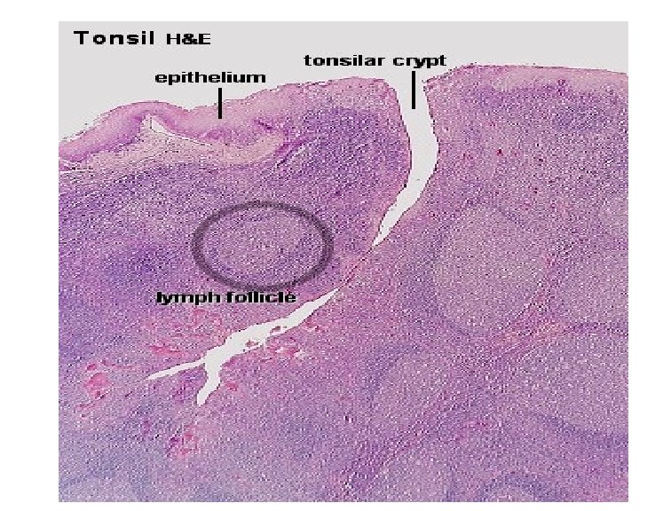
Palatine Tonsil (H&E)
Pharyngral tonsil Single lymphoid mass Site: Under mucous membrane of nasopharynx pseudostratified ciliated columnar epithelium No crypts but folds • • contain diffuse lymphoid tissue