Thrombotic Disorders Virchows triad Rudolph Virchow 1821 1902

Thrombotic Disorders

Virchow’s triad Rudolph Virchow, 1821 -1902 Vessel wall injury Hypercoagulability of blood Stasis of blood

Arterial Thrombosis - 1 • Interruption of blood flow causes ischemic necrosis of involved organ/limb • Vessel wall injury (eg, atheroma) initiates most events • Emboli may cause downstream small vessel infarction (ie. embolic stroke) • Examples: MI, stroke, limb ischemia/gangrene

Arterial Thrombosis - 2 • High shear in arterial circulation → platelets play greater role than thrombin/fibrin formation in initiating thrombosis Ø Antiplatelet agents preferred for prevention of arterial events in most cases Ø Inherited conditions that promote thrombin formation (inherited thrombophilia) are not major risk factors for arterial thrombosis
")
Arterial Thrombosis (From: Fishback, J. , Dept of Pathology, University of Kansas Medical Center)

Pathophysiology of Arterial Thrombosis • Atherosclerotic vessel disease is major risk factor • Atherosclerosis is a complex process associated with endothelial injury and inflammation

Atherogenesis is a complex process Arteriosclerosis, Thrombosis, and Vascular Biology 2008

Plaque Rupture and Arterial Thrombosis in Atheromatous Disease From Hansson, NEJM 2005

Risk Factors for Atherosclerosis • • Smoking Hypertension Hypercholesterolemia Diabetes/metabolic syndrome Family history Age Obesity Sedentary life style

Venous Thrombosis - 1 • • • Obstruction to venous outflow causes edema, swelling, pain, and inflammation in the area drained by the affected vein Most common in deep veins of the legs/pelvis, associated with venous stasis. Complicated by pulmonary embolism (PE), where thrombus breaks free and follows the venous return through the right side of the heart into the pulmonary arteries. Massive PE (typically from large proximal leg or pelvic veins) can cause hypoxemia, shock and death. Mortality rate from PE averages about 5%. Rarely, paradoxical embolus enters arterial circulation via patent ductus arteriosus or atrial septal defect causing arterial occlusion (stroke, limb ischemia, etc).

Venous Thrombosis - 2 • Low shear and stasis allows accumulation of activated clotting factors, so clots are fibrin-rich. Ø Drugs that slow or block thrombin generation preferred over antiplatelet drugs for prevention and treatment of venous thrombosis Ø Inherited or acquired conditions that promote thrombin formation increase risk of venous thrombosis
")
Deep Venous Thrombosis (image from NLM)
")
DVT causing leg ischemia (Phlegmasia cerulea dolens)

Pulmonary Embolism

Pulmonary Embolism
 Stasis of blood")
Vessel wall injury Hypercoagulability of blood AKA “Thrombophilia” (acquired or inherited) Stasis of blood

Some Causes of Venous Stasis • Surgery or major trauma • Cancer (vessel obstruction by tumor) • Immobilization (prolonged bedrest, paralysis, leg cast, long airplane trips, etc) • Obesity • Pregnancy • Congestive heart failure • Venous insufficiency or obstruction

Acquired Hypercoagulability - 1 • Age • Surgery or trauma • Cancer (and cancer chemotherapy) • Myeloproliferative disorders (e. g. , polycythemia vera) • Pregnancy and post-partum period • Estrogens (oral contraceptives, postmenopausal hormone replacement)

Acquired Hypercoagulability - 2 • Antiphospholipid syndrome • Heparin-induced thrombocytopenia
 Rises with Age Figure 2. Average annual sex-")
The Incidence of Venous Thromboembolism (VTE) Rises with Age Figure 2. Average annual sex- and age-specific incidence of venous thromboembolism among community residents of Olmsted County, Minnesota, from 1980 through 1990. (From: Heit, J MAYO CLIN PROC. 2001)
 Antiphospholipid antibodies (APA) • A general term for antibodies directed against")
Antiphospholipid Antibodies 1) Antiphospholipid antibodies (APA) • A general term for antibodies directed against a variety of epitopes present on phospholipid/protein complexes. – Examples: anticardiolipin Ab, anti-beta-2 glycoprotein I Ab • These are relatively common. Many patients asymptomatic, more likely to be associated with disease when present in high titers 2) Lupus anticoagulants (LAC) • Subset of antiphospholipid antibodies that inhibit in vitro coagulation. • Misleading name – many patients don’t have systemic lupus erythematosous (SLE), and these antibodies much more likely to be associated with arterial or venous thrombosis than bleeding. • Acts as an anticoagulant in vitro only – prolongs a. PTT and other phospholipid-dependent clotting tests.

Antiphospholipid Syndrome • Laboratory: persistently positive tests for antiphospholipid antibodies (LAC and other high titer APA) • Clinical manifestations: • Arterial and/or venous thrombosis • Recurrent fetal loss • Autoimmune thrombocytopenia or hemolytic anemia • Uncertain pathophysiology • May be associated with SLE, or with no associated condition (primary antiphospholipid syndrome). • Occasional “catastrophic” presentation with multiple organ failure • Some patients require lifelong anticoagulation

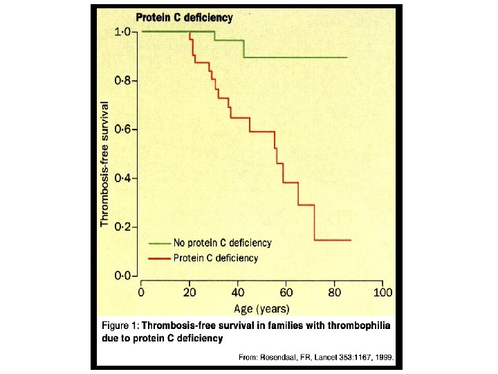
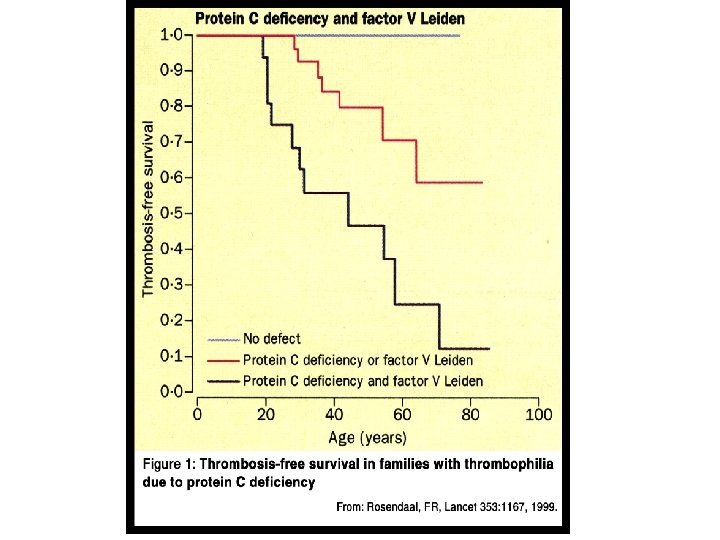
Genetic Risk Factors for VTE • Deficiency of major coagulation Inhibitors: uncommon, genetically heterogeneous conditions associated with up to 20 fold increased VTE risk in some families 1) Antithrombin Deficiency 2) Protein C Deficiency 3) Protein S Deficiency • Other conditions- more common but less risky (3 -5 fold increased VTE risk) 1) Factor V Leiden (~ 5% of US population) 2) Prothrombin G 20210 A polymorphism (~ 2%) Ø There are many other candidate genes! Risks associated with mutations in these are unknown

VTE is a multigenic disease • Gene defect ≠ disease! • Most people with single gene defects are asymptomatic • The most common known gene defects are only present in a minority of VTE cases. • FV Leiden is present in 20 -40% of VTE cases • It is likely that multiple gene defects contribute in most cases but can only test for a handful of these at present • Multiple defects interact, often synergistically. • Ascertainment bias profoundly affects the relative risk estimates for specific gene defects. • General population vs. thrombophilic family

Absolute vs Relative Risk

THE RELATIVE RISK OF VENOUS EVENTS IN RELATIVES OF PATIENTS WITH THROMBOPHILIA SEEMS HIGH… Vossen et al, J Thromb Haemost 2004; 2: 1526

BUT THE ABSOLUTE RISK IS LOW! Vossen et al, J Thrombos Haemost 2005; 3: 459 Bleeding risk with long term anticoagulation estimated at 1 -3%/year

VTE risk is determined by multiple inherited and acquired factors

Family history predicts thrombotic risk just as well as laboratory testing for thrombophilia A case-control study Fam Hx VTE % VTE pts % controls Odds Ratio for VTE (95% CI) Negative 68. 5 82. 7 1 Any relative 31. 5 17. 3 2. 2 (1. 9 -2. 6) Relative < 50 6. 7 2. 7 (2. 2 -3. 4) 1. 9 3. 9 (2. 7 -5. 7) 15. 0 > 1 Relative 6. 0 ARCH INTERN MED 2009; 169: 610

Thrombophilic mutations do not predict thrombotic risk well in the absence of a family hx of VTE Thromb Haemost 2011; 106: 646

Idiopathic VTE is more likely to recur than provoked VTE • Any event that occurs in the absence of a precipitating event or clinical risk factor for VTE • Associated with recurrence rates in the 20 -30% range • The occurrence of idiopathic VTE is a better predictor of recurrence than laboratory evaluation for “hypercoagulable states”

The risk of recurrent venous thromboembolism is higher in patients with idiopathic events Lancet 2003; 362: 523– 26 Idiopathic VTE Other risk factor Postop VTE

The risk of recurrent VTE is not strongly affected by the presence of inherited thrombophilia Lancet 2003; 362: 523– 26 Hazard ratio 1. 50 (95% CI = 0. 822. 77) p=0. 187

Diagnosis of Venous Thromboembolism • Symptoms & physical examination are not sufficiently sensitive or specific to diagnose VTE • D-dimer level- sensitive but not specific (negative test makes VTE unlikely) Ø Objective documentation of thrombus is essential to establishing the diagnosis

Objective Testing for VTE Deep Venous Thrombosis • Duplex Doppler ultrasonography • Contrast or MR venography Pulmonary Embolism • Spiral computed tomography (CT) • Ventilation-perfusion scan (V/Q scan) • MR angiography
 compressible - no DVT Vein not")
DVT - DOPPLER ULTRASOUND A V Vein (V) compressible - no DVT Vein not completely compressible - DVT

DVT - DOPPLER ULTRASOUND

DVT- MR vs Contrast Venography MR Contrast

Pulmonary Embolism-Spiral CT
 Normal High probability of PE Ventilation without perfusion")
Ventilation-Perfusion Scans (From: www. massgeneralimaging. org/newsletter/july_2003/) Normal High probability of PE Ventilation without perfusion suggests pulmonary embolus
- Slides: 42